Computer aided diagnostic system incorporating shape analysis for diagnosing malignant lung nodules

a computer-aided diagnostic system and shape analysis technology, applied in image analysis, image enhancement, instruments, etc., can solve the problems of reducing mortality rates, difficult to distinguish true nodules from shadows, and difficulty in computer-aided image data search schemes,

Active Publication Date: 2013-10-03
UNIV OF LOUISVILLE RES FOUND INC
View PDF9 Cites 31 Cited by
  • Summary
  • Abstract
  • Description
  • Claims
  • Application Information

AI Technical Summary

Problems solved by technology

Detecting pulmonary nodules, however, is a complicated task.
The difficulty for computer aided image data search schemes is distinguishing true nodules from (overlapping) shadows, vessels and ribs.
Regular chest radiography and sputum examination programs have proven ineffective in reducing mortality rates.
Although screening for lung cancer with chest X-rays can detect early lung cancer, such screening can also possibly produce many false-positive test results, causing needless extra tests.
Furthermore, while large (e.g., greater than 1 cm in diameter) malignant nodules are often relatively easy to detect with conventional screening equipment and can be diagnosed with needle biopsy or bronchoscopy techniques, these techniques are typically unsuitable for detecting smaller nodules, particularly if such nodules are located deep in the lung tissue or away from large airways.
As may be imagined, waiting a period of time to determine whether a lung nodule is malignant provides a possibly malignant nodule time to grow and become a larger health risk.
Thus, many of these techniques have been found to be unsuitable for early stage lung cancer detection.
However, these spherical, cylindrical, or circular assumptions are typically not adequate for describing the general geometry of the lesions.
The drawbacks to these approaches are the difficulties in detecting lung wall nodules.
Tracking temporal nodule behavior is a challenging task, however, because of changes in the patient's position at each data acquisition, as well as effects of heart beats and respiration.
In order to accurately measure how the nodules are developing in time, all of these motions need to be compensated by registering the LDCT data sets taken at different times. Several methods have been proposed for solving medical image registration problems and excluding the lung motions; however, the accuracy of nodule classification based on the growth rate still remains below clinical requirements.

Method used


Image

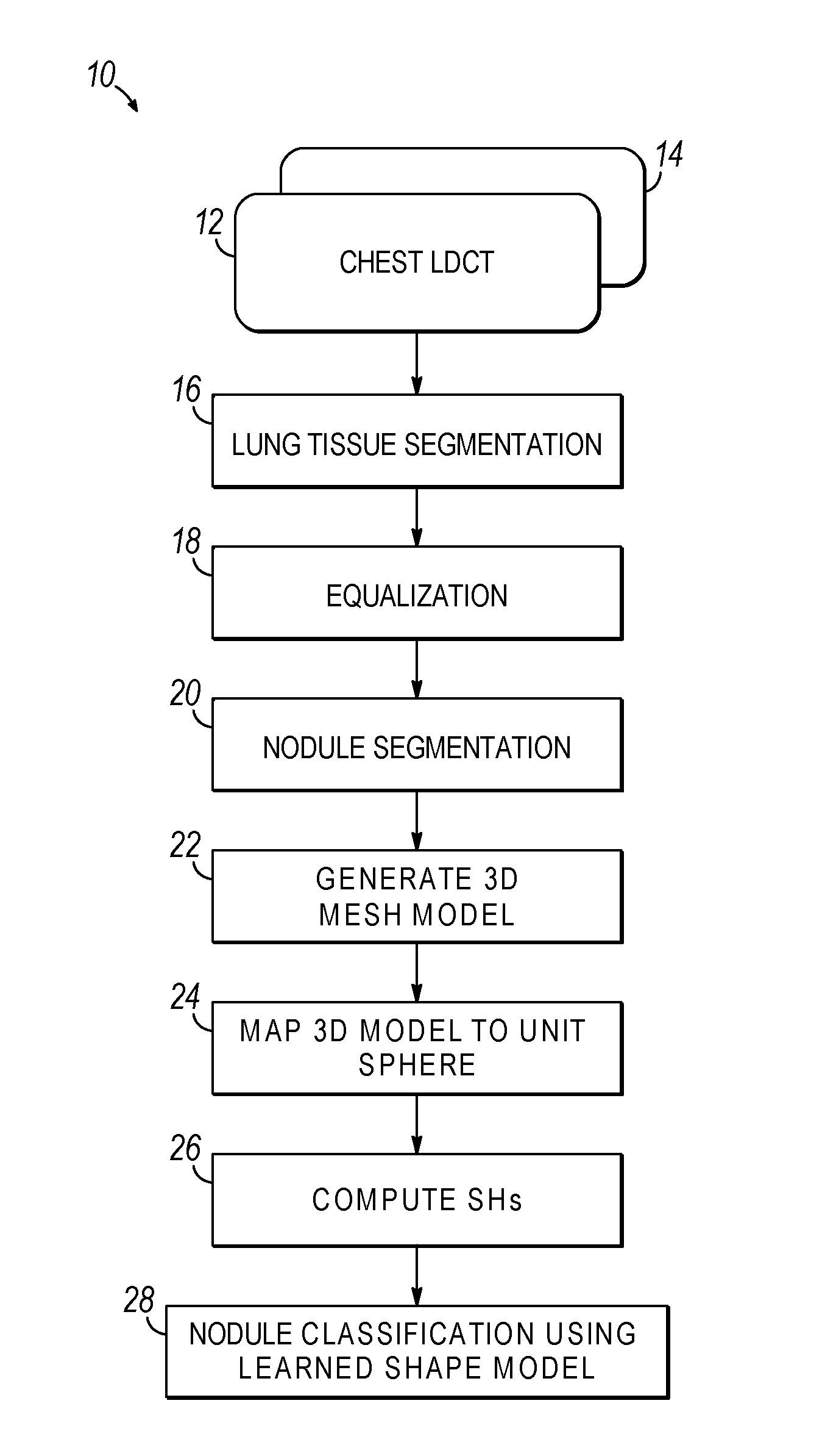
  • Computer aided diagnostic system incorporating shape analysis for diagnosing malignant lung nodules
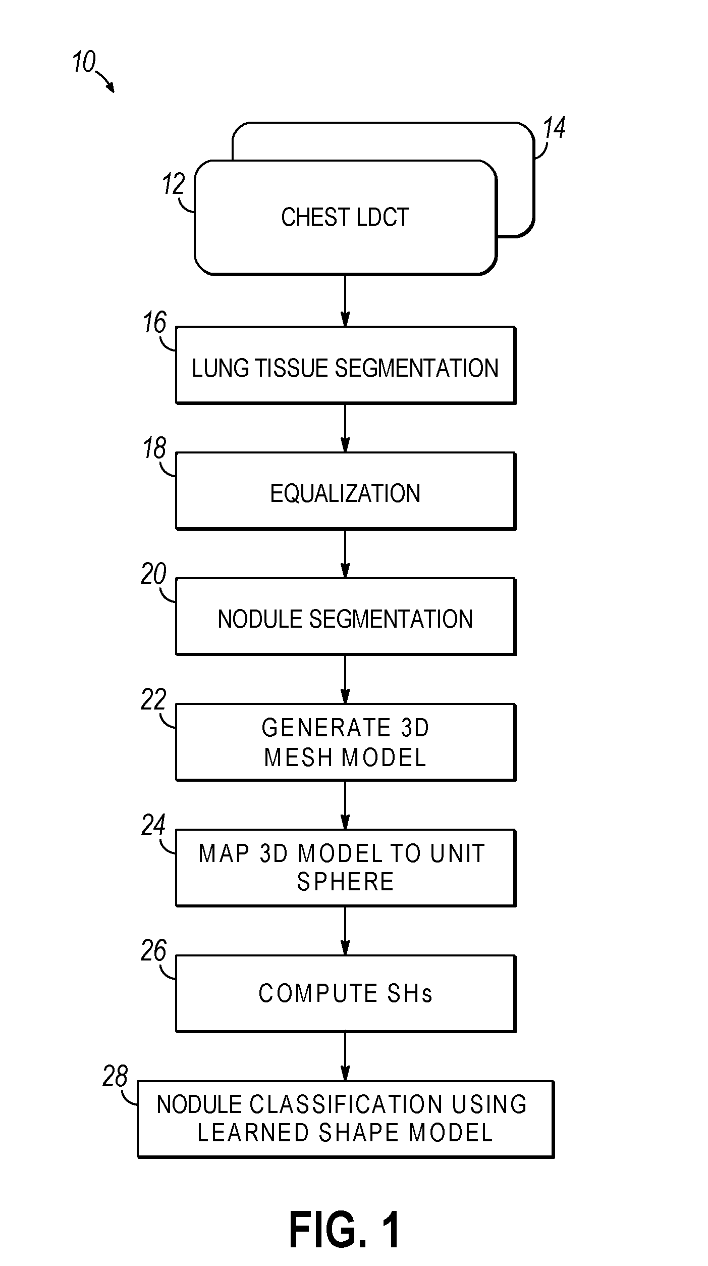
  • Computer aided diagnostic system incorporating shape analysis for diagnosing malignant lung nodules
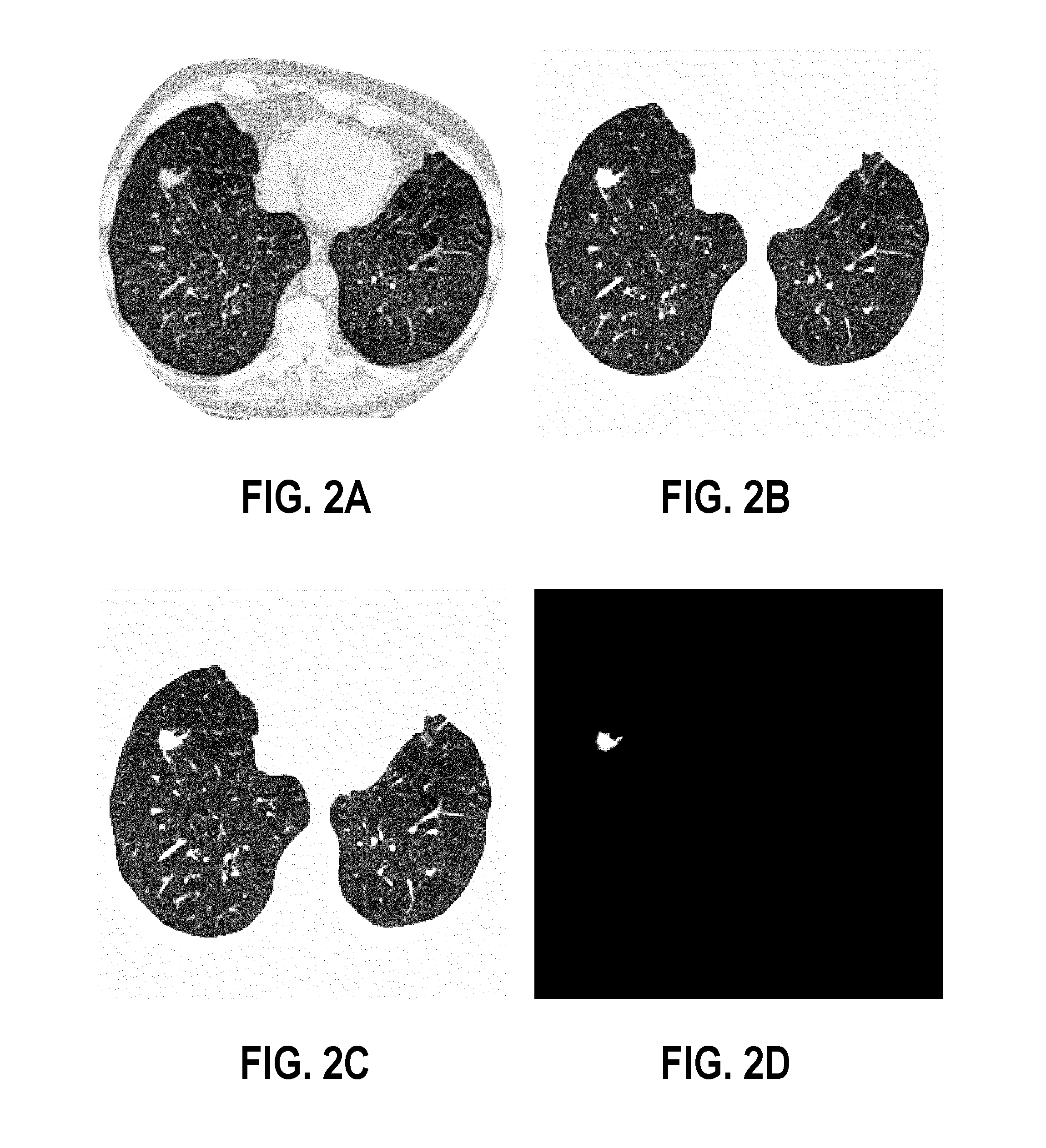
  • Computer aided diagnostic system incorporating shape analysis for diagnosing malignant lung nodules

Examples


working example

[0058]To justify the proposed methodology of analyzing the 3D shape of both malignant and benign nodules, the above proposed shape analysis framework was pilot-tested on a database of clinical multislice chest LDCT scans of 327 lung nodules (153 malignant and 174 benign). The CT data sets each have 0.7×0.7×2.0 mm3 voxels, with nodule diameters ranging from 3 mm to 30 mm. Note that these 327 nodules were diagnosed using a biopsy (the ground truth).

[0059]FIGS. 8A-L illustrate results of segmenting pleural attached nodules shown by axial (FIGS. 8A-D), sagittal (FIGS. 8E-H) and coronal (FIGS. 8I-L) cross sections. The pixel wise Gibbs energies in each cross section are higher for the nodules than for any other lung voxels including the attached artery. Therefore, the approach separates accurately the pulmonary nodules from any part of the attached artery. The evolution terminates after 50 iterations because the changes in the total energy become close to zero. The error of segmentation ...


PUM


Abstract

A computer aided diagnostic system and automated method diagnose lung cancer through modeling and analyzing the shape of pulmonary nodules. A model used in such analysis describes the shape of pulmonary nodules in terms of spherical harmonics required to delineate a unit sphere corresponding to the pulmonary nodule to a model of the pulmonary nodule.

Description

CROSS-REFERENCE TO RELATED APPLICATIONS[0001]This application claims the benefit of U.S. Provisional Application No. 61 / 617,865, filed on Mar. 30, 2012 by Ayman S. El-Baz et al., the entire disclosure of which is incorporated by reference herein.FIELD OF THE INVENTION[0002]The invention is generally related to computer analysis of medical image data, and in particular to the detection of malignant lung nodules in medial image data, e.g., low dose computed tomography (LDCT) chest scans.BACKGROUND OF THE INVENTION[0003]Lung cancer remains the leading cause of mortality cancer. In 1999, there were approximately 170,000 new cases of lung cancer in the U.S., where approximately one in every eighteen women and approximately one in every twelve men develop lung cancer. Lung tumors (visible on chest film as pulmonary nodules) are the most common manifestation of lung cancer being the principal cause of cancer-related deaths. A pulmonary nodule is an approximately spherical volume of higher ...

Claims


Application Information

Patent Timeline
Patent Type & Authority Applications(United States)
IPC IPC(8): G06T7/00
CPCG06T7/0012G06T2207/30064G06T2207/20081G06T2207/10081
Inventor EL-BAZ, AYMAN S.NITZKEN, MATTHEW
Owner UNIV OF LOUISVILLE RES FOUND INC
Why Eureka
Try Eureka