Pulmonary nodule edge rebuilding and partitioning method based on computed tomography (CT) image

A technology for CT images and pulmonary nodules, which is applied in the field of image processing, can solve the problems that restrict the segmentation of pulmonary nodules, and achieve the effects of overcoming segmentation difficulties, getting rid of speckle noise, and strengthening edge detection

Inactive Publication Date: 2013-04-10
CHANGCHUN UNIV OF TECH
View PDF2 Cites 30 Cited by
  • Summary
  • Abstract
  • Description
  • Claims
  • Application Information

AI Technical Summary

Problems solved by technology

Since these missing signals do not completely disappear, but are reduced, so to solve the problem of restricting the segmentation of pulmonary nodules, this part of the weak edge signal can be amplified

Method used


Image

  • Pulmonary nodule edge rebuilding and partitioning method based on computed tomography (CT) image
  • Pulmonary nodule edge rebuilding and partitioning method based on computed tomography (CT) image

Examples


Embodiment Construction

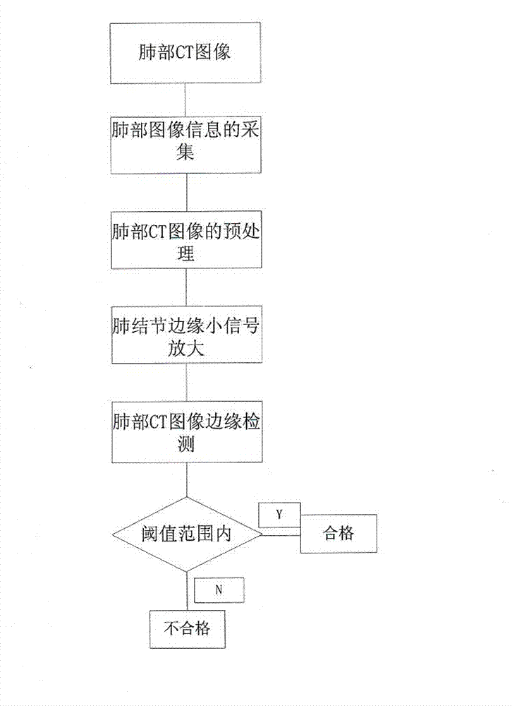
[0041] like figure 1 Shown, the steps of the present invention are as follows:

[0042] (1) Data collection: it can load and display multiple data formats at the same time; the user graphical interface consists of five parts: toolbar, graphical interface group, 3D display window, 3D and slice operation controller;

[0043] (2) Image preprocessing:

[0044] 1. Image Registration

[0045] Due to differences in image shooting time and external objective conditions, each image has its own scope of application and limitations. Image registration in multiple modes can give full play to the characteristics of the image itself and complement information. It is the basis of image fusion and prerequisites;

[0046] 2. Image Fusion

[0047] The images of the same target collected by multi-source channels are processed through image processing, and the information of each channel is extracted, and finally synthesized into the same image for observation or further processing;

[0048...


PUM


Abstract

The invention discloses a pulmonary nodule edge rebuilding and partitioning method based on a computed tomography (CT) image. According to the pulmonary nodule edge rebuilding and partitioning method, the image is subjected to spatial transformation by using a transformation method which has a sparse representation ability on gradient characteristics; a high energy transformation coefficient is reserved through shrinkage of a transformation domain; the image is rebuilt through inverse transformation to realize strengthening of the gradient characteristics; and amplification of small signals of the gradient characteristics is realized through multistage strengthening of the signals, a pulmonary nodule edge is rebuilt, and important edge information is provided for subsequent partitioning. The pulmonary nodule edge rebuilding and partitioning method provides a clustering-based pulmonary nodule partitioning algorithm, does not have the process of a training classifier, has a self-training ability, and can be used for strengthening edge detection, overcoming partitioning difficulty caused by uneven gray levels, and eliminating influence by speckle noise. The pulmonary nodule edge rebuilding and partitioning method can also be used for establishing a CT image partitioning algorithm evaluation system and combining contours drawn manually by different clinical medical experts into optimum partitioning standards so that the partitioning algorithm can be compared systematically, and the effectiveness of the partitioning algorithm can be revealed.

Description

technical field [0001] The invention relates to an image processing method, in particular to a method for reconstructing and segmenting the edge of pulmonary nodules based on CT images. Background technique [0002] In recent years, the morbidity and mortality of lung cancer in all countries in the world have continued to rise, especially in industrial cities with high population density, and the number of lung cancer patients in my country also ranks first in the world. According to the statistics of the World Health Organization (WHO), lung cancer ranks first in the cause of cancer death in men, and ranks second in female tumors after breast cancer. An important measure to reduce the mortality of lung cancer patients is early diagnosis and early treatment. [0003] Imaging diagnostic methods are mainly used to diagnose early lung cancer, including X-ray diagnosis and CT diagnosis. CT diagnosis is superior to X-ray diagnosis in detecting lung cancer, and can detect tumors...

Claims


Application Information

Patent Timeline
Patent Type & Authority Applications(China)
IPC IPC(8): G06T7/00
Inventor 邵向鑫林晓梅田野商婷婷
Owner CHANGCHUN UNIV OF TECH
Why Eureka
Try Eureka