Supervoxel sequence lung image 3D pulmonary nodule segmentation method based on multimodal data

A technology of sequential images and pulmonary nodules, applied in the field of medical image processing, can solve problems such as relatively mature solutions for three-dimensional images of lung lesions

Active Publication Date: 2017-10-03
TAIYUAN UNIV OF TECH
View PDF0 Cites 32 Cited by
  • Summary
  • Abstract
  • Description
  • Claims
  • Application Information

AI Technical Summary

Problems solved by technology

At present, with the maturity of 3D reconstruction technology, its application in clinical diagnosis and treatment is becoming more an

Method used


Image

  • Supervoxel sequence lung image 3D pulmonary nodule segmentation method based on multimodal data
  • Supervoxel sequence lung image 3D pulmonary nodule segmentation method based on multimodal data
  • Supervoxel sequence lung image 3D pulmonary nodule segmentation method based on multimodal data

Examples


Embodiment Construction

[0090] The present invention will be described in detail below in conjunction with specific embodiments.

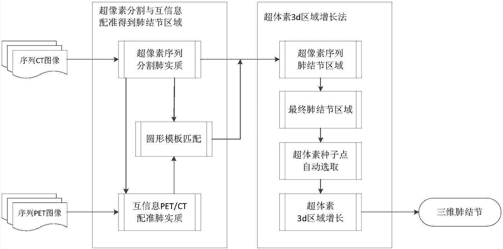
[0091] refer to figure 1 , the main process includes: superpixel segmentation sequence lung parenchyma, mutual information registration multimodal PET / CT sequence lung parenchyma data, variable circular template matching sequence lung nodule area, supervoxel 3D area growth and other steps, the present invention The specific implementation of the method is as follows:

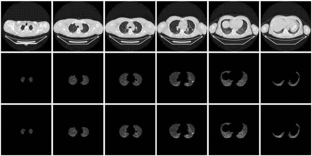
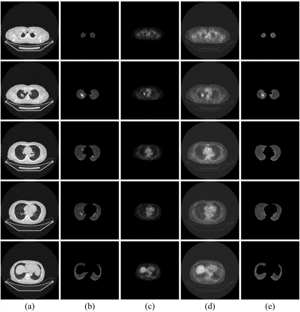
[0092] A. Superpixel segmentation sequence lung parenchyma: use the superpixel sequence image segmentation algorithm to obtain the superpixel samples of the ROI sequence image, then use the self-generated neural forest algorithm to cluster the superpixel samples, and finally according to the clustered superpixel set The grayscale feature and position feature identify the nodular lung parenchyma area, and prepare for the accurate extraction, segmentation, and three-dimensional reconstruction of pulmonary no...


PUM


Abstract

The invention discloses a supervoxel sequence lung image 3D pulmonary nodule segmentation method based on multimodal data. The method comprises the following steps: A) extracting sequence lung parenchyma images through superpixel segmentation and self-generating neuronal forest clustering; B) registering the sequence lung parenchyma images through PET/CT multimodal data based on mutual information; C) marking and extracting an accurate sequence pulmonary nodule region through a multi-scale variable circular template matching algorithm; and D) carrying out three-dimensional reconstruction on the sequence pulmonary nodule images through a supervoxel 3D region growth algorithm to obtain a final three-dimensional shape of pulmonary nodules. The method forms a 3D reconstruction area of the pulmonary nodules through the supervoxel 3D region growth algorithm, and can reflect dynamic relation between pulmonary lesions and surrounding tissues, so that features of shape, size and appearance of the pulmonary nodules as well as adhesion conditions of the pulmonary nodules with surrounding pleura or blood vessels can be known visually.

Description

technical field [0001] The invention belongs to the field of medical image processing, and relates to segmentation and three-dimensional reconstruction of pulmonary nodules, in particular to a method for supervoxel three-dimensional segmentation and reconstruction of pulmonary nodules based on multimodal data of medical PET and CT images. Background technique [0002] Three-dimensional reconstruction can reflect the dynamic relationship between lung lesions and surrounding tissues, and it is convenient to intuitively understand the shape, size, appearance and other forms of pulmonary nodules and the pulling situation with the surrounding pleura or blood vessels, which is helpful for doctors to intuitively understand the condition It can also guide the operation of complex anatomical areas to improve the quality of surgery and reduce the risk of surgery, which is worthy of further clinical application. The current clinical application of medical imaging mostly produces two-di...

Claims


Application Information

Patent Timeline
IPC IPC(8): G06T7/00G06T7/12G06T7/33
CPCG06T7/0014G06T2207/30064G06T7/12G06T7/337
Inventor 强彦崔强杨晓兰强薇赵涓涓王华
Owner TAIYUAN UNIV OF TECH
Why Eureka
Try Eureka