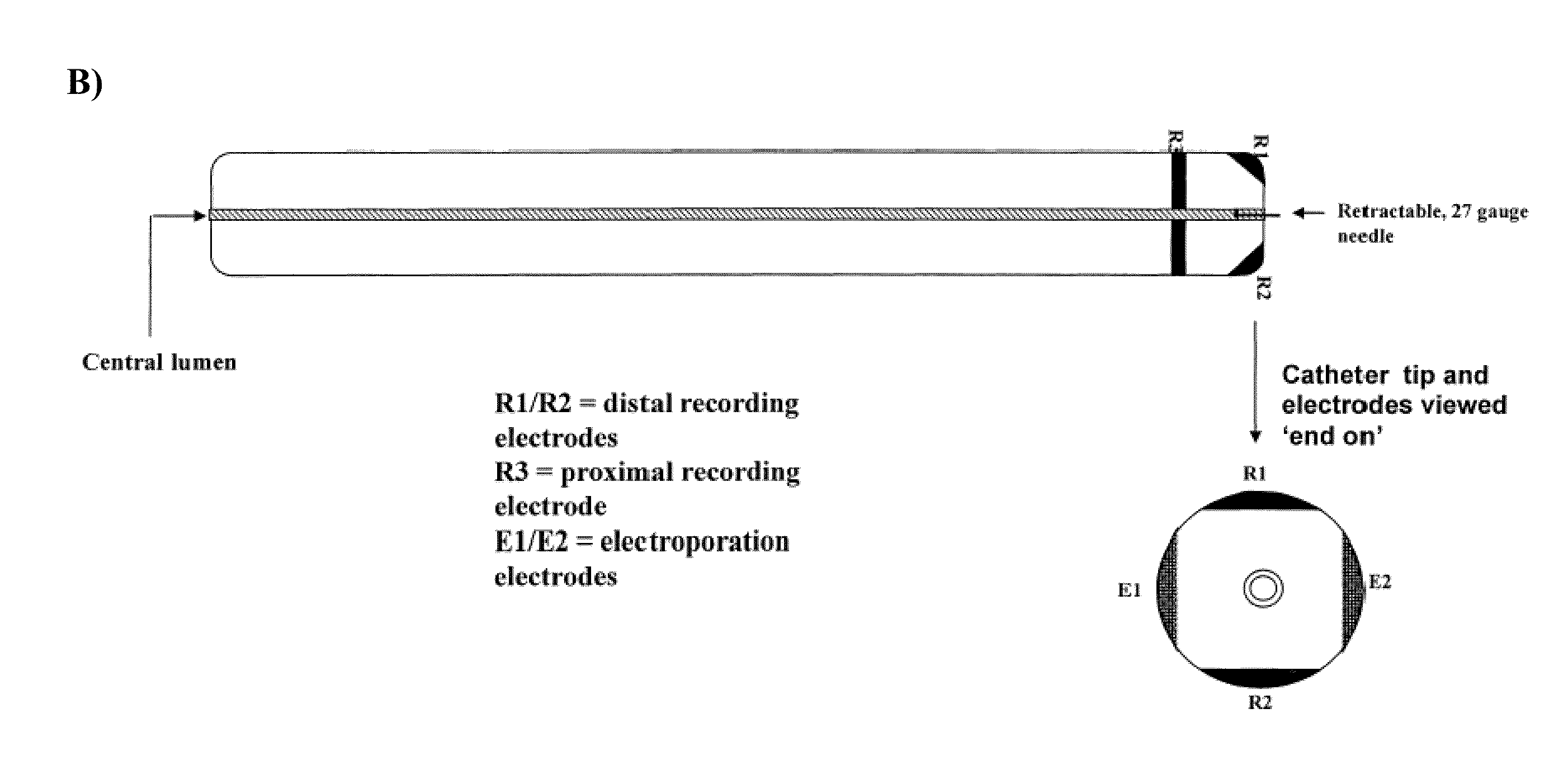
[0007]In some embodiments, the present invention provides a device comprising: (a) an elongate member with an inner lumen configured for delivery of a therapeutic agent to a treatment site within a subject, (b) an electroporation element configured to deliver electric current to the treatment site within a subject, and (c) an electrophysiology monitoring element configured to monitor electrical signals (e.g. at or around the treatment site within said subject, such as the heart). In some embodiments, the present invention provides a device comprising: (a) an elongate member with an inner lumen configured for delivery of a therapeutic agent to a treatment site within a subject, and (b) an electroporation element configured to deliver electric current to the treatment site within a subject. In some embodiments, the present invention provides a device comprising: (a) an elongate member with an inner lumen configured for delivery of a therapeutic agent to a treatment site within a subject, and (b) an electrophysiology monitoring element configured to monitor and / or record electrical signals. In some embodiments, the present invention provides a device comprising: (a) an electrophysiology monitoring element configured to monitor electrical signals and (b) an electroporation element configured to deliver electric current to the treatment site within a subject. In some embodiments, the electroporation element is located at the distal tip of the device (e.g., at or near the distal end of the elongate member). In some embodiments, the electroporation element comprises a plurality of electroporation electrodes (e.g., which may be at or near the end of the elongate member). In some embodiments, the electrophysiology monitoring element comprises a plurality of recording electrodes. In some embodiments, the plurality of monitoring electrodes comprises one or more distal monitoring electrodes and one or more proximal monitoring electrodes. In some embodiments, the device further comprises a handle located at the proximal end of the device. In some embodiments, the handle comprises one or more control elements. In some embodiments, the handle comprises one or more injection ports in fluid communication with the inner lumen. In some embodiments, the injection ports are configured for the loading therapeutic agents into the inner lumen. In some embodiments, a device comprises an inflatable and deflatable balloon element located at the distal tip of the elongate member. In some embodiments, the electroporation element is located on the balloon element. In some embodiments, the electroporation element comprises piezoelectric crystals configured to generate ultrasound energy. In some embodiments, the electroporation element comprises electrodes mounted and / or housed in and / or on the balloon element. In some embodiments, the electrophysiology monitoring element is located in and / or on said balloon element.
[0008]In some embodiments, the present invention provides a method of treating a disease or condition in a subject comprising: (a) inserting a catheter into the subject and placing the distal end of the catheter at or near a treatment site, (b) delivering a therapeutic agent to the treatment site through the lumen of the catheter, and (c) electroporating the treatment site with electrodes located on the distal end of the catheter (e.g., such that cells at the treatment site are transfected with reagents delivered via the catheter). In some embodiments, the method further comprises an initial step of monitoring electrical signals at the treatment site with an electrophysiology monitoring element. In some embodiments, the method further comprises (d) monitoring electrical signals at the treatment site with an electrophysiology monitoring element. In some embodiments, the method further comprises (e) comparing electrical signals from the initial step with electrical signals of step (d). In some embodiments, the method further comprises (f) determining the effectiveness of the treating based on comparison of the electrical signals from the initial step with electrical signals of step (d). In some embodiments, the therapeutic agent comprises gene therapy reagents. In some embodiments, the gene therapy reagents comprise nucleic acids (e.g., plasmids or AAV vectors comprising a gene of interest). In some embodiments, the nucleic acids comprise DNA. In some embodiments, the DNA comprises one or more mini-genes.
[0009]In some embodiments, the present invention comprises a system comprising: (a) an elongate member comprising an inner lumen, wherein said inner lumen is configured for delivery of a therapeutic agent to a treatment site within a subject, (b) an electroporation element, wherein said electroporation element is configured to deliver electric current to said treatment site within a subject, and (c) an electrophysiology monitoring element, wherein said electrophysiology monitoring element is configured to monitor electrical signals (e.g., in an around the treatment site in order to guide the device). In some embodiments, a system provides an elongate member with an inner lumen and electroporation element. In some embodiments, a system comprises an elongate member with an inner lumen and an electrophysiology monitoring element. In some embodiments, a system comprises an electroporation element and an electrophysiology monitoring element. In some embodiments, the electroporation element is located at the distal tip of the system. In some embodiments, the electroporation element comprises a plurality of electroporation electrodes. In some embodiments, the electrophysiology recording element comprises a plurality of monitoring electrodes. In some embodiments, the plurality of monitoring electrodes comprises one or more distal monitoring electrodes and one or more proximal monitoring electrodes. In some embodiments, the system further comprises a handle located at the proximal end of the system. In some embodiments, the handle comprises one or more control elements. In some embodiments, the handle comprises one or more injection ports in fluid communication with the inner lumen. In some embodiments, the injection ports are configured for loading therapeutic agents into the inner lumen.
