Enhanced animal cell growth using ultrasound

a technology of animal cells and ultrasound, applied in the field of increasing animal cell growth, can solve the problems of taking scientists many years of trial and error to learn to derive and maintain stem cells in the laboratory, and stem cells cannot work with their neighbors to pump blood through the body, etc., and achieve the effect of increasing the rate of cell proliferation

Active Publication Date: 2011-11-10
HIDACA
View PDF4 Cites 5 Cited by
  • Summary
  • Abstract
  • Description
  • Claims
  • Application Information

AI Technical Summary

Benefits of technology

[0019]The present invention provides a method of increasing the rate of cell proliferation in animal cells grown in cell culture, by stimulating them with calibrated ultrasound.
[0020]In one aspect, the invention comprises a method of enhancing the rate of cell growth in an animal cell culture through exposure to ultrasound of specified frequencies and intensities. These methods are beneficial to cells in the vast majority of environments, creating turbulence on the microscopic scale in the area immediately adjacent to the walls of the cells and other solid surfaces.
[0021]The ultrasound may have a frequency between about 1 MHz and about 2 MHz, depending inter alia on the species and type of cell used in culture. In one embodiment, the ultrasound has a frequency between about 1.4 MHz to about 1.6 MHz. In one embodiment, it consists of a pulsed ultrasound, which assists in minimizing temperature increase of the environment. In one embodiment, pulses generated at a duty cycle of approximately 4:1 (off:on), with a pulse period of approximately 1 second, are effective. In one embodiment, the ultrasound is calibrated to achieve a balance of the harmful effects of “collapse cavitations” caused by the ultrasound and the beneficial turbulence it affords the cells, allowing increased nutrient uptake and metabolic byproduct expulsion.
[0023]In another aspect, the invention comprises a method of correcting the emitted output to maximize the effectiveness of the ultrasound, based on the sensed intensities, if said sensor is employed. This assists in maximizing cell growth rates.
[0025]In one embodiment, the ultrasound can be directed such that reflections and interference are minimized, or tuned to give maximum effectiveness to the ultrasonic emission.

Problems solved by technology

Although many simpler proteins can be produced using rDNA in bacterial cultures, more complex proteins that are glycosylated (carbohydrate-modified) currently must be made in animal cells.
It has taken scientists many years of trial and error to learn to derive and maintain stem cells in the laboratory without them spontaneously differentiating into specific cell types.
For example, a stem cell cannot work with its neighbors to pump blood through the body (like a heart muscle cell), and it cannot carry oxygen molecules through the bloodstream (like a red blood cell).
However, unspecialized stem cells can give rise to specialized cells, including heart muscle cells, blood cells, or nerve cells.
Today, donated organs and tissues are often used to replace ailing or destroyed tissue, but the need for transplantable tissues and organs far outweighs the available supply.
In some cases, this sound wave is powerful enough to collapse the cavities, which causes even more extreme turbulence, high temperatures, and free radicals in the vicinity of the former cavity.
This can strip harmful bacteria off of a surface, and even kill a large number of them.
However, the method provides no solution for the use of ultrasound outside the confines of a wound.
Unfortunately, though increased cell growth is beneficial to the fermentation process, the parameters investigated by this group do not provide the optimal rate of protein expression in fermentation processes.

Method used


Image

  • Enhanced animal cell growth using ultrasound
  • Enhanced animal cell growth using ultrasound
  • Enhanced animal cell growth using ultrasound

Examples


example 1

Isolation of CD34+ Hematopoietic Stem / Progenitor Cells (HSPC)

[0057]Leukophoresis product (LP) was obtained with the patients' informed consent (in accordance with the institutional guidelines approved by the Human Research Ethics Board of the University of Alberta).

[0058]LP was used to isolate CD34+ hematopietic stem / progenitor cells (HSPC) using immunomagnetic beads (Miltenyi-Biotec, Auburn, Calif.) according to the manufacturer's instructions. Briefly, light-density mononuclear cells were separated by density gradient centrifugation over 60 percent Percoll (GE Healthcare, Quebec, Canada), labeled with a CD34+ antibody (QBEND 10) and passed through a positive selection column. The purity of isolated CB CD34+ cells was more than 92% as determined by fluorescence-activated cell sorter (FACS) analysis.

example 2

Stimulation with Low-Intensity Pulsed Ultrasound (LIPUS)

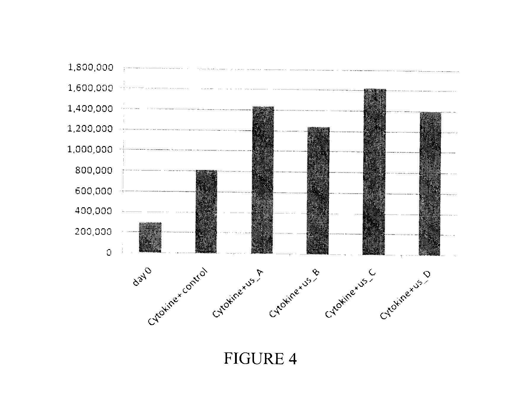
[0059]All cells were then maintained in Iscove's modified Dulbecco's medium (IMDM, GibcoBRL, Long Island, N.Y., USA) supplemented with 20% bovine growth serum (BGS; Hyclone, Logan, Utah, USA) and combination of cytokines (SCF: 100 ug / ml, TPO: 50 ug / ml, Flt3-ligand: 50 ug / ml) for four days as described below.

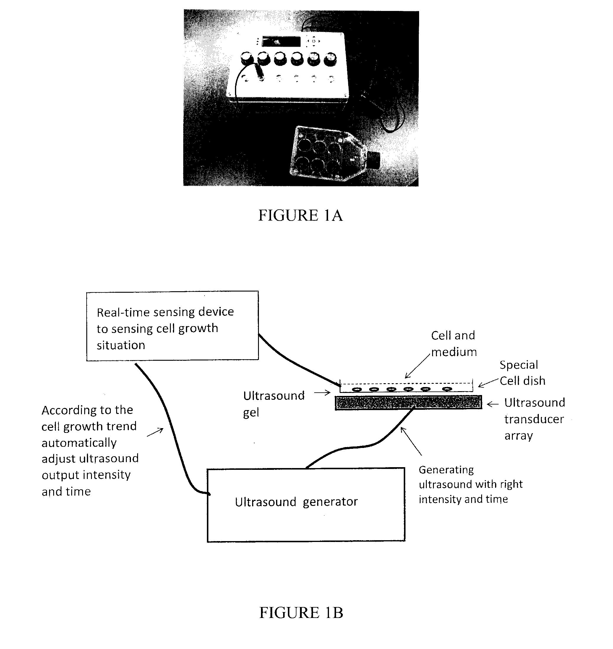
[0060]To stimulate the cultured cells with low-intensity pulsed ultrasound (LIPUS), a device producing a 1.5-MHz ultrasound wave, 20% duty cycle, with adjustable output intensity between 30 mW / cm2 to 100 mW / cm2 was used To enable cultured cells to be treated by ultrasound, four ultrasound transducers were fitted on a plastic frame and connected to the control panel of the signal generator via four independent cables. The cells (3×10⁵) were seeded in 12-well plates for all the experiments. The operation of the transducers was checked before each experiment. The plates were placed on ultrasound transducers using a coupling gel...

example 3

Cell Proliferation and Viability

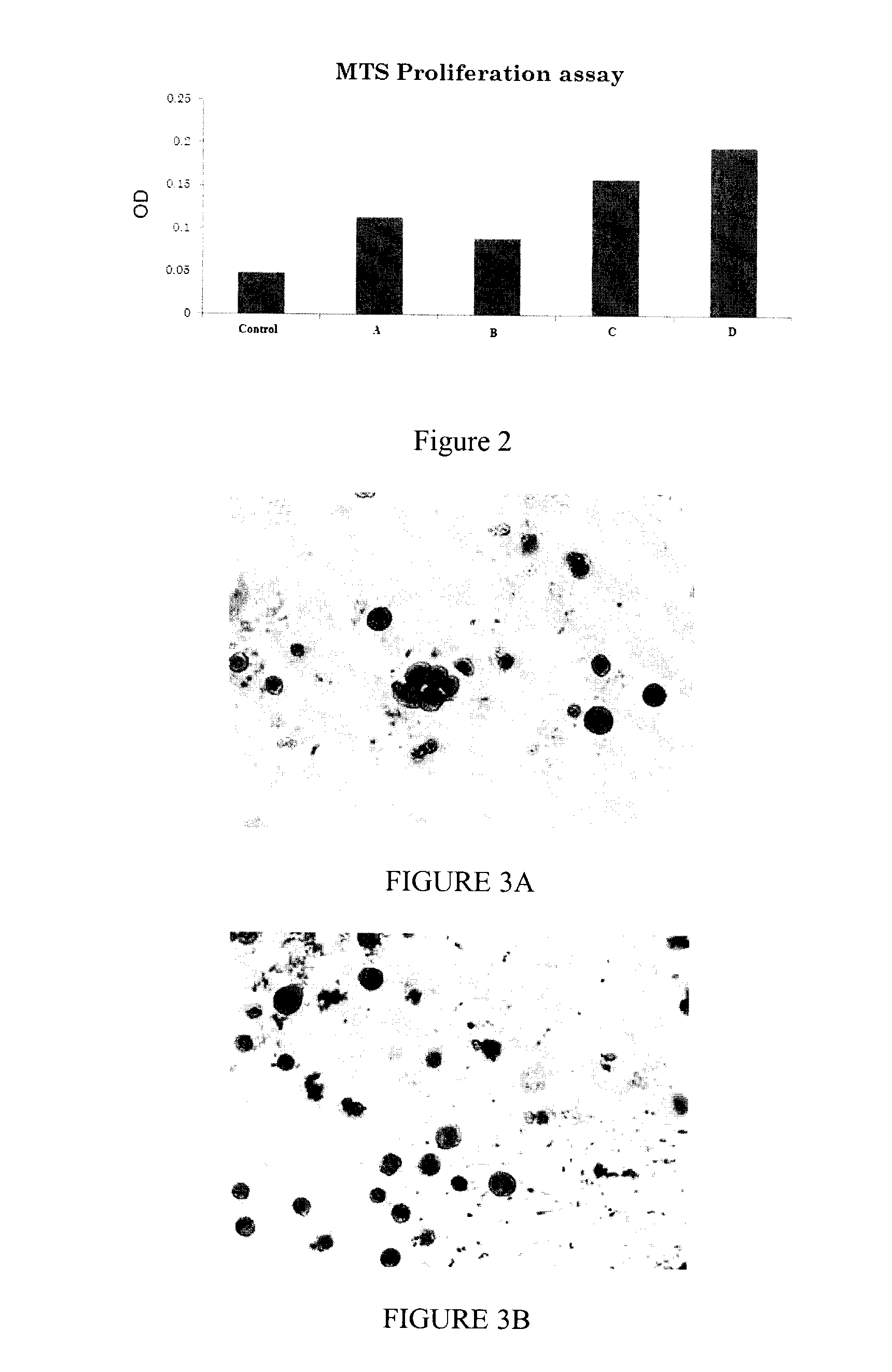
[0061]Cell proliferation and viability was assessed at day 5 by trypan blue exclusion and 3-(4,5-dimethylthiazol-2-yl)-5-(3carboxymethoxyphenyl)-2-(4-sulfophenyl)-2H-tetrazolium (MTS) cytotoxicity assay, using CellTiter 96 AQueous One Solution (Promega, Madison, Wis.). Briefly, 3×10⁵ cells per well were stimulated by LIPUS in combination with a cytokine cocktail (SCF: 100 ug / ml, TPO:50 ug / ml, Flt3-ligand: 50 ug / ml) for four days. For trypan blue exclusion assay, cells were resuspended in culture medium were stained by addition an equal volume of 0.4% trypan blue (Sigma, St. Louis, Mo.) in PBS and counted using a Neubauer haemocytometer.

[0062]In order to perform MTS assay, CellTiter 96 AQueous One Solution (20 μl per 100 μl medium) was added into each well, and cells were incubated at 37° C. for 1-4 h. The absorbance (490 nm) was measured, and cell proliferation ratios were calculated as the ratio of absorbance in treated cells versus that in untreated...


PUM

PropertyMeasurementUnit
frequencyaaaaaaaaaa
frequencyaaaaaaaaaa
frequencyaaaaaaaaaa

Abstract

A method of increasing the rate of growth of an animal cell or cell culture include the use of ultrasound at a frequency greater than about 1 MHz.

Description

CROSS REFERENCE TO RELATED APPLICATIONS[0001]This application claims the priority benefit of U.S. Provisional Application No. 61 / 091,830 filed on Aug. 26, 2008 entitled “METHOD TO INCREASE THE RATE OF PROTEIN EXPRESSION IN CELL”, the contents of which are incorporated herein by reference.FIELD OF THE INVENTION[0002]The present method relates to methods of increasing animal cell growth in cell culture by exposing the culture to ultrasonic stimulation.BACKGROUND OF THE INVENTION[0003]It is commonplace to grow animal cells in cell culture. Mass culture of animal cell lines is fundamental to the manufacture of viral vaccines and many products of biotechnology. Biological products produced by recombinant DNA (rDNA) technology in animal cell cultures include enzymes, synthetic hormones, immunobiologicals (monoclonal antibodies, interleukins, lymphokines), and anticancer agents. Although many simpler proteins can be produced using rDNA in bacterial cultures, more complex proteins that are ...

Claims


Application Information

Patent Timeline
Patent Type & Authority Applications(United States)
IPC IPC(8): C12Q3/00C12N13/00
CPCC12N5/0647C12M35/04C12N13/00
Inventor CHEN, JIEXING, JAMESANG, WOON T.GUL, HILAL
Owner HIDACA
Why Eureka
Try Eureka