
Device for tissue extraction

Active Publication Date: 2012-01-26
SIEMENS HEALTHCARE GMBH
View PDF22 Cites 2 Cited by
  • Summary
  • Abstract
  • Description
  • Claims
  • Application Information

AI Technical Summary

Benefits of technology

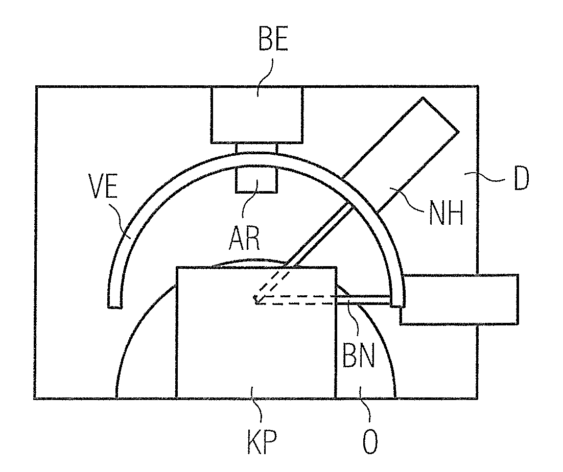
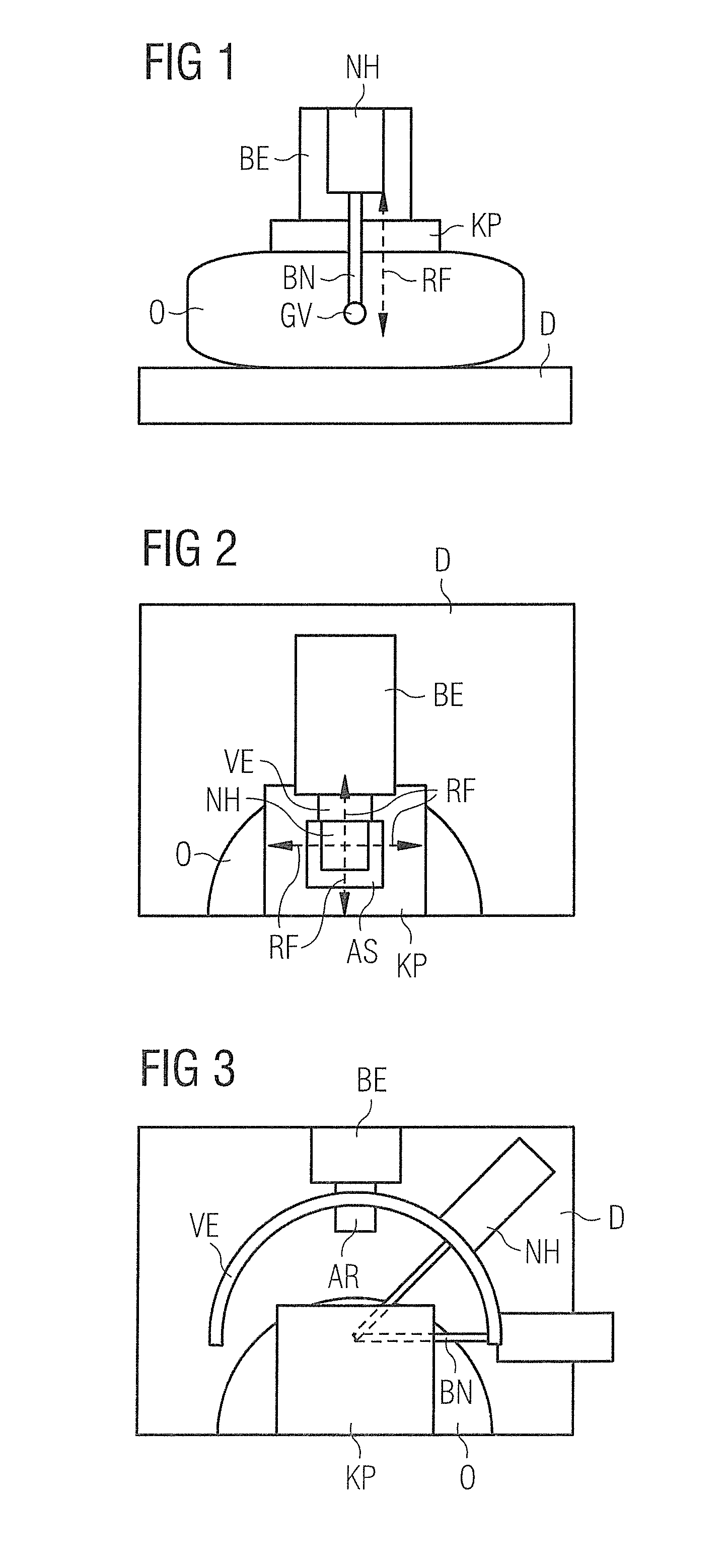
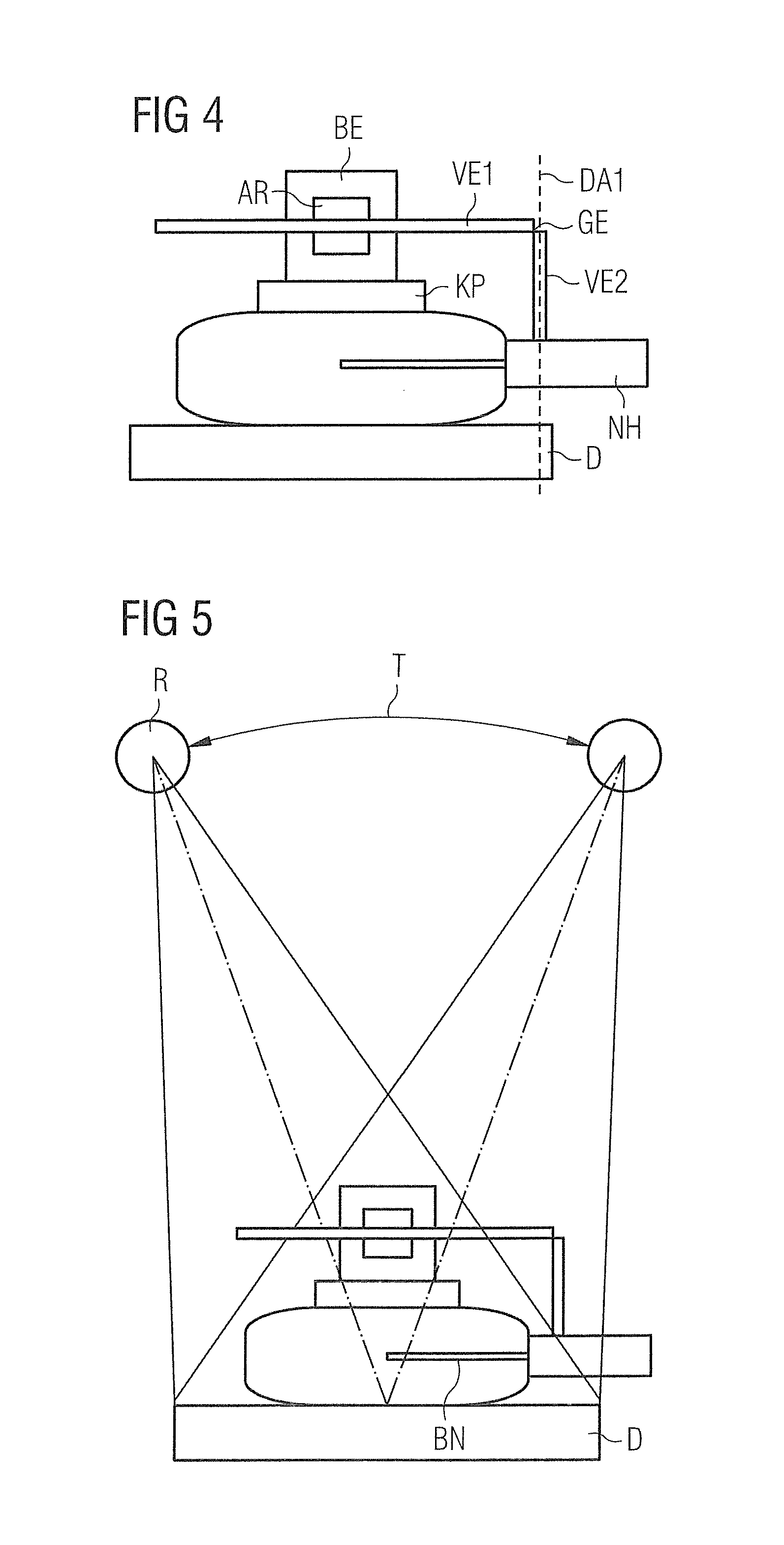
[0006]This device for tissue extraction has an alignment unit, a needle mount accommodating at least one biopsy needle and a connection unit. The connection unit is fashioned such that the needle mount can be arranged to the side of a subject. The connection unit is connected via an alignment arm with the alignment unit and can be positioned by means of this, among other things. The connection unit can be displaced vertically and / or horizontally with the alignment arm of the alignment unit. In one embodiment, the connection unit can be fashioned as one piece. The alignment unit is arranged in a boundary region of the detector unit. The needle mount for tissue extraction can be arranged along a circle segment around the subject to be biopsied. The needle mount can be aligned in that the biopsy needle integrated into this can be arbitrarily introduced into a subject.
[0007]The device has the advantage that a needle mount accommodating at least one biopsy needle for tissue extraction can be aligned via a connection element such that no regions to be examined are occluded by this during an x-ray acquisition.
[0008]The invention has the advantage that out-of-plane artifacts are avoided.
[0009]The invention also the advantage that the presentation of the breast tissue is clear and distinct.
[0010]The invention has the further advantage that the extraction of the tissue sample takes place with minimization of the penetration depth in the subject.

Problems solved by technology

However, the arrangement entails the disadvantage that parts of the biopsy unit occlude x-ray image information and generate additional artifacts, known as out-of-plane artifacts.

Method used


Image

  • Device for tissue extraction
  • Device for tissue extraction
  • Device for tissue extraction

Examples


Embodiment Construction

[0016]In the device for tissue extraction according to the invention, the needle mount accommodating the biopsy needle can be placed via a connection element such that no regions to be examined are occluded by parts of the device during an x-ray acquisition.

[0017]FIGS. 1 and 2 show a known biopsy unit BE. With this biopsy unit the biopsy needle is introduced vertically into a breast. The biopsy unit BE is thereby arranged directly above the subject O to be examined. In this known embodiment the subject O is compressed and fixed between compression plate KP and surface of a detector D. For example, this subject O can be the breast of a patient. The breast O remains captive from an overview acquisition until the extraction of the tissue in the compression unit of the mammography apparatus. A biopsy needle BN is attached in a biopsy needle mount NH, and this is connected via a connection unit VE with an alignment arm of an alignment unit BE. The biopsy needle tip BN is aligned on a loc...


PUM


Abstract

In a device for tissue extraction from a breast, a needle mount accommodating a biopsy needle can be placed by a connection element such that no regions of the breast that are to be examined by x-rays are occluded by parts of the device during an x-ray image acquisition.

Description

BACKGROUND OF THE INVENTION[0001]1. Field of the Invention[0002]The present invention relates to devices for extracting a tissue sample from a subject, such as for implementing a biopsy of a breast.[0003]2. Description of the Prior Art[0004]In a biopsy a tissue sample is extracted—for example from the tissue of a breast of a patient—for histological examination. The extraction of the tissue sample can take place with the assistance of imaging methods. An overview volume set of the breast is created with a tomosynthesis method. X-ray images of the breast are acquired to create the overview volume set. For x-ray image acquisition, an x-ray tube is moved (for example in an orbit) over a detector. For example, in a tomosynthesis procedure the x-ray tube moves in an angle range from +25° to −25°. An x-ray radiation in the x-ray source is then triggered at regular intervals, and the respective x-ray image is read out and buffered by the detector. An overview volume set is subsequently cre...

Claims


Application Information

Patent Timeline
IPC IPC(8): A61B10/02
CPCA61B10/0233A61B2019/205A61B17/3403A61B90/17
Inventor FISCHER, DANIEL
Owner SIEMENS HEALTHCARE GMBH
Learn More