
Method to configure an imaging device

a technology for imaging devices and configuration methods, applied in measurement devices, medical practices/guidelines, instruments, etc., can solve the problems of time-consuming and error-prone manual conversion, complicated and extensive settings of cited imaging devices, etc., to achieve automatic conversion of protocols, reduce manual post-processing, and improve efficiency and quality of conversion

Inactive Publication Date: 2012-02-16
SIEMENS HEALTHCARE GMBH
View PDF15 Cites 23 Cited by
  • Summary
  • Abstract
  • Description
  • Claims
  • Application Information

AI Technical Summary

Benefits of technology

The invention provides a method for configuring an imaging device by using a database that contains variants of protocols for the operation of imaging devices and types of imaging devices. The computer uses a data-driven learning procedure to determine relationships between the parameters and create a protocol plan for a new imaging device with a target type. This method improves the automatic conversion of protocols, reducing the need for manual post-processing and achieving a higher consistency of the protocols among one another and between product families and individual configurations. Overall, the invention reduces time, cost, and error in the conversion of protocols for new imaging devices.

Problems solved by technology

The cited imaging devices require complex and extensive settings in order to acquire images in a desired quality or with desired properties with regard to resolution, contrast, section, size, etc.
Due to the high number of protocols, this manual conversion is time-consuming and error-prone.

Method used


Image

  • Method to configure an imaging device
  • Method to configure an imaging device
  • Method to configure an imaging device

Examples


Embodiment Construction

[0023]Among other things, the following exemplary embodiments refer to magnetic resonance tomography and the respective protocols necessary therefor. However, the invention is not limited to magnetic resonance tomography; but can be applied to arbitrarily different imaging devices that must be configured by protocols.

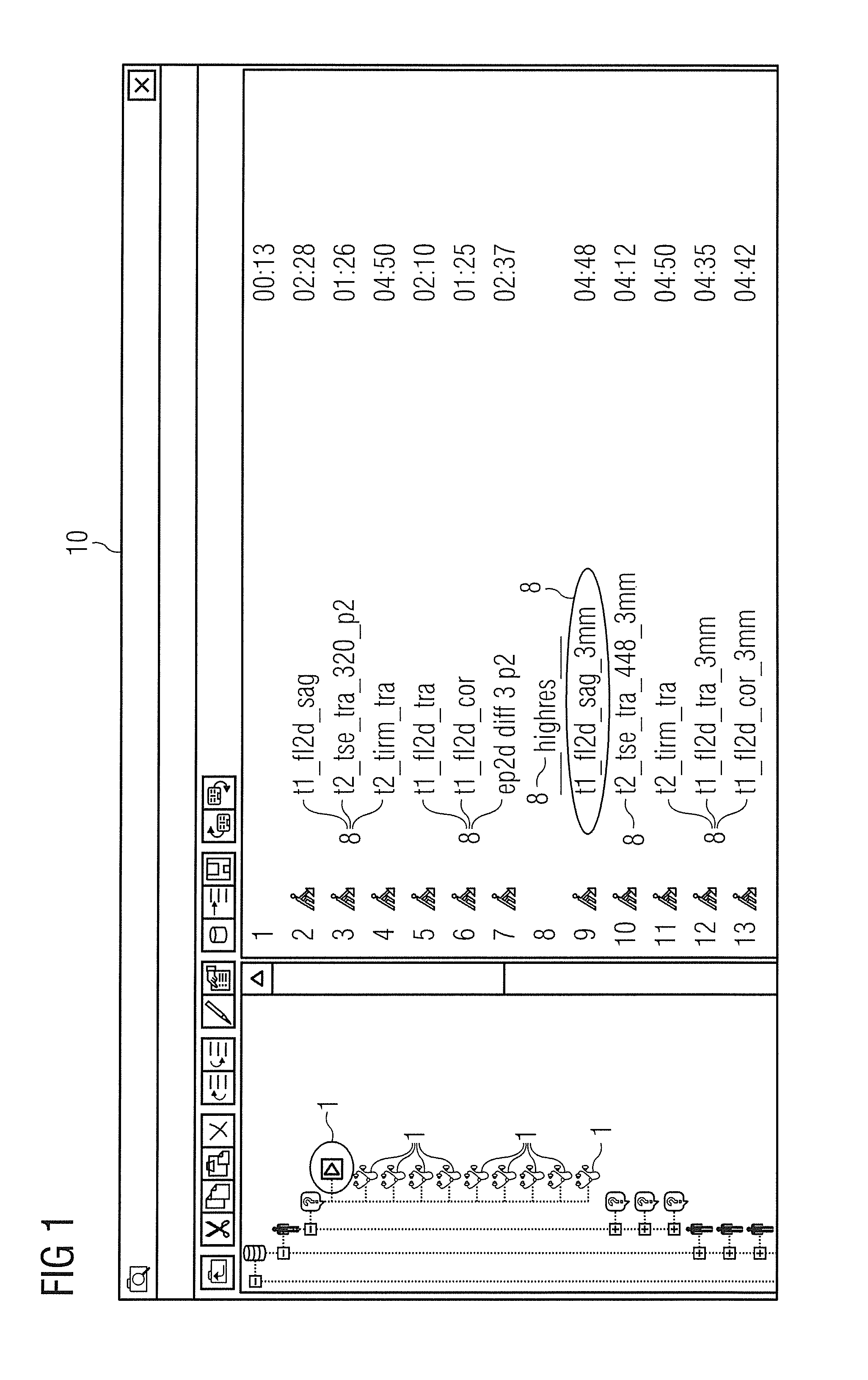
[0024]FIG. 1 shows a user interface 10 as is known for processing of protocols to configure imaging devices. Protocol categories 1 in a tree structure are shown in a left portion of the user interface 10. In the right portion of the user interface 10, protocols 2 are shown which are contained in the protocol category 1 marked by an ellipse in the left portion. For example, the protocols 2 respectively define a sequence for image acquisition by a magnetic resonance tomograph whose duration is likewise indicated in FIG. 1.

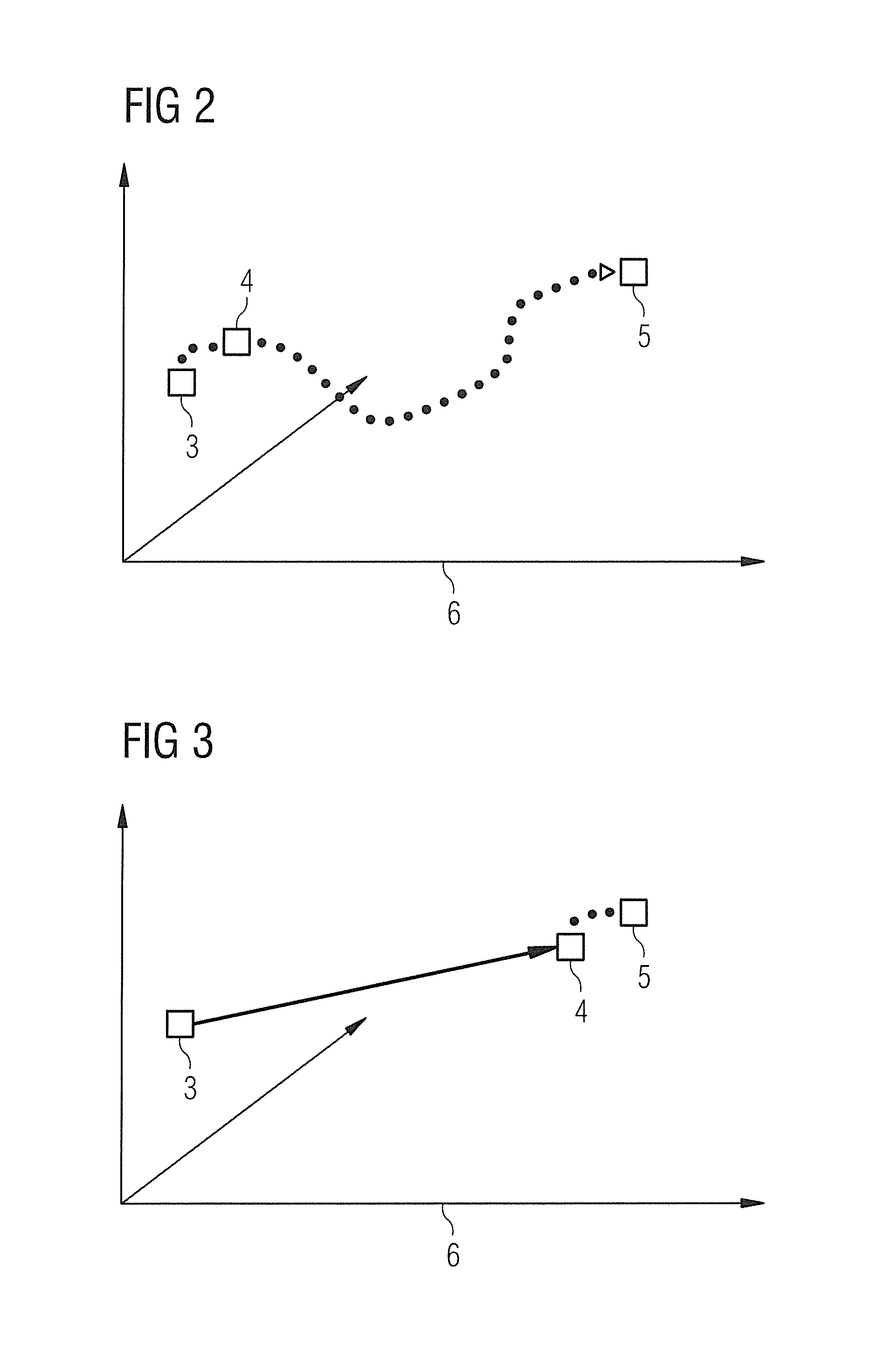
[0025]FIG. 2 shows a simple method for configuration of an imaging device with a new type. A source parameter vector 3 is provided in a parameter space ...


PUM


Abstract

A database contains variants of protocols for the operation of magnetic resonance tomographs as well as different types of magnetic resonance tomographs. Each variant contains parameter values and is associated with one of the types. In a training phase, relationships are determined between the parameters among one another and / or between the parameters and the associated types and are stored as patterns in a knowledge base. A protocol plan for the operation of a new magnetic resonance tomograph is created later in an application phase using the determined pattern. The method offers the advantage that the efficiency and quality of the automatic conversion of the protocols is improved. The improved quality of the protocol plan reduces operating time and costs for a manual post-processing of the protocols. Furthermore, a higher consistency of the protocols among one another is achieved both between product families and between individual configurations.

Description

BACKGROUND OF THE INVENTION[0001]1. Field of the Invention[0002]The present invention concerns a method for configuring an imaging device, as well as a computer programmed to implement such a method, and a non-transitory computer-readable storage medium encoded with programming instructions that cause a computer to implement such a method, when the storage medium is loaded into the computer.[0003]2. Description of the Prior Art[0004]Different imaging devices—for example magnetic resonance tomography systems, computed tomography systems, ultrasound apparatuses and x-ray systems—are known from medical technology. In order to achieve an uncomplicated use of these complex systems, it is necessary to configure the imaging devices. A number of measurement instructions and other settings that are specific to the respective imaging device (but possibly also to a specific use case) must be established for this purpose. The cited imaging devices require complex and extensive settings in order...

Claims


Application Information

Patent Timeline
Patent Type & Authority Applications(United States)
IPC IPC(8): G06F15/18
CPCG01R33/543G06F19/325G01R33/546G16H30/20G16H70/20
Inventor GLASER-SEIDNITZER, KARLHEINZHAUPTMANN, WERNERKIEFER, BERTHOLDOTTE, CLEMENS
Owner SIEMENS HEALTHCARE GMBH
Learn More