Method and system for analysing tissue from images

a tissue analysis and image technology, applied in image data processing, instruments, character and pattern recognition, etc., can solve the problems of inherently unreliable approaches, errors in breast density of 2-3%, and error correction performed via breast thickness estimation might not be correcting for error in the correct way

Active Publication Date: 2012-07-26
HIGHNAM RALPH +3
View PDF5 Cites 22 Cited by
  • Summary
  • Abstract
  • Description
  • Claims
  • Application Information

AI Technical Summary

Benefits of technology

[0080]In a further embodiment, the method and system described herein is applied repeatedly to images gathered over a period of time, thus enabling automatic temporal compar...

Problems solved by technology

However, errors and unknown values in the image physics make such approaches inherently unreliable.
Problematically, film mammograms rarely have breast thickness recorded and this is why an estimate from the image is usually made for film images.
In fact, academic work (Highnam& Brady 1999, Blot 2005) has suggested that errors of 1 mm in the breast thickness can lead to errors in breast density of 2-3%.
Although successful in generating valid hint values and reasonable breast densities against visual assessment, the approach has several drawbacks, especially for digital images where a recorded breast thickness is known from the digital machine itself.
Thus the error correction performed via estimation of breast thickness might not be correcting ...

Method used


Image

  • Method and system for analysing tissue from images
  • Method and system for analysing tissue from images
  • Method and system for analysing tissue from images

Examples


Embodiment Construction

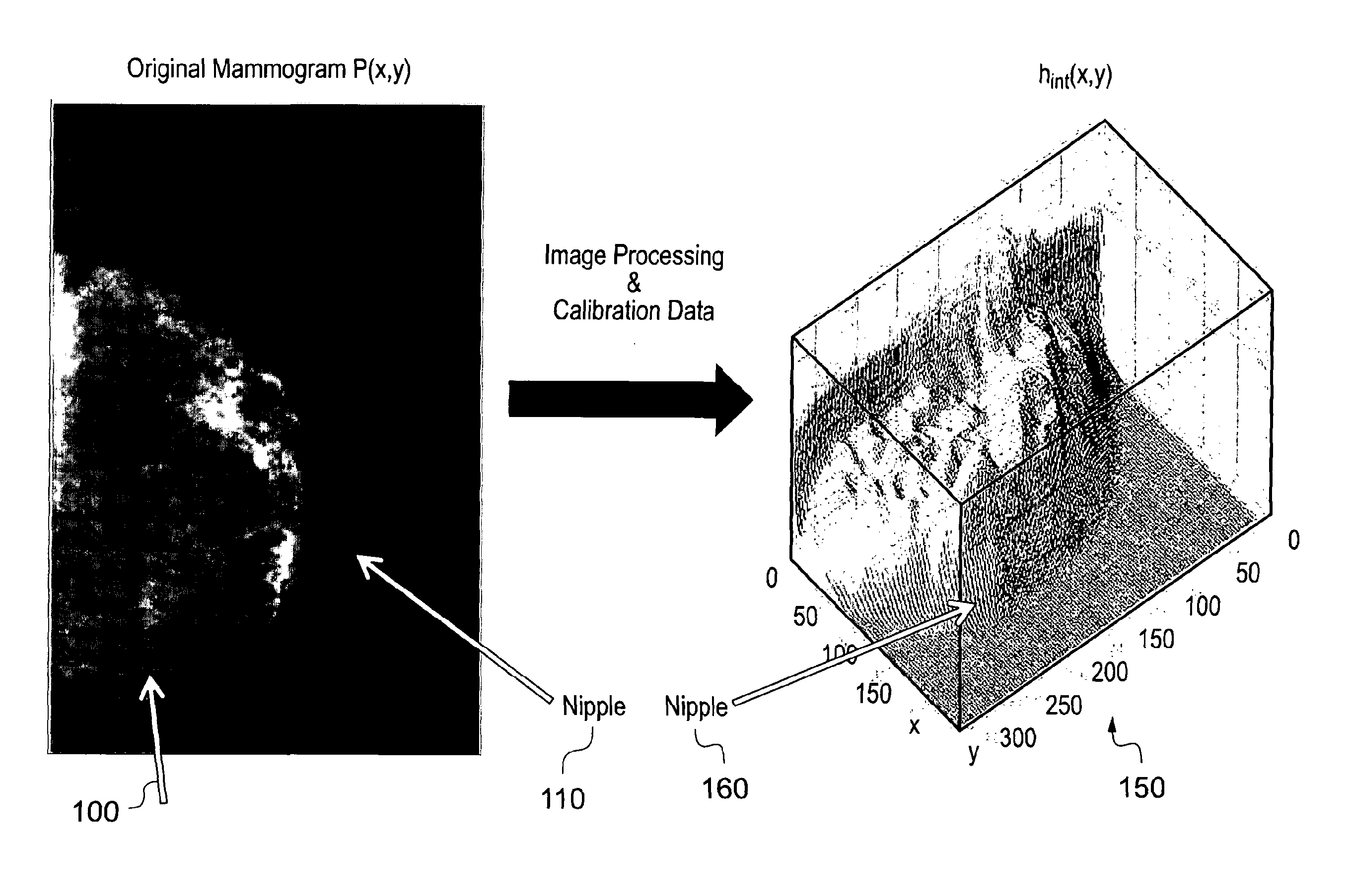
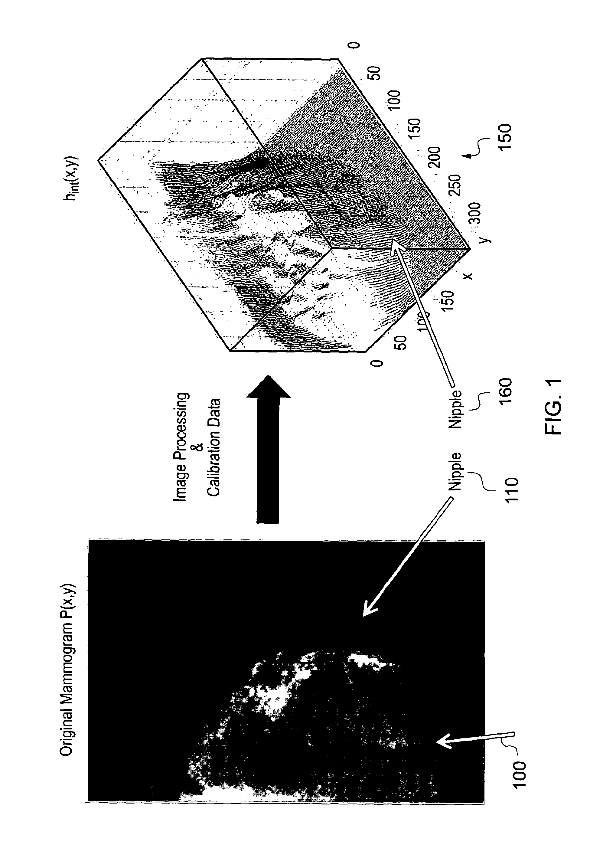
[0095]In FIG. 1, the mammogram shows a breast 100 which is relatively fatty (as indicated by the relative darkness of the image) with some denser tissue notable towards the nipple 110 (the whiter parts). In what is referred to as the hint representation 150 (on the right of the picture), peaks towards the nipple 160 indicate high values of hint, i.e. denser tissue.

[0096]Once the hint representation is generated, numerous clinically useful tasks can be performed, including, but not limited to: automatic estimation of breast composition by summing up the hint and hfat values and subsequently computing breast density; optimized display of the image ready for the radiologist to view; tracking an object's development over time to ascertain whether it is growing; computer-aided detection, whereby the computer marks onto the image suspect areas for the radiologist to consider; generally analyzing multiple images (temporal, dual energy, views etc).

[0097]The first embodiment notes that dense...


PUM


Abstract

Disclosed is a method of analysing tissue from an image comprising providing an electronic image of tissue (100, 400, 450, 600, 800, 1100), determining a reference value from the image (1070, 1170, 1270), establishing an hint representation (500,700) of the image, and using the hint representation in analysis of the tissue to quantify the breast and compute a calibration error. Also disclosed is a system which runs an inner breast edge detection algorithm (1310) on the electronic image to detect the inner breast edge on the image (1315), and refined the inner breast edge location (1340) if a calibration error is not acceptable (1324). Also disclosed is automatic estimation of breast composition and temporal analysis of images.

Description

FIELD OF THE INVENTION[0001]The present invention relates to the analysis of an image. A reference value on the image is established and the image can be self-calibrated and thus quantified.BACKGROUND OF THE INVENTION[0002]Many in the field are attempting to quantify images for various purposes but encounter issues relating to errors or unknown values in the imaging physics data. For example, trying to quantify the image based on an absolute model of the physics along with assumed properties of tissue needs all the imaging physics data to be known and accurate. Even the inclusion of a quantification step wedge into each image still requires much of the imaging physics data and assumes it is accurate. Hence the need to use in-image reference values.[0003]For example, a mammogram is created by sending x-ray photons towards the breast and then detecting how many x-ray photons make it through. The smaller the number of x-ray photons that make it through, the denser the breast tissue.[00...

Claims


Application Information

Patent Timeline
IPC IPC(8): G06K9/00
CPCG06T7/0012G06T2207/30068G06T7/403G06T7/44
Inventor HIGHNAM, RALPHBRADY, JOHN MICHAELKARSSEMEIJER, NICOYAFFE, MARTIN
Owner HIGHNAM RALPH
Why Eureka
Try Eureka