Medical report generation apparatus, method and program

a report generation and medical technology, applied in the field of medical report generation apparatus, a medical report generation method and a medical report generation program, can solve the problems of imposing time and work on the insufficient information for a doctor or the like, and insufficient information for a patient, etc., to achieve the effect of easy and appropriate selection

Inactive Publication Date: 2012-10-04
FUJIFILM CORP
View PDF2 Cites 43 Cited by
  • Summary
  • Abstract
  • Description
  • Claims
  • Application Information

AI Technical Summary

Benefits of technology

[0009]In view of the foregoing circumstances, the present invention provides a medical report generation apparatus, method and program in which a supplementary image that can assist a user in understanding a key image, which has been judged to be important for diagnosis, is easily and appropriately selected when a medical report to which the key image is attached is generated.
[0034]According to a medical report generation apparatus, method and program of the present invention, a slice image generated based on three-dimensional medical image data obtained by imaging a subject is selected, as a key image representing a diagnostic characteristic feature, and a selected position in the key image is input, and a slice image that includes the selected position and represents a cross section different from a cross section represented by the key image is determined, as a supplementary image, in the three-dimensional medical image data based on the key image and the selected position, and a medical report including the key image and information for identifying the supplementary image is generated. Therefore, it is possible to easily generate the supplementary image representing the selected position by a different cross section, and to efficiently generate a medical report. Further, since a doctor or the like who uses the medical report for diagnosis can easily refer to the supplementary image, the doctor or the like can easily understand the medical report.

Problems solved by technology

However, when only the key image attached to the medical report and the position information about the region of interest described in the medical report are provided, the information is insufficient for a doctor or the like in some cases to diagnose a patient while accurately recognizing the region of interest, such as the position and the size of a diseased part or the like.
That may impose time and work on the doctor or the like.
Therefore, the position of an actual region, skeleton or the like of a patient and an anatomical position in the schematic diagram do not sufficiently match with other.
Hence, comparison has been difficult in some cases.
Therefore, there has been a problem that selection of the image related to the key image is not easy for a user who is not used to an operation for appropriately changing an image processing parameter set and an operation for selecting the image related to the key image.

Method used


Image

  • Medical report generation apparatus, method and program
  • Medical report generation apparatus, method and program
  • Medical report generation apparatus, method and program

Examples


first embodiment

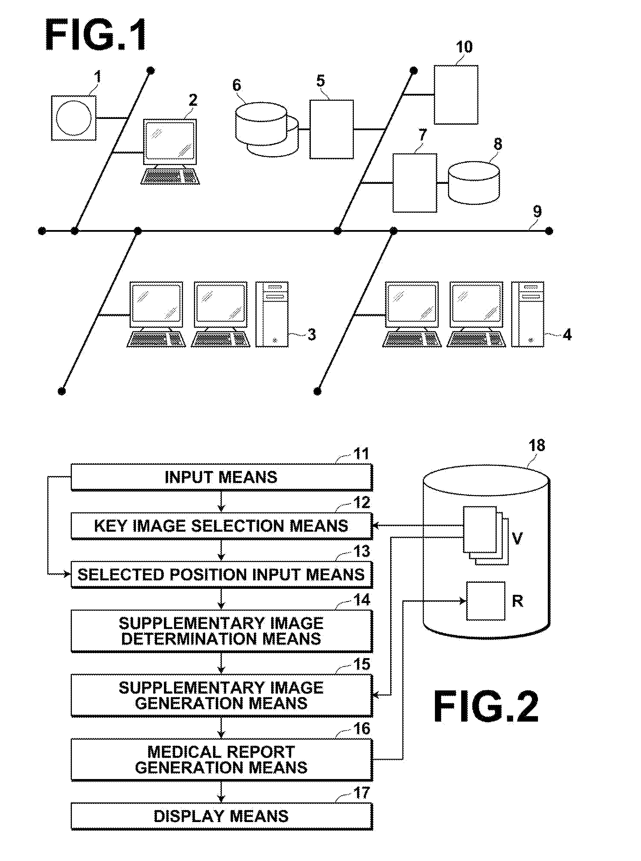
[0061]FIG. 2 is a schematic block diagram illustrating the configuration of a medical image display system to which the medical image processing apparatus according to the present invention installed in the medical information system has been applied.

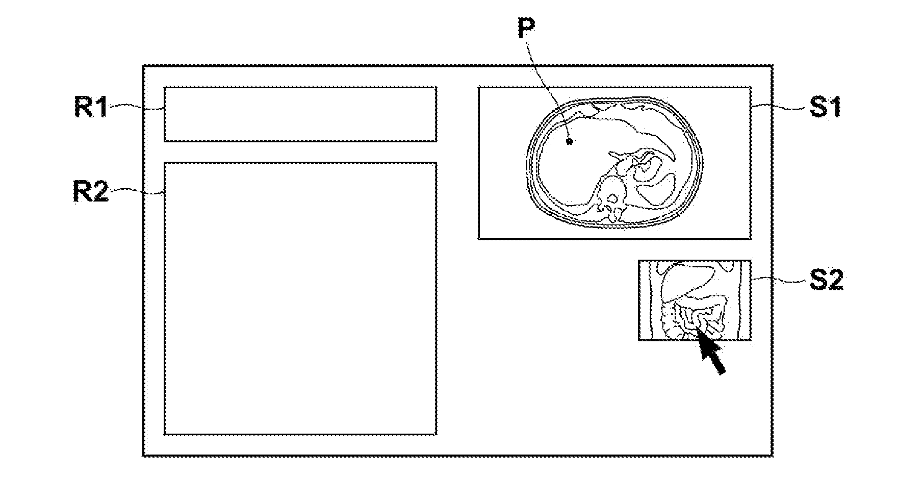
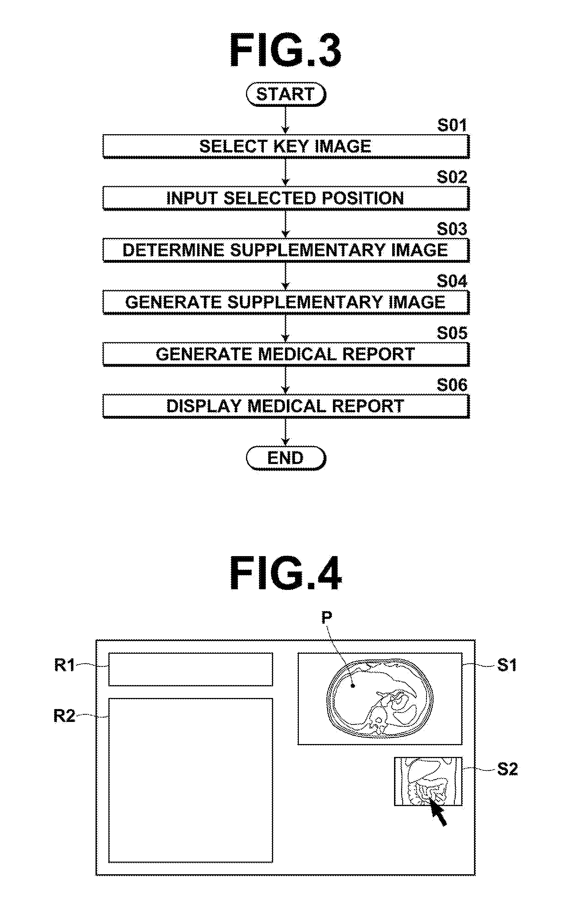
[0062]In this embodiment, the workstation 3 for the radiology department (a report generation apparatus) includes an input means 11 (input device), such as a mouse and a keyboard, a storage means 18 including a storage device, such as a hard disk and a memory, and a key image selection means 12. The key image selection means 12 selects, as key image S1 representing a diagnostic characteristic feature, a slice image generated from three-dimensional medical image data V obtained by imaging a subject. Further, the workstation 3 for the radiology department includes a selected position input means 13 that inputs selected position P in the key image, and a supplementary image determination means 14. The supplementary image determination mean...

second embodiment

[0087]In correspondence table T2 used in the second embodiment, the inclination of a supplementary image corresponding to the inclination of a key image differs for each region. Here, when a region to which a selected position belongs is a vertebral column, an aorta or the like, which curves toward the front direction of the subject, if a coronal image or the like is used as the supplementary image for example, only a part of a region including the selected position is included in the supplementary image. Therefore, it is highly likely that the region including the selected region is hard to be recognized. To prevent such problems, when the region to which the selected position belongs is a region, such as a vertebral column and an aorta, which curves toward the front direction of the subject, a slice image representing a cross section including a curved line of the curved region should be determined as the supplementary image. Such a supplementary image is desirable to effectively ...


PUM


Abstract

A medical report generation apparatus includes a key image selection unit that selects, as a key image representing a diagnostic characteristic feature, a slice image generated based on three-dimensional medical image data obtained by imaging a subject, a selected position input unit that inputs a selected position in the key image, a supplementary image determination unit that determines, based on the key image and the selected position, a slice image that includes the selected position and represents a cross section different from a cross section represented by the key image, as a supplementary image, in the three-dimensional medical image data, and a medical report generation unit that generates a medical report including the key image and information for identifying the supplementary image.

Description

BACKGROUND OF THE INVENTION[0001]1. Field of the Invention[0002]The present invention relates to a medical report generation apparatus, a medical report generation method and a medical report generation program for generating a medical report about image-based diagnosis of a patient.[0003]2. Description of the Related Art[0004]In image-based diagnosis in medical fields, a person in charge of generating a medical report reads and interprets a medical image obtained by imaging a patient, and generates the medical report that describes position information about a region of interest judged to be important for diagnosis, and to which a key image including the region of interest is attached. Further, a doctor or the like checks the generated medical report to diagnose the patient.[0005]However, when only the key image attached to the medical report and the position information about the region of interest described in the medical report are provided, the information is insufficient for a...

Claims


Application Information

Patent Timeline
Patent Type & Authority Applications(United States)
IPC IPC(8): G06K9/00G16H10/60G16H15/00G16H30/40
CPCG06F19/321G06F19/322G06F19/3406A61B2576/00G06F19/345G06F19/3487A61B5/748G06F19/3443G16H40/63G16H10/60G16H50/70G16H50/20G16H15/00G16H30/40
Inventor IWASAKI, TAIJI
Owner FUJIFILM CORP
Why Eureka
Try Eureka