
Mechanism Of Quantitative Dual-Spectrum IR Imaging System For Breast Cancer

a breast cancer and imaging system technology, applied in the field of quantitative dual-spectrum ir imaging system for breast cancer, can solve the problems of not having a sufficient spatial resolution, not being sensitive enough to detect breast cancer, and being both very costly

Active Publication Date: 2013-01-31
NAT TAIWAN UNIV
View PDF12 Cites 9 Cited by
  • Summary
  • Abstract
  • Description
  • Claims
  • Application Information

AI Technical Summary

Benefits of technology

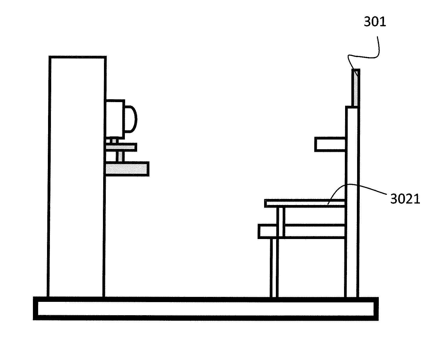
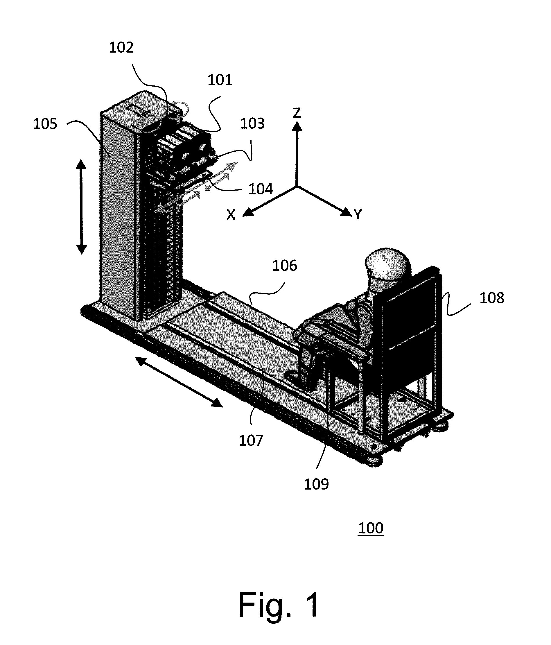
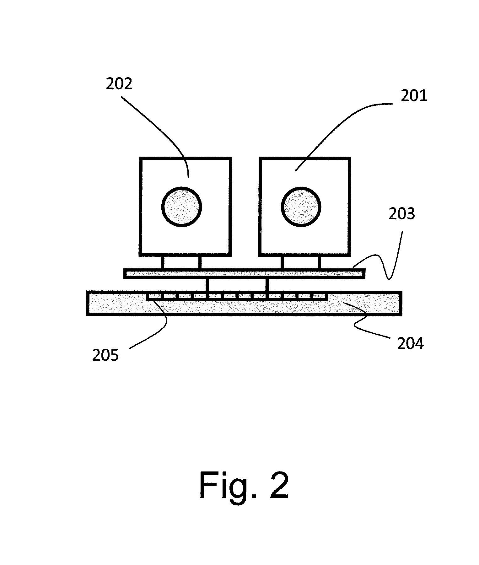
The technology in this patent allows for the ability to capture infrared images of the human body at different times and can be used for non-invasive detection of humans, assessing chemotherapy treatment response, and early detection of certain diseases. Additionally, the patent describes a method for obtaining a Dual-Spectrum spectrogram where two cameras are used to capture images of an object at different times and the image data is calibrated to ensure accurate results. The method also involves using two armrests to help maintain the object's posture during the examination.

Problems solved by technology

However, they both do not have a sufficient spatial resolution and are not sensitive enough to detect breast cancers.
Although the MRI and the PET have the potential for early detection of breast cancers, they are both very costly and not usually used as the first-line defense.
Nevertheless, the usefulness of IR images in detecting breast cancers at a single time is not usually enough to assess the chemotherapy treatment response and early detection due to the physiological and environmental influence on the skin temperature distribution.

Method used


Image

  • Mechanism Of Quantitative Dual-Spectrum IR Imaging System For Breast Cancer
  • Mechanism Of Quantitative Dual-Spectrum IR Imaging System For Breast Cancer
  • Mechanism Of Quantitative Dual-Spectrum IR Imaging System For Breast Cancer

Examples


embodiment 1

[0029]2. The system as described in Embodiment 1, wherein the first camera and the second camera are a long-wave Infra-red (LIR) camera and a middle-wave Infra-red (MIR) camera respectively.

[0030]3. The system as described in Embodiment 1, wherein the platform has a first plane, the first camera and the second camera are configured on the first plane to move along a first direction and to be rotated on the first plane, and the platform is configured to move along one of a second direction and a third direction.

embodiment 3

[0031]4. The system as described in Embodiment 3, wherein the first direction is a horizontal direction, and the first plane is a horizontal plane.

[0032]5. The system as described in Embodiment 3, wherein the track has a forth direction which is perpendicular to the first direction.

[0033]6. The system as described in Embodiment 3, wherein the first direction and the forth direction are parallel to the first plane.

[0034]7. The system as described in Embodiment 3, wherein the second direction is perpendicular to the third direction.

[0035]8. The system as described in Embodiment 3, wherein the third direction is vertical to the first plane.

[0036]9. The system as described in Embodiment 1, wherein the first calibration marker and the second calibration marker are cross line centers.

[0037]10. The system as described in Embodiment 1 further comprises a slab supporting the first camera and the second camera, wherein the slab is disposed on the platform, and after calibrating the first came...

embodiment 12

[0040]13. The method as described in Embodiment 12 further comprising a step of disposing a first calibration marker and a second calibration marker on the seat for respectively calibrating the first optical axis and the second optical axis.


PUM


Abstract

A mechanism of quantitative dual-spectrum IR imaging (QDS-IR) system for examining the breast cancer is reported. The major mechanism of the system is a pair of long-wave Infra-red (LIR) and middle-wave Infra-red (MIR) cameras with the keen temperature sensitivity and the high spatial resolution. The optical axes of cameras are calibrated by the help of two calibration makers set up on the seat for carrying an object to make them parallel to each other and locate on the same level. The design provides an imaging system with the high reproducibility supported by 7 free degrees and the high adjustability. The proposed system could ensure the positions of the object and two cameras are the same at the different time points and find the best relative positions between the seat and two cameras for the objects with different body types. Therefore, it has potential ability to detect breast cancer or monitor the effect of chemotherapy.

Description

FIELD OF THE INVENTION[0001]The invention relates to a mechanism of quantitative dual-spectrum IR imaging system for breast cancer. More specifically, this invention relates to an image system with MIR (middle-wave Infra-red) and LIR (long-wave Infra-red) cameras for accepting longitudinal infrared images over several time points in order to obtain more usable information of detecting the breast cancer.BACKGROUND OF THE INVENTION[0002]Breast cancer has been ranked the leading cause of cancer deaths for females. Common imaging modalities, such as X-ray mammograms, MRI images and Ultrasound, used to detect breast cancers at present are mammogram and breast sonography. However, they both do not have a sufficient spatial resolution and are not sensitive enough to detect breast cancers. Although the MRI and the PET have the potential for early detection of breast cancers, they are both very costly and not usually used as the first-line defense.[0003]Due to the advantages of non-invasion,...

Claims


Application Information

Patent Timeline
Patent Type & Authority Applications(United States)
IPC IPC(8): A61B6/00
CPCA61B5/706A61B5/708A61B5/015A61B5/0091A61B5/0077G01J2005/0077G01J5/0025G01J5/53G01J5/80
Inventor CHUNG-MING, CHENSI-CHEN, LEEWAN-JOU, LEECHE-WEI, CHANGCHING-YEN, LEEYU-CHUN, CHIENCHIA-YEN, LEE
Owner NAT TAIWAN UNIV
Learn More