Methods and systems for analyzing, prioritizing, visualizing, and reporting medical images

a medical image and system technology, applied in the field of medical imaging, can solve the problems of limited reporting capabilities of reviewing physicians, inability of hospitals to timely and inability of reviewing physicians to review each patient's medical images. to achieve the effect of improving reporting capabilities

Active Publication Date: 2013-02-14
RADLOGICS +1
View PDF6 Cites 33 Cited by
  • Summary
  • Abstract
  • Description
  • Claims
  • Application Information

AI Technical Summary

Problems solved by technology

While current medical image retrieval and processing systems have provided physicians tremendous capabilities in storing and retrieving medical images, there are limitations associated with these systems.
For instance, for a typical scan, a hospital may obtain hundreds of images, and a reviewing physician might not have time to review each of the images to determine whether a patient has a particular type of medical condition.
In cases in which a hospital scans several patients in a relatively short period of time, the hospital might not have the resources to timely review each patient's medical images to determine whether a physician should review the image further, and whether the patient has a particular type of medical condition.
In addition, modern medical imaging systems can operate much more quickly than older systems, which has led to a decrease in the time it takes to generate a scan.
As the time to generate scans decreases and the number of scans (and images obtained) per patient increases, hospitals without sufficient resources might not be able to review each image and provide patients with medical care in an accurate and efficient manner.
Further, while some hospitals might have medical imaging, processing and retrieval systems for handling scans, current systems are not capable of accurately and efficiently prioritizing scans.
In addition, current systems do not provide scan reviewing and patient treating physicians with the capability to acquire accurate patient-specific diagnostic information from each of the images or scans.

Method used


Image

  • Methods and systems for analyzing, prioritizing, visualizing, and reporting medical images
  • Methods and systems for analyzing, prioritizing, visualizing, and reporting medical images
  • Methods and systems for analyzing, prioritizing, visualizing, and reporting medical images

Examples


Embodiment Construction

[0028]While various embodiments of the invention have been shown and described herein, it will be obvious to those skilled in the art that such embodiments are provided by way of example only. Numerous variations, changes, and substitutions will now occur to those skilled in the art without departing from the invention. It should be understood that various alternatives to the embodiments of the invention described herein may be employed in practicing the invention.

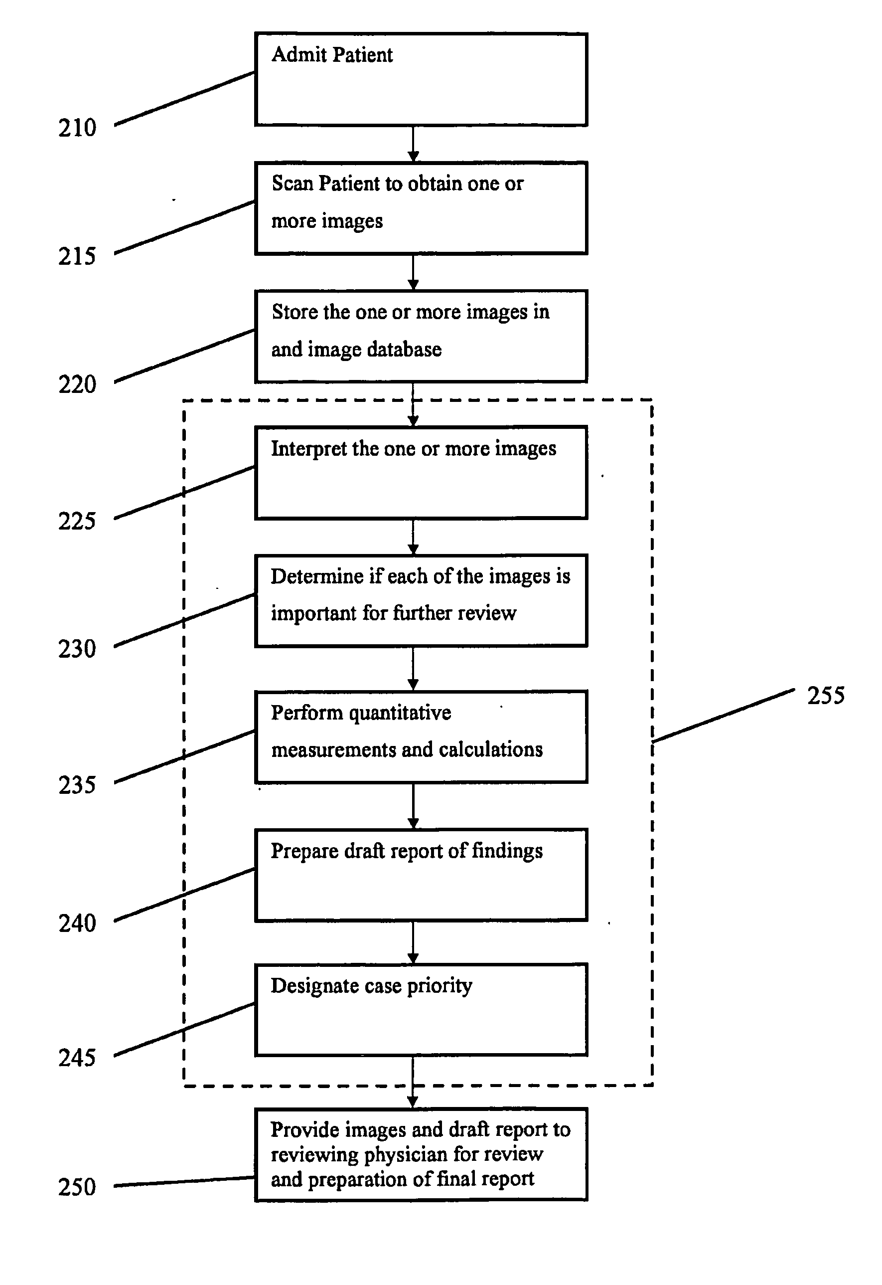
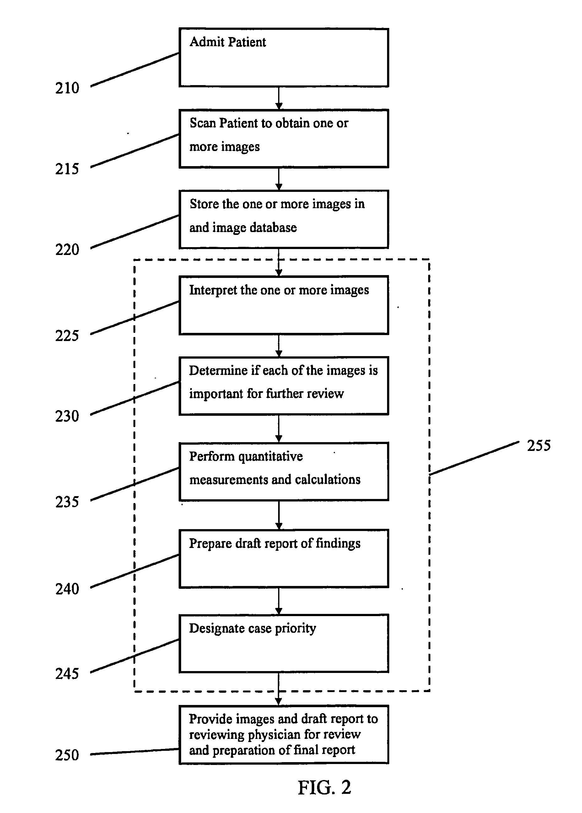
[0029]The invention provides methods and systems for analyzing and prioritizing medical images, and for reporting medical findings. For example, an analysis of medical images according to some aspects of the system and methods disclosed herein may be used to identify critical medical conditions, and, based on this analysis, said system and method may further be used to organize a work list for a reviewing physician based on the severity of the medical findings and to then create a text document that lists the medical findi...


PUM


Abstract

Methods and systems for retrieving and processing medical diagnostic images are provided, comprising using picture analysis prioritization visualization and reporting system (“PAPVR system”) to determine whether each of one or more images from an image database or imaging device is of medical interest to a reviewing physician, determine whether one or more of the images is representative of the images, and provide the one or more images to a display and analysis system for review by a reviewing physician. The PAPVR system can provide the one or more images with a Key Image that is representative of the images. In addition, the PAPVR system can detect whether a patient suffers from a particular ailment, and provide a reviewing physician quantitative information that is relevant to the patient's condition.

Description

CROSS-REFERENCE[0001]This application claims the benefit of U.S. Provisional Patent Application Ser. No. 61 / 299,309, filed on Jan. 28, 2010, which is entirely incorporated herein by reference.FIELD OF THE INVENTION[0002]This application generally relates to medical imaging; more particularly, this invention relates to systems and methods for analyzing, prioritizing, visualizing and reporting medical images.BACKGROUND OF THE INVENTION[0003]Medical imaging systems, such as computerized tomography (“CT”) scanners and magnetic resonance imaging (“MRI”) scanners, allow a physician to examine a patient's internal organs and areas of the patient's body that require a thorough examination for medical treatment. In use, a visualizing scanner outputs two-dimensional (“2D”) and three-dimensional (“3D”) medical images that can include a sequence of computerized cross-sectional images of a certain body organ, which is then interpreted by reviewing physician, such as a specialized radiologist.[00...

Claims


Application Information

Patent Timeline
Patent Type & Authority Applications(United States)
IPC IPC(8): G06K9/36G06F17/30G16H10/60G16H30/20
CPCG06F19/3487G06F19/321G16H10/60G16H15/00G06F16/51G16H30/20G06F18/22G06T7/0012
Inventor BECKER, MOSHEGREENSPAN, HAYITKONEN, ELIAHUMAYER, ARNALDO
Owner RADLOGICS
Why Eureka
Try Eureka