Image segmentation of organs and anatomical structures

a technology for applied in the field of image segmentation of organs and anatomical structures, to achieve the effect of facilitating the further image registration procedures and reducing image nois

Active Publication Date: 2013-02-21
PATHFINDER THERAPEUTICS +1
View PDF3 Cites 30 Cited by
  • Summary
  • Abstract
  • Description
  • Claims
  • Application Information

AI Technical Summary

Benefits of technology

[0007]In one exemplary embodiment, the present invention utilizes the similarity of organ morphological structures on nearby neighboring image slices, and builds a deformation field between two nearby neighboring image slices by conducting image registration between these two image slices. Accordingly, when the target areas on one image slice are defined either by the user manually or from the previous segmentation step, the corresponding areas on the nearby neighboring image slice can be defined by applying the deformation field, even where the target and background areas are of the same or similar image intensities. In other words, the image registration between two slices helps distinguish the target and background areas even when they show same or similar image intensities. The user defines the target and background on one image slice, and the morphological structures on all other image slices can be deduced from it based on the deformation fields built with the image registration procedure.
[0010]FIG. 2 shows a chart of the steps of a method for organ segmentation using a deformation field in 3-D images. The first step is acquiring 3-D images of the organ of interest from the same or similar imaging modalities, such as (but not limited to), computed tomography (CT) images with or without contrast, magnetic resonance (MR) images of a same or similar pulse sequence, and positron emission tomography (PET) images. An image smoothing or filtering procedure may be applied if necessary to reduce image noise and facilitate the further image registration procedures.

Problems solved by technology

While registration-based segmentation is known, the prior art does not conduct registration procedures to explore the similarity between neighboring cross section slices and use it to perform image segmentation.

Method used


Image

  • Image segmentation of organs and anatomical structures
  • Image segmentation of organs and anatomical structures
  • Image segmentation of organs and anatomical structures

Examples


Embodiment Construction

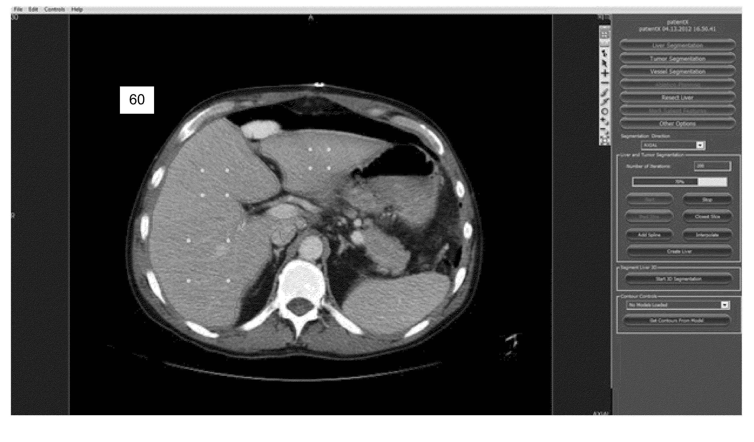
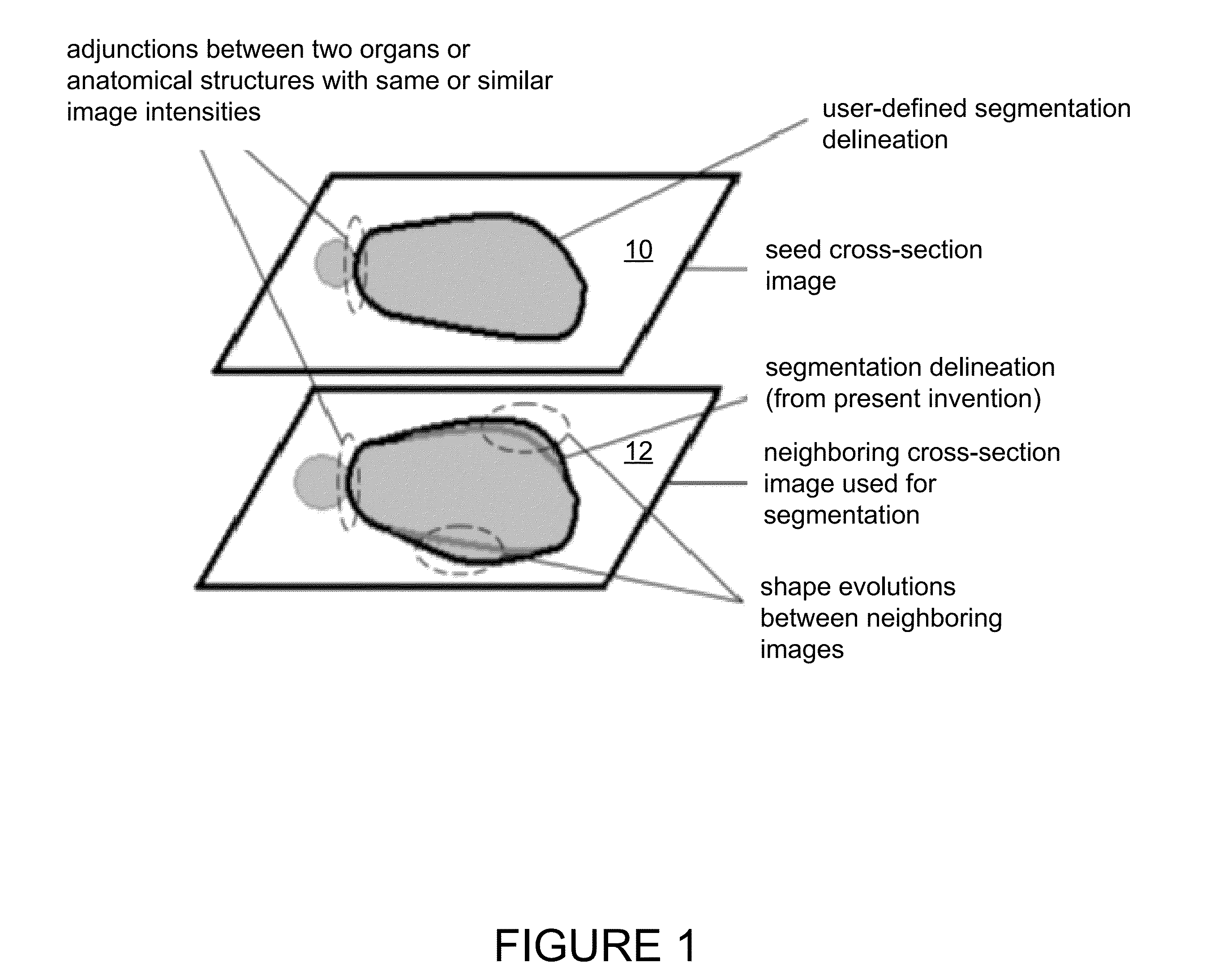
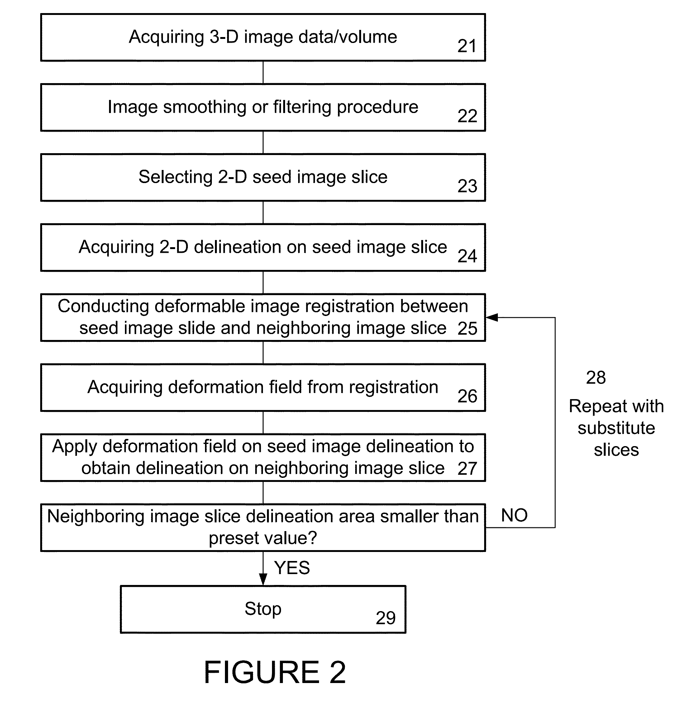
[0017]Image registration provides a method to define the corresponding points or elements between two images. In various exemplary embodiments, the present invention comprises methods to conduct image segmentation by imaging target morphological shapes evolving from one 2-dimension (2-D) image slice to one or more nearby neighboring 2-D images taken from a 3-dimension (3-D) image. One area defined by a user as a target on an image slice can be found in a corresponding area on a nearby neighboring image slice by using a deformation field generated with deformable image registration procedure between these two image slices. It provides a solution to distinguish target and background areas with the same or similar image intensities, which is one of most difficult issues in the prior art, such as intensity-based region growing methods.

[0018]In one exemplary embodiment, as shown in FIG. 1, the present invention utilizes the similarity of organ morphological structures on nearby neighbori...


PUM


Abstract

A system and method to conduct image segmentation by imaging target morphological shapes evolving from one 2-dimension (2-D) image slice to one or more nearby neighboring 2-D images taken from a 3-dimension (3-D) image. One area defined by a user as a target on an image slice can be found in a corresponding area on a nearby neighboring image slice by using a deformation field generated with deformable image registration procedure between these two image slices. It allows the user to distinguish target and background areas with the same or similar image intensities.

Description

[0001]This application claims benefit of and priority to U.S. Provisional Application No. 61 / 476,744, filed Apr. 18, 2011, by Senhu Li, et al., and is entitled to that filing date for priority. The specification, figures and complete disclosure of U.S. Provisional Application No. 61 / 476,744 are incorporated herein by specific reference for all purposes.FIELD OF INVENTION[0002]This invention relates to a method and apparatus to distinguish the target and background areas with the same or similar image intensities for medical image segmentation purposes.BACKGROUND OF THE INVENTION[0003]Organ and anatomical structure segmentation is of importance in several medical applications, including the creation of surfaces used in image-guided surgical systems. A variety of prior art organ segmentation methods and systems are disclosed in Dawant, et al., U.S. Pat. No. 7,519,209, which is incorporated herein in its entirety by specific reference for all purposes.[0004]One of the most difficult is...

Claims


Application Information

Patent Timeline
Patent Type & Authority Applications(United States)
IPC IPC(8): G06K9/34
CPCA61B5/055G06T2207/20144A61B6/5235A61B6/5258A61B6/032A61B6/037A61B6/501A61B6/5211G06T7/0012G06T7/0089G06T2207/10081G06T2207/10088G06T2207/10104G06T2207/20036G06T2207/20108A61B6/469G06T7/149G06T7/194G16H50/20
Inventor LI, SENHULENNON, BRIANWAITE, JONSTEFANSIC, JAMES
Owner PATHFINDER THERAPEUTICS
Why Eureka
Try Eureka