
System and device for characterizing cells

a cell and cell technology, applied in the field of cell analysis, can solve the problems of inability to identify reliable epigenetic, proteomic signature, and inability of genetic profiling to stage tumors and predict clinical outcomes, and achieve the effect of reducing the difficulty of detecting tumors and detecting tumors

Active Publication Date: 2013-03-21
THE JOHN HOPKINS UNIV SCHOOL OF MEDICINE
View PDF28 Cites 23 Cited by
  • Summary
  • Abstract
  • Description
  • Claims
  • Application Information

AI Technical Summary

Benefits of technology

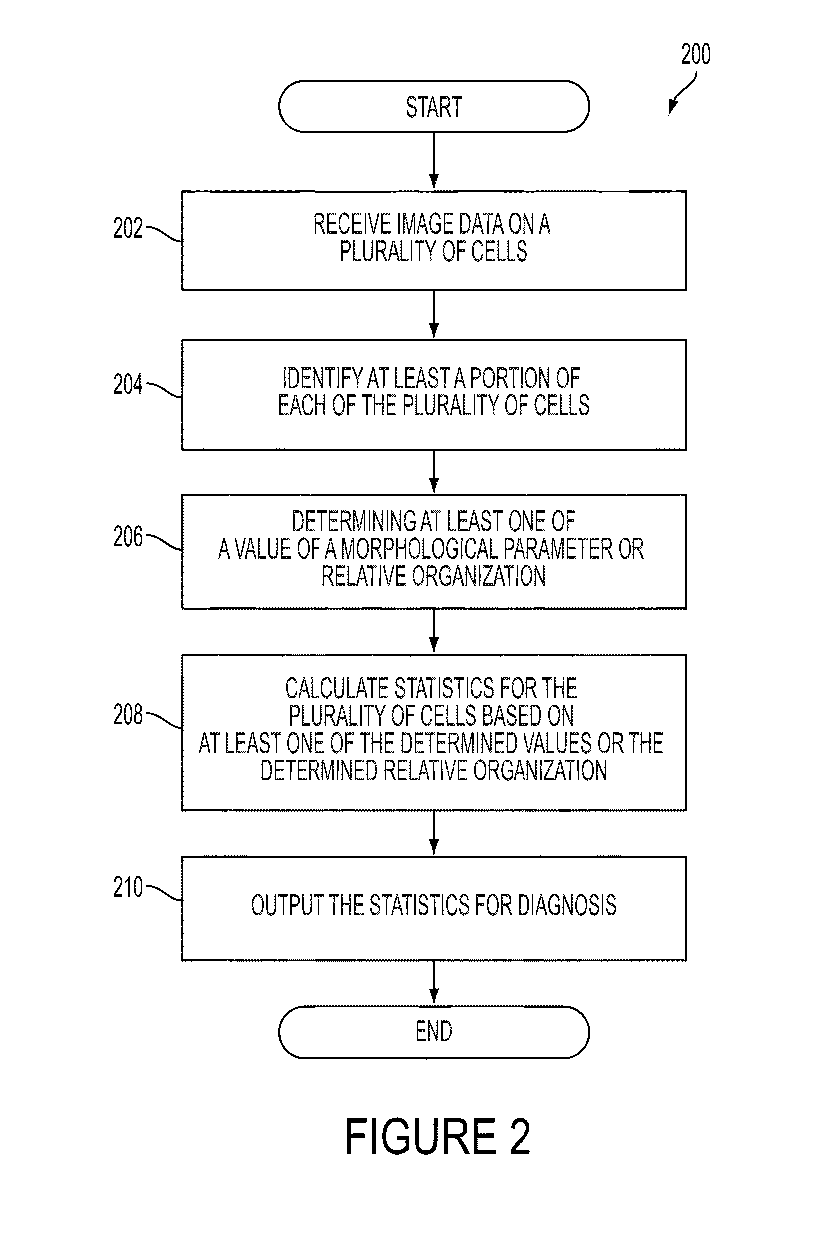
The patent describes a diagnostic device and method for identifying and distinguishing metastatic cells from non-metastatic cells based on the morphological characteristics of cells. The device includes a microscope and a computing device that receives image data of cells and calculates statistics based on the identified cells. The device can also determine the value of a morphological parameter for each cell and output the information for diagnosis. The method involves receiving image data on cells in different cell cycle phases, normalizing the data, identifying the cells, and determining the value of a morphological parameter for each cell. The device and method can be used for the diagnosis and treatment of cancer.

Problems solved by technology

However, despite research efforts, no reliable genetic, epigenetic, or proteomic signature of cancer metastasis has been identified so far.
In particular, a recent systematic genetic analysis of cells from primary pancreatic tumors of early-stage patients and liver metastatic sites of late-stage patients, while displaying shared signaling pathways, revealed highly heterogeneous genetic mutational sets, potentially limiting the ability of genetic profiling to stage tumors and predict clinical outcomes.

Method used


Image

  • System and device for characterizing cells
  • System and device for characterizing cells
  • System and device for characterizing cells

Examples


Embodiment Construction

[0047]Some embodiments of the current invention are discussed in detail below. In describing embodiments, specific terminology is employed for the sake of clarity. However, the invention is not intended to be limited to the specific terminology so selected. A person skilled in the relevant art will recognize that other equivalent components can be employed and other methods developed without departing from the broad concepts of the current invention. All references cited anywhere in this specification are incorporated by reference as if each had been individually incorporated.

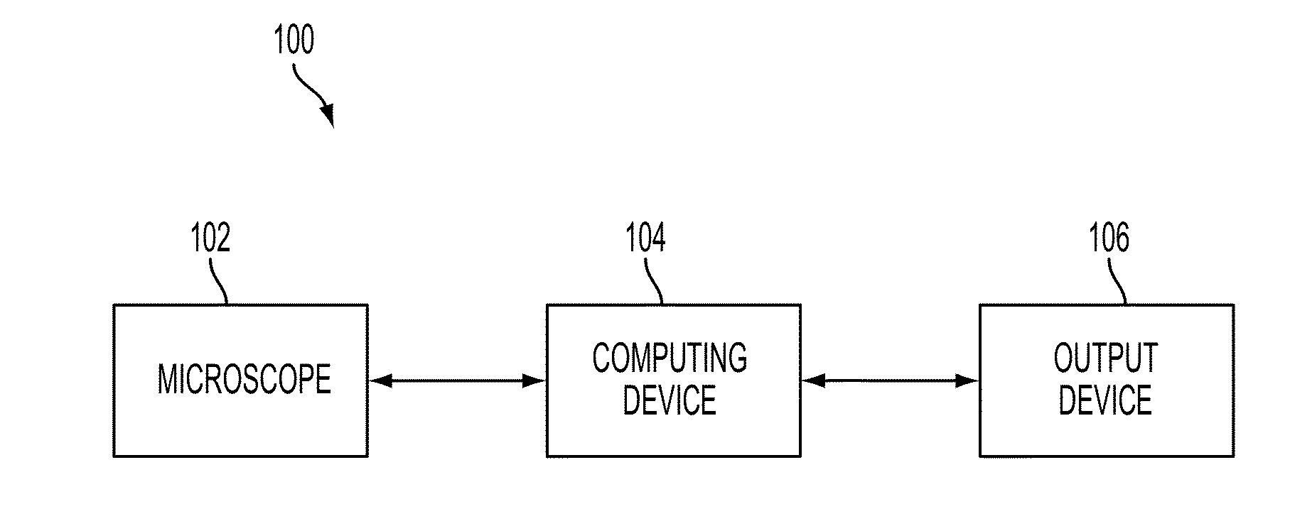
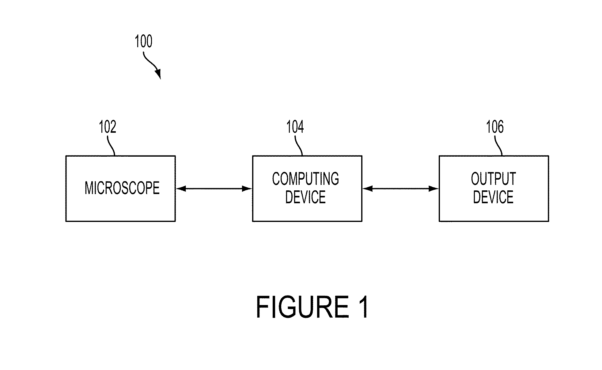
[0048]FIG. 1 illustrates a block diagram of system 100 according to an embodiment of the current invention. System 100 may include microscope 102, computing device 104, and output device 106. Microscope 102 may be configured to obtain image data on a plurality of cells. Microscope 102 may include a camera, a motorized stage, and motorized excitation and emission filters. The image data obtained by microscope 10...


PUM


Abstract

A diagnostic device includes a microscope configured to obtain image data on a plurality of cells and a computing device. The computing device is configured to receive the image data, identify at least a portion of each of the plurality of cells based on the received image data, determine at least one of a value of a morphological parameter for each identified at least a portion of the plurality of cells or a relative organization among the identified at least a portion of the plurality of cells, and calculate statistics for the plurality of cells based on the at least one of the determined values of the morphological parameter or the determined relative organization, the statistics including information suitable for distinguishing metastatic cells from non-metastatic cells. The diagnostic device further includes an output device configured to output the statistics for diagnosis.

Description

CROSS-REFERENCE OF RELATED APPLICATION[0001]This application claims priority to U.S. Provisional Application No. 61 / 499,955 filed Jun. 22, 2011, U.S. Provisional Application No. 61 / 500,234 filed Jun. 23, 2011, U.S. Provisional Application No. 61 / 500,244 filed Jun. 23, 2011, U.S. Provisional Application No. 61 / 533,446 filed Sep. 12, 2011, and U.S. Provisional Application No. 61 / 645,930 filed on May 11, 2012, which are hereby incorporated by reference in their entireties.[0002]This invention was made with Government support of Grant No. 54 CA-143868, awarded by the National Institute of Health / National Cancer Institute. The U.S. Government has certain rights in this invention.BACKGROUND[0003]1. Field of Invention[0004]The currently claimed embodiments of this invention relate to analyzing cells.[0005]2. Discussion of Related Art[0006]The contents of all references, including articles, published patent applications and patents referred to anywhere in this specification are hereby incor...

Claims


Application Information

Patent Timeline
IPC IPC(8): G06K9/00G06V10/28
CPCG06K9/00127G06T7/0012G06T2207/10056G06K9/38G06K9/00134G06K9/00147G06T2207/30024G01N33/48728G01N33/48792G06V20/693G06V20/698G06V10/28G06V20/69G01N15/14G01N21/6458G01N21/6486G01N2201/06113G01N2201/10
Inventor WIRTZ, DENIS GASTONWU, PEI-HSUNKHATAU, SHYAM BHARATCHEN, WEI-CHIANGPHILLIP, JR., JUDE MARVIN GEORGEBINDER, ZEV ARITSENG, YIIDER
Owner THE JOHN HOPKINS UNIV SCHOOL OF MEDICINE
Learn More