Automated fish reader using learning machines

Active Publication Date: 2014-03-13
NEOGENOMICS LAB
View PDF6 Cites 24 Cited by
  • Summary
  • Abstract
  • Description
  • Claims
  • Application Information

AI Technical Summary

Benefits of technology

[0009]A method and system for automated analysis of fluorescent in situ hybridization (FISH) images based is provided. The inventive FISH reader program pre-processes the digitized FISH image to select analysis regions, segment objects within selected regions, separate color signals and measure signals for each separated color. Prior to classification of the data extracted from the FISH images, analysis regions are selected, spot colors are separated and signals are measured for each separated color. Different components of the data may be analyzed separately using different learning machines, such as neural networks, decision trees, regression or support vectors machines. For example, a first learning machine may be used to classify spots according to color, while a second learning machine may be used to distinguish signals where the cell in the image is partially obscured by another cell overlapping the cell. The separation of the classification functions allows the learning machine to be optimized for the specific attribute of interest. The results are then merged to produce an output that is based on the most useful (determinative) features within the image and optimized to overcome issues that are particularly problematic in FISH analyses. An exemplary disclosure of multiple SVM architecture and analysis is provided in U.S. Pat. No. 6,996,549 of Zhang, et al., the disclosure of which is incorporated herein by reference. In one embodiment, separate learning machines perform their analyses sequentially then merge the results to produce the analysis for output (or further processing). In another embodiment, the learning machines may perform their respective operations in parallel in a first round of classification, and an additional learning machine may be used to combine the first round outputs in a second round of classification to generate a single conclusion, e.g., a diagnosis, based on the individual test results.
[0010]In one aspect of the invention, an automated reader for reading fluorescence in-situ hybridization signals includes one or more computer processors for receiving a digitized FISH image comprising a plurality of spots having colors, wherein the one or more processors is programmed to execute the steps of: converting colors within the FISH image to a hue value; separately for each color: extracting a plurality of quantitative values to detect the presence of signals corresponding to spots; and applying a plurality of algorithms to extract features from the signals to determine cell shapes and segment cells within the FISH image; rec

Problems solved by technology

A major limitation of the FISH technique for dot counting is the need to examine large numbers of cells.
Visual evaluation by a trained cytogeneticist of large numbers of cells and enumeration of hybridization signals is tedious, time consuming and expensive.
Unfortunately, there are many obstacles to be overcome before automated analysis of FISH images can be implemented on a widespread basis.
Additional obstacles include that the cells are not usually well defined, there are no clear boundaries between cells, overlapping of cells is co

Method used


Image

  • Automated fish reader using learning machines
  • Automated fish reader using learning machines
  • Automated fish reader using learning machines

Examples


Embodiment Construction

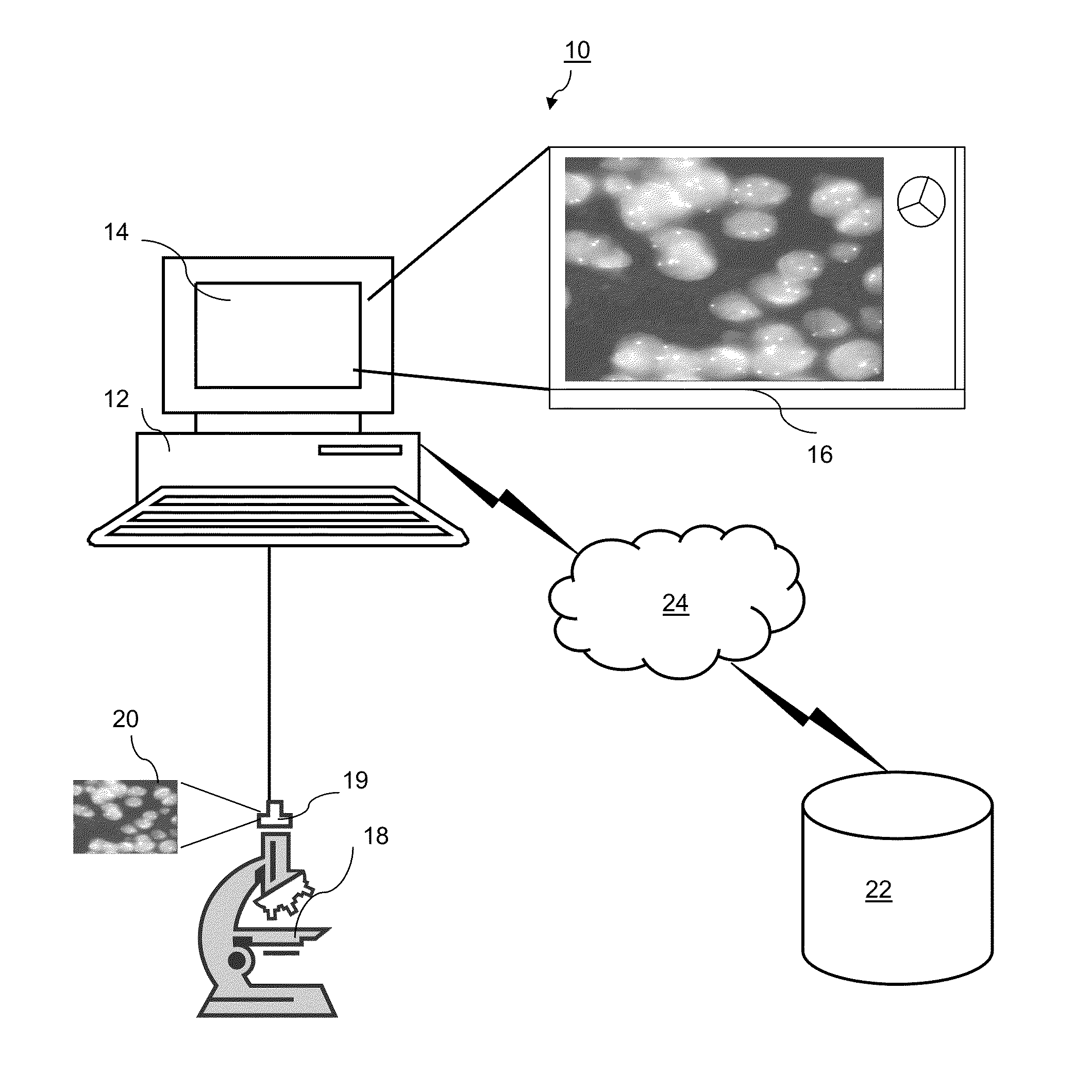
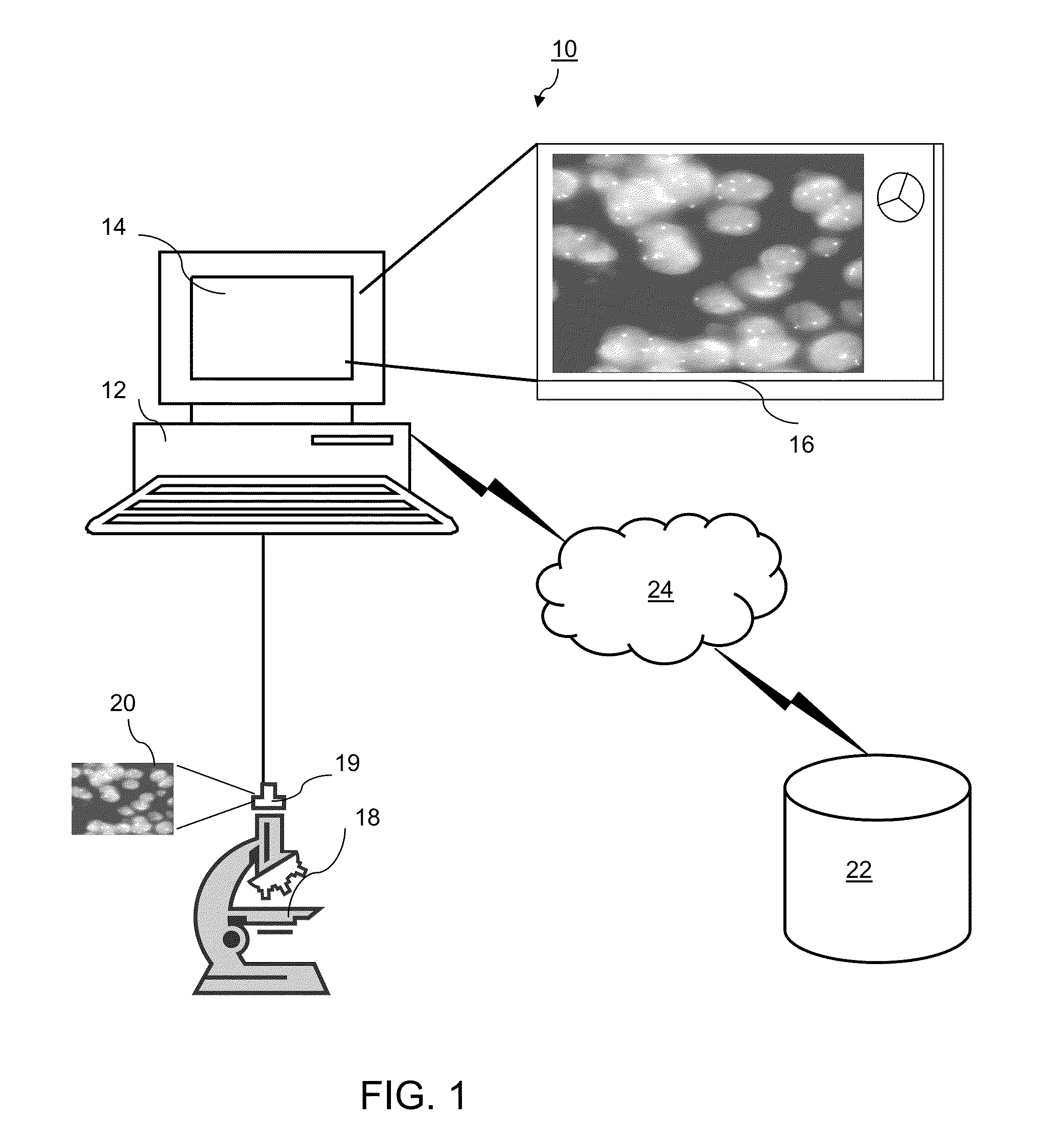
[0023]For purposes of the description of the invention, the terms “dot” or “spot”, or pluralities thereof, may be used interchangeably to refer to the detected fluorescence signal(s) resulting from hybridization of tagged probes with target sequences for the detection of abnormalities using tissue sections or cell samples.

[0024]FIG. 1 is a block diagram illustrating an exemplary automated fluorescence in situ hybridization (FISH) analysis system 10. The exemplary system 10 includes one or more computers 12 with a display 14 (one of which is illustrated). The display 14 presents a windowed graphical user interface (“GUI”) 16 to a user. The display 14 may be a touch screen, allowing entry of selections using the screen, or a user interface in the form of a mouse, stylus, touch pad, trackball, or other device may be connected by cable or wireless link to the computer 12.

[0025]The system 10 further includes a microscope 18 with a digital detector 19 such as a camera or other optical det...


PUM


Abstract

An automated reader for reading fluorescence in-situ hybridization signals includes one or more computer processors for receiving a digitized FISH image and executing the steps of converting colors within the image to a hue value, separately for each color extracting quantitative values to detect the presence of signals corresponding to spots and applying a plurality of algorithms to extract features from the signals to determine cell shapes and segment cells within the FISH image. After recombining the signals, the extracted features for the colors learning machines are used to classify the spots according to the color and separate merged signals of classified spots that are in close proximity to each other within the image. The classified spots are counted to determine relative frequency of colors and a report is generated providing the number of classified spots of each color.

Description

RELATED APPLICATIONS[0001]This application claims the benefit of the priority of U.S. Provisional Application No. 61 / 699,301, filed Sep. 11, 2012.FIELD OF THE INVENTION[0002]The present invention relates to a computer-implemented method for automatically identifying and quantifying cellular structures tagged using fluorescence in-situ hybridization (FISH).BACKGROUND OF THE INVENTION[0003]In recent years, fluorescence in-situ hybridization (FISH) has emerged as one of the most significant new developments in the analysis of human chromosomes. FISH offers numerous advantages compared with conventional cytogenetics techniques because it allows detection of numerical chromosome abnormalities during normal cell interphase.[0004]Historically, FISH and other in situ hybridization results played a primary role in mapping genes on human chromosomes. Results from these experiments were collected and compiled in databases, and this information proved useful during the annotation phase of the H...

Claims


Application Information

Patent Timeline
IPC IPC(8): G06K9/00
CPCG06K9/00147G06T7/0012G06T2207/10024G06T2207/10056G06T2207/20061G06T2207/20072G06T2207/20076G06T2207/20081G06T2207/30024G06T2207/30242G06V20/695G06V20/698
Inventor ZHANG, HONGALBITAR, MAHER
Owner NEOGENOMICS LAB
Why Eureka
Try Eureka