Method and compositions for detection and enumeration of genetic variations

a technology of genetic variation and composition, applied in the field of genetic analysis, can solve the problems of complex analysis, limited digital technology to counting tens to thousands of molecules, and limited accuracy and sensitivity of instruments and experimental nois

Active Publication Date: 2007-03-22
THE JOHN HOPKINS UNIV SCHOOL OF MEDICINE
View PDF35 Cites 296 Cited by
  • Summary
  • Abstract
  • Description
  • Claims
  • Application Information

AI Technical Summary

Benefits of technology

[0014] In a sixth embodiment of the invention a method is provided for isolating nucleotide sequence variants. Microemulsions comprising one or more species of analyte DNA molecules are formed. Analyte DNA molecules in the microemulsions are amplified in the presence of reagent beads. The reagent beads are bound to a plurality of molecules of a primer for amplifying the analyte DNA molecules. Product beads

Problems solved by technology

Such mutations occur in only a small fraction of the cells in a tissue, thereby complicating their analysis.
But digital technologies have so far been limited to counting tens to thousands of molecules, ei

Method used


Image

  • Method and compositions for detection and enumeration of genetic variations
  • Method and compositions for detection and enumeration of genetic variations
  • Method and compositions for detection and enumeration of genetic variations

Examples


example 1

Materials and Methods

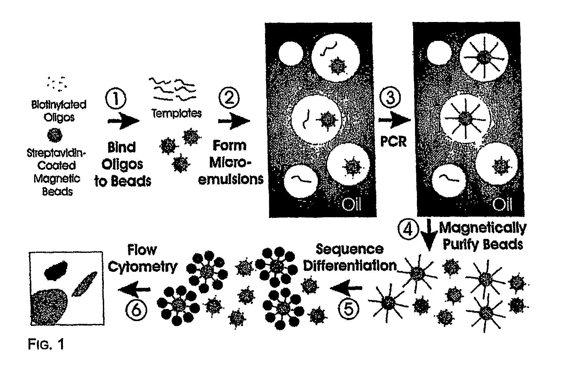
[0045] Step 1—Coupling oligonucleotides to beads. Superparamagnetic beads of 1.05+ / −0.1 um in diameter, covalently bound to streptavidin, were purchased from Dynal Biotech, Inc. (650.01, Lake Success, N.Y.). Beads were washed once with 1×PCR buffer (53286, Invitrogen, Carlsbad, Calif.) then suspended in Bind and Wash Buffer (BWB) (5 mM Tris-HCl, 0.5 mM EDTA, 1.0 M NaCl, pH 7.5). Beads were incubated in BWB for 30 min at room temperature in the presence of 10 uM oligonucleotides (FIG. 8). These oligonucleotides were modified with a dual biotin group at the 5′ end with the biotin groups separated by a six-carbon linker (IDT, Coralville, Iowa). After binding, the beads were washed 3 times with 1×PCR buffer to thoroughly remove unbound oligonucleotides.

[0046] Step 2—Preparing microemulsions. Microemulsions for PCR were prepared by slight modifications of previously described methods (14) (15). The oil phase was composed of 4.5% Span 80 (S6760, Sigma, St. Louis, M...

example 2

Results

[0053] Step 1—Coupling oligonucleotides to beads. We used streptavidin-beads because of the simplicity of coupling biotinylated oligonucleotides to them. Oligonucleotides with just a single 5′ biotin group were found to dissociate from the beads during temperature cycling, while oligonucleotides labeled with dual biotin groups at their 5′ end (separated by a six-carbon linker) were stable to cycling. As determined by fluoroscopic measurements of oligonucleotides doubly labeled with 6-FAM and biotin, ˜105 oligonucleotide molecules were bound to each bead. We found that short oligonucleotides (20 bases) did not work as well for priming as longer ones (41 bp), perhaps because of steric hindrance at the bead surface. It is likely that amino-, sulfhydryl-, or carboxyl-modified oligonucleotides covalently coupled to beads modified with corresponding reactive groups could also function as bead-bound primers for BEAMing.

[0054] Step 2—Preparing microemulsions. The size of the indiv...

example 3

Characteristics of Microemulsions.

[0059] Pilot experiments demonstrated that simply stirring the water-oil mixtures described in Materials and Methods produced very stable microemulsions of a size compatible with that of the beads. In the experiment shown in FIG. 2, the aqueous compartment contained a blue dye and 1 micron magnetic beads that were labeled by binding to an oligonucleotide that was biotinylated at its 5′ end and labeled with fluorescein at its' 3′ end The appearance of emulsions immediately after their formation is shown in FIG. 2. As expected, this appearance was unchanged after temperature cycling during PCR (15). Most aqueous compartments contained no beads, as expected from the figures provided in the previous section. Those compartments that did contain beads generally contained only one, though a fraction contained more, as expected from a Poisson distribution and non-uniform aqueous compartment sizes. “Heterozygous” beads containing PCR products representing ...


PUM

PropertyMeasurementUnit
Fractionaaaaaaaaaa
Fractionaaaaaaaaaa
Fractionaaaaaaaaaa

Abstract

Many areas of biomedical research depend on the analysis of uncommon variations in individual genes or transcripts. Here we describe a method that can quantify such variation at a scale and ease heretofore unattainable. Each DNA molecule in a collection of such molecules is converted into a single particle to which thousands of copies of DNA identical in sequence to the original are bound. This population of beads then corresponds to a one-to-one representation of the starting DNA molecules. Variation within the original population of DNA molecules can then be simply assessed by counting fluorescently-labeled particles via flow cytometry. Millions of individual DNA molecules can be assessed in this fashion with standard laboratory equipment. Moreover, specific variants can be isolated by flow sorting and employed for further experimentation. This approach can be used for the identification and quantification of rare mutations as well as to study variations in gene sequences or transcripts in specific populations or tissues.

Description

[0001] This application claims the benefit of applications Ser. No. 60 / 485,301 filed Jul. 5, 2003 and Ser. No. 60 / 525,859, filed Dec. 1, 2003, the contents of both of which are expressly incorporated herein.[0002] The invention disclosed herein was made using funds from the National Institutes of Health grants CA 43460, CA 57345, and CA62924. The United States government therefore retains certain rights in the invention.[0003] A portion of the disclosure of this patent document contains material which is subject to copyright protection. The copyright owner has no objection to the facsimile reproduction by anyone of the patent document or the patent disclosure, as it appears in the Patent and Trademark Office patent file or records, but otherwise reserves all copyright rights whatsoever. FIELD OF THE INVENTION [0004] The invention relates to the field of genetic analysis. In particular, it relates to methods and compositions for analyzing variations in individual genes or transcripts...

Claims


Application Information

Patent Timeline
IPC IPC(8): C12Q1/68C12P19/34C12M3/00C07H21/04C12NC12N15/10
CPCC07H21/04C12N15/1075C12Q1/6858C12Q2565/537C12Q2563/149C12Q2563/143C12Q1/686
Inventor DRESSMAN, DEVINYAN, HAIKINZLER, KENNETHWVOGELSTEIN, BERT
Owner THE JOHN HOPKINS UNIV SCHOOL OF MEDICINE
Why Eureka
Try Eureka