Biopsy incision closure device

a biopsy incision and closure technology, applied in the field of medical devices and methods, can solve the problems of insufficient closure force, and achieve the effects of promoting eversion, reducing unwanted tissue inversion effects, and improving healing

Inactive Publication Date: 2014-03-13
ZIPLINE MEDICAL
View PDF12 Cites 23 Cited by
  • Summary
  • Abstract
  • Description
  • Claims
  • Application Information

AI Technical Summary

Benefits of technology

[0019]The reinforcement members may provide structural benefits as well. The members may minimize unwanted tissue inversion effects which could result from torque applied to the legs of the frame by the closing mechanism. Since the latch mounts above the skin, a moment arm is created wherever the latch(es) attaches to the device which can twist the mounting point and the frame resulting in inverted incision edges. The “spider leg” geometry of the reinforcement members can act as a struts or ribs to counteract this torque because they convert torque forces into normal forces (perpendicular to the skin plane) under each reinforcement member. Since the reinforcement member extends relatively far from the incision, twisting of the frame and subsequent wound inversion is inhibited.
[0020]In an additional aspect, the present invention can provide biopsy closure devices which can evert the edges of the tissue as they are brought together in order to improve healing. In such embodiments, an eversion lip will be provided along the edges of the first and second legs of the frame so that the lips engage the tissue and extend inwardly from the elliptical periphery of the frame when present on the tissue. The eversion lip is attached to the frame with a living hinge or otherwise so that it will evert upwardly as the frame is closed, thus lifting the tissue to provide the desired tissue edge eversion. Such tissue eversion promoting may also be achieved by deflecting the frame legs so that they are “normally” in a lifted state, so that when the base is adhered to the skin (it must be pressed down a bit to fully contact the skin), the upwardly deflected inner portions of the base will lift the skin slightly to promote eversion upon closure. This approach may be in addition to or an alternative to the hinged approach described above.

Problems solved by technology

While the legs will be biased to closing, usually the closure force is not sufficient to close the tissue after biopsy, and a further latching or other closure device will be needed to close the tissue opening, as described below.

Method used


Image

  • Biopsy incision closure device
  • Biopsy incision closure device
  • Biopsy incision closure device

Examples


Embodiment Construction

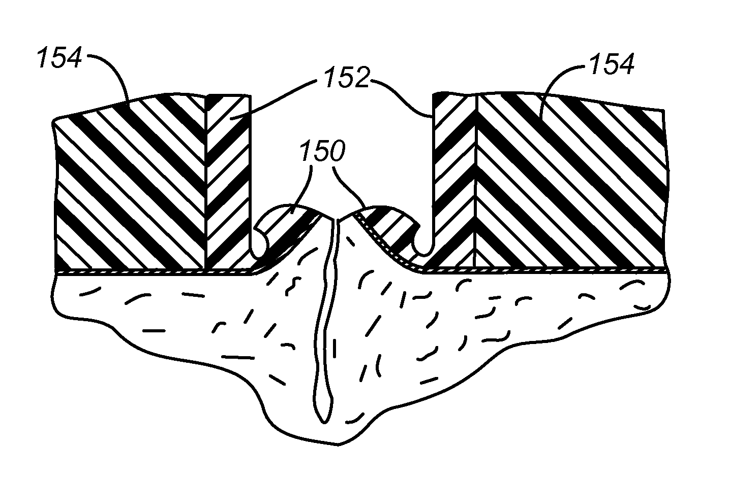
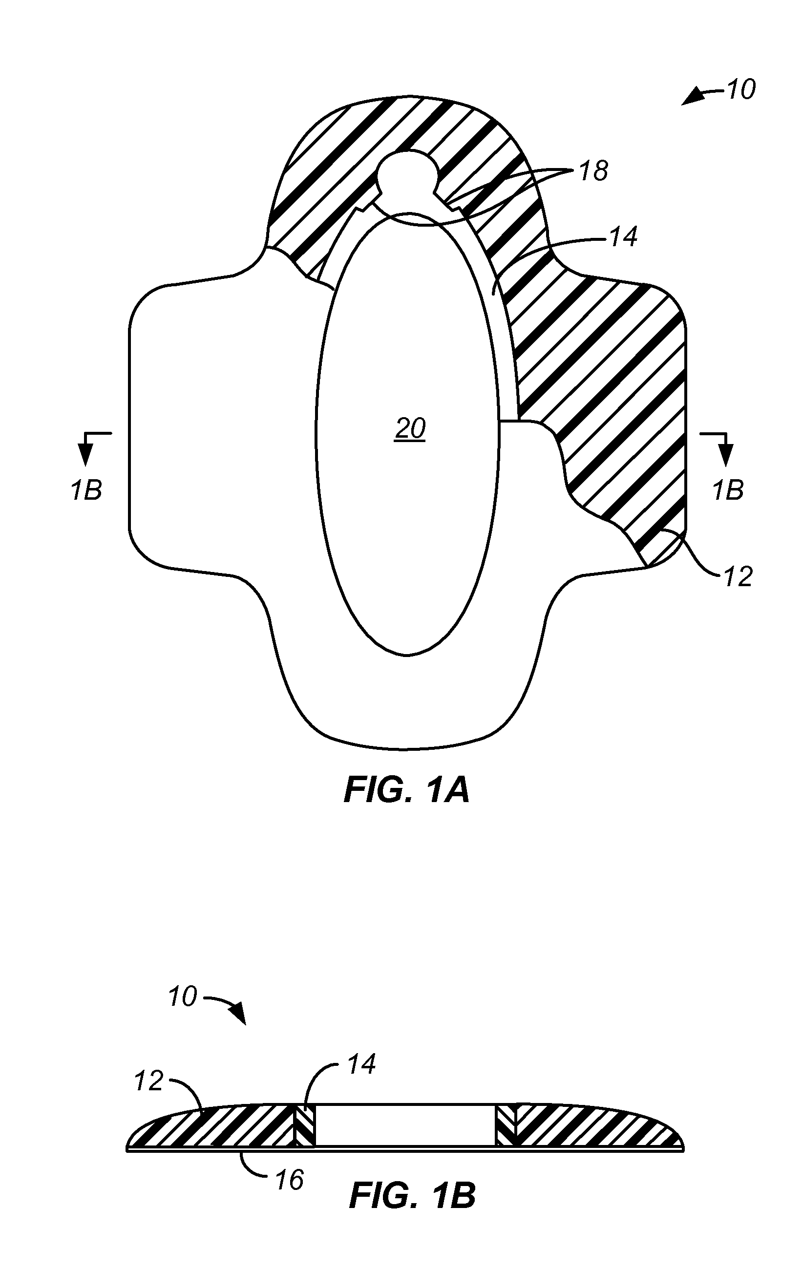
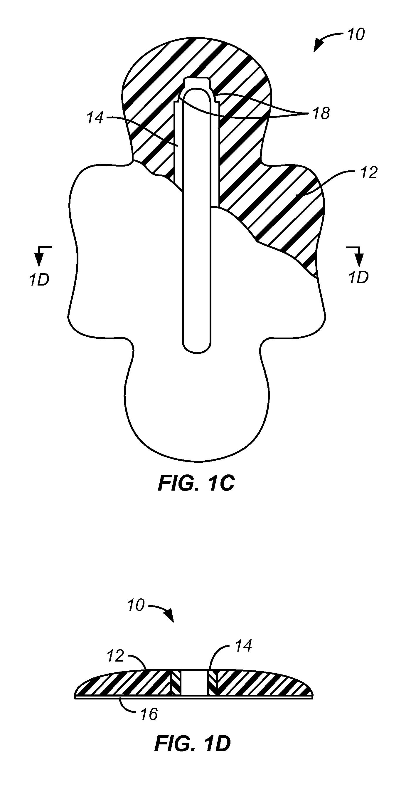
[0048]Referring to FIGS. 1A through 1D, a biopsy incision closure device 10 comprises a base 12 having an integrated or embedded frame 14, typically formed by overmolding a soft polymeric base material over a preformed metal or hard plastic frame. The frame 14 is resilient and, when free from biasing forces, assumes the elliptical or oval configuration seen in FIG. 1A. The frame 14 has living hinges 18 at each end (only one of which is visible in the broken-away section of the base) which allow the base to be closed by applying laterally inward forces to the frame, as shown in FIGS. 1C and 1D. Laterally inward forces may be provided by any one of a variety of external closure devices which could be simple tapes, patches, sutures, or the like. Closure devices could be more complex, including zippers, clips, and other structures as taught in copending PCT Application PCT / US2010 / 00430, the full disclosure of which has been previously incorporated herein by reference. Regardless of the ...


PUM


Abstract

A biopsy incision closure device includes a base having a frame incorporated therein. Together, the base and frame define an opening for performing a biopsy incision when the device is placed over a tissue surface. The base is typically composed of an elastomeric material and the frame comprises resilient inelastic members which can be used to close the opening in a highly uniform manner with minimum distortion and stress introduced into the tissue edges being drawn together.

Description

CROSS REFERENCE TO RELATED APPLICATIONS[0001]This application is a continuation U.S. Patent Application No. 13 / 096,602, filed Apr. 28, 2011, which claims the benefit of the following provisional applications: Provisional Application No. 61 / 343,916, filed on May 3, 2010; Provisional Application No. 61 / 397,604, filed on Jun. 14, 2010; and Provisional Application No. 61 / 462,329, filed on Feb. 1, 2011, the full disclosures of which are incorporated herein by reference.BACKGROUND OF THE INVENTION[0002]1. Field of the Invention[0003]The present invention relates generally to medical devices and methods. More particularly, the present invention relates to a device and method for closing a wound resulting from tissue biopsy.[0004]Excisional biopsy typically removes an elliptical section of tissue, usually containing the full dermis and in some cases the subcutaneous fatty layer as well. Such biopsies typically leave an elliptical opening in the skin that requires closing. Such elliptical bi...

Claims


Application Information

Patent Timeline
Patent Type & Authority Applications(United States)
IPC IPC(8): A61B17/08
CPCA61B17/085A61B17/0293A61B2017/00407A61B2017/00862A61B2050/008
Inventor BELSON, AMIRSTORNE, ERICBECKEY, BRIAN
Owner ZIPLINE MEDICAL
Why Eureka
Try Eureka