Stent for a vascular meniscal repair and regeneration

a meniscal and vascular technology, applied in the field of surgical devices for repairing or regenerating body tissue, can solve the problems of meniscal tears and defects, white zone and red/white zone tears and defects that typically do not heal, and achieve the effects of facilitating healing and/or regeneration, facilitating its fixation to the meniscal tissue, and facilitating the healing of an avascular

Active Publication Date: 2007-03-22
HOWMEDICA OSTEONICS CORP
View PDF98 Cites 191 Cited by
  • Summary
  • Abstract
  • Description
  • Claims
  • Application Information

AI Technical Summary

Benefits of technology

[0011] It is an objective of the present invention to provide a channel for blood, blood components and / or nutrients, and cells to travel from the vascular (red) zone such as in knee meniscus or synovium (i.e. knee capsule) to the avascular (white) or partially vascular (red / white) zone to facilitate healing and / or regeneration in these zones.
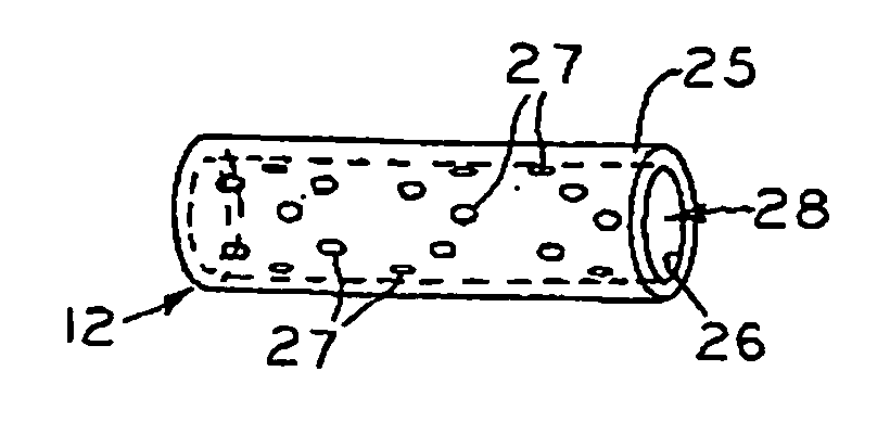
[0012] It is also an objective of the present invention to provide a biocompatible tube. The tube can have a stopping brim to prevent it from being inserted completely through the tissue. The tube is intended to be located within meniscal tissue such that it provides a channel from the vascular zone of the tissue (meniscus or synovium) to the avascular (white) or partially vascular (red / white) region. The tube wall can have openings, perforations, holes, or porosity that allow for blood, nutrients, and cells to enter the tube through the walls of the tube or stent. The tube wall exterior can be roughened or have protrusions or threads that will facilitate its fixation to the meniscal tissue. The “tube” could be a cylinder with a porous configuration such that blood, nutrients, and cells could travel within and through the device.
[0013] It is also an objective of the present invention to provide a pathway through which blood, nutrients, and cells can pass to facilitate healing of an avascular (or partially vascular) tear / defect or to facilitate regeneration of avascular (or partially vascular) tissue when an implant is placed in addition to the tube(s) after performing a partial meniscectomy. In the case of a partial meniscectomy, the channel could function to deliver a blood or fibrin clot to the volume space of meniscus that was removed such that the clot acts as a scaffold in which cells can travel and propagate, thus, facilitating regeneration of that portion of the meniscus. In this case the open channel would also provide the access of the vascular area components to the in situ scaffold (i.e. blood or fibrin clot).
[0014] It is also an objective of the present invention to be comprised of a network of biocompatible tubes that are either attached to, integral with, or in close proximity to a meniscus implant. The implant is also comprised of a biocompatible material and can have interconnected porosity. The tube can have a stopping brim to prevent it from being inserted completely through the tissue. The meniscus implant / tube(s) device is located adjacent to avascular (white or red / white) meniscal tissue such that the tubes protrude into the meniscal tissue to or through the vascular tissue (meniscus or synovium). The tube(s) provides a channel from the vascular zone of the tissue (meniscus or synovium) to the avascular or partially vascular region into or onto the meniscus implant. This meniscus implant / tube(s) device (i.e. tubes integrated or attached to a scaffold) provides a pathway through which blood, nutrients, and cells can pass to the meniscus implant so that healing and regeneration of an avascular (or partially vascular) defect or tear can be accomplished. The tube portion of the meniscus / tube(s) device can have any or all of the same features as described in the tube device alone.
[0015] It is also an objective of the present invention to provide a method for repairing damaged or diseased fibrocartilage tissue (i.e. meniscus of the knee, labrum of the shoulder, acetabular labrum of the hip, articular disc of the wrist, spinal disc, temporomandibular disc, etc.). After locating the tear or degeneration in the avascular or partially vascular zone, one of two tasks can be performed. The tissue can be removed from the inner portion of the tear (i.e. perform a partial meniscectomy) or the tear can be repaired using a number of standard repair techniques (vertical or horizontal mattress suturing, Mitek's RapidLoc™ Meniscal Repair Device for the meniscus, Bionx Arrow™ for the meniscus, etc.). If a partial meniscectomy is to be performed followed by implantation of a meniscus regenerating or replacing device or implant, then the stent can be placed into the remnant native meniscal tissue such that it provides an open channel through which blood, nutrients, and cells can flow from the vascular region of the tissue to the implant, thus facilitating healing or regeneration. After a partial meniscectomy, a meniscus / tube(s) device (i.e. tubes integrated or attached to a scaffold) could be implanted and fixed to the remaining native meniscal tissue such that the tube portion of the device protrudes into and / or through the vascular zone of the meniscus and / or synovium. The delivery of the meniscus / tube(s) device could be accomplished arthroscopically with standard techniques. The fixation could be accomplished arthroscopically as well using standard devices and techniques (suture, meniscal repair devices such as DePuy Mitek's RapidLoc, Linvatec's Bionx Arrow, etc.). If the tear is to be repaired (instead of removing the tissue via a partial meniscectomy), then the stent can be arthroscopically placed either using an all inside or inside-out technique from the outer tear surface to or through the vascular region (providing a channel) or through both tear surfaces to the vascular region (providing a channel and a fixation for the tear), thus, facilitating repair of the avascular or partially vascular tear. Alternatively, the stent could be arthroscopically delivered using an outside-in technique.
[0016] It is also an objective of the present invention to provide a method for delivering biological treatments [i.e. blood, platelet rich plasma, bone marrow, stem cells, fibroblast cells, synoviocyte cells, other cells, angiogenic factors (new blood vessel formation growth factors such as VEGF, IGF, etc. . . . ), other growth factors, hyaluronic acid, gene therapies, other biologic molecules etc.], drugs [analgesic, anti-clotting, clotting, anti-inflammatory, anti-infectives, etc. . . . ], and other substances to the tear or defect area through the tube. After the tube device is positioned or during / before the insertion process, a substance could be delivered through the tube either to the tear / defect area or to the vascular area. The substance could enhance or initiate healing, increase blood flow, improve angiogenesis, induce clotting in the duct and tear / defect, deliver cells, deliver growth factors, deliver biologic elements, etc.

Problems solved by technology

Similar actions in the other joints can result in similar defects and tears in the similar structures present in those joints.
Meniscus degeneration can also occur as a result of aging so that soft or hard areas develop in the tissue such that even common activities such as squatting can cause meniscal tears and defects.
Meniscectomies typically provide immediate pain relief and restoration of knee function to the patient; however, with the absence of the meniscus, the long term effect on the knee can be cartilage wear on the condylar or tibial plateau surfaces and the eventual development of an arthritic condition such as osteoarthritis.
Clinical experience indicates that white zone and red / white zone tears and defects typically do not heal even if they are stabilized with standard repair techniques.
The option of not treating these types of defects is known to result in propagation of tears and defects and degeneration of the meniscus and subsequent degeneration of the articular cartilage and development of osteoarthritis.
Most surgeons, however, do not attempt to deliver and fix blood / fibrin clots to facilitate the repair of these types of tears because of the technical challenges.
With the fluid turned off, the surgeon has the technical challenge of not being able to see the surgical site clearly; therefore, a technical dilemma exists: in order to see more clearly the fluid needs to be turned on, but in order to deliver and fix the clot the fluid needs to be turned off.
Therefore, the technical challenges are too difficult to overcome in an arthroscopic environment; the surgeon therefore typically excises injured or degenerated white zone and red / white zone tissue (i.e. performs a partial or total meniscectomy).
Performing these procedures in a non-arthroscopic setting (i.e. open condition) is not a viable option due to patient expectations, increased morbidity, and increased risks associated with larger incisions.
These implants are being developed to regenerate meniscal tissue; however, they are effective only when the implant is placed in direct contact with the vascular (red) zone of the meniscus.
Therefore, if the defect area is confined to the avascular zone only, then one of the meniscal implants referred to above will not regenerate that tissue.
Unfortunately for the patient who receives the partial meniscectomy, the long term prognosis includes chronic knee pain, break down of the articular cartilage, osteoarthritis, and even eventual total knee replacement.
Since much of the nutrients for the spinal disc are delivered via diffusion through the vertebral endplates, the sclerotic bone could tend to decrease the amount of nutrients delivered to the disc, thus contributing to the diseased state of the disc.

Method used


Image

  • Stent for a vascular meniscal repair and regeneration
  • Stent for a vascular meniscal repair and regeneration
  • Stent for a vascular meniscal repair and regeneration

Examples


Embodiment Construction

[0054]FIG. 1 shows a view of a normal meniscus. The meniscus has a triangular cross section as shown in FIG. 2. The top of the articulating surface 1 interfaces with the femoral condyle and the bottom of the articulating surface 4 interfaces with the tibial plateau. The inner edge 8 and the outer rim 2 are indicated in the figure. The outer wall 3 defines the outermost boundary of the meniscus tissue.

[0055]FIG. 2 shows a cross sectional view of a normal meniscus showing approximate locations of the vascular (red) zone 7, avascular (white) zone 5, and partially vascular (red / white) zone 6 in an adult human.

[0056]FIG. 3 shows a cross sectional view of a meniscus with a vertical tear 11 in the red / white zone 6. The inner and outer tear faces, 9 and 10, are indicated in this figure.

[0057]FIG. 4 shows a cross sectional view of a meniscus with a vertical tear 11 in the red / white zone 6 with a stent 12 inserted at the outer tear face 10 through the vascular (red) zone 7. The tear 11 has...


PUM


Abstract

A surgical stent made of biocompatible material for implantation in human tissue to enable blood and nutrients to flow from an area of vascular tissue to an area of tissue with little or no vasculature.

Description

FIELD OF INVENTION [0001] The present invention relates generally to surgical devices for repairing or regenerating body tissue and more specifically to surgical devices for repairing or regenerating soft tissues (i.e. articular cartilage, fibrocartilage, collagenous structures, ligaments, tendons, meniscus, spinal disc, TMJ disc etc . . . ) of the joints (knee, hip, shoulder, temporomandibularjoint, spine, fingers, ankle, toes, etc . . . ), and to surgical methods using such devices. BACKGROUND OF INVENTION [0002] Meniscus tissue is comprised of a type of tissue known as fibrocartilage. Fibrocartilage is present in the form of a disc (spine, temporo-mandibular joint), meniscus (knee), labrum (shoulder, hip), etc. In the knee, as shown in FIG. 1, the meniscus is a semi-lunar, wedge shaped tissue that sits on top of the tibia and articulates with the tibia and femur during gait activities. It acts as a shock absorber between the femur and tibia and distributes the compressive and she...

Claims


Application Information

Patent Timeline
Patent Type & Authority Applications(United States)
IPC IPC(8): A61F2/06A61F2/02A61F2/30A61F2/38A61F2/82
CPCA61F2/02A61F2/2493A61F2002/30766A61F2/82A61F2002/30761A61F2/3872B33Y80/00A61F2/06
Inventor SCHWARTZ, HERBERT EUGENE
Owner HOWMEDICA OSTEONICS CORP
Why Eureka
Try Eureka