
Imaging and lighting optics of a contact eye camera

a technology of contact eye and imaging camera, which is applied in the field of ophthalmic examination equipment, can solve the problems of limited design of light conditioning optics, unique manufacturing process difficulties, and difficulty in achieving reliable sealing of contact optics

Inactive Publication Date: 2014-03-20
VISUNEX MEDICAL SYST
View PDF6 Cites 28 Cited by
  • Summary
  • Abstract
  • Description
  • Claims
  • Application Information

AI Technical Summary

Benefits of technology

The patent describes a special assembly process for an imaging system that uses a gap between the optical window and the imaging lens to ensure proper alignment and optical performance. The process involves aligning and sealing the optical window with the shell of the system and then mounting the rest of the lenses and lighting fixtures separately. This alignment and gap between the optical window and lens are critical for optimal optical performance. The use of the gap also helps to reduce thermal stress and allows for more sophisticated sealing techniques.

Problems solved by technology

However, such design has exhibited its unique difficulty in the manufacturing process and was not materialized in the real products.
Although the improvement in the design is significant, it also comes with its own problems, including for example the limitation on the design of light conditioning optics and the difficulty in achieving reliable sealing for the contact optics.

Method used


Image

  • Imaging and lighting optics of a contact eye camera
  • Imaging and lighting optics of a contact eye camera
  • Imaging and lighting optics of a contact eye camera

Examples


first embodiment

[0047]Solid state light emitting devices are used as the light sources in the imaging apparatus discussed in this invention. In the imaging apparatus, as shown in FIG. 16, the light sources 1603 are placed directly against the light conditioning device 1602. The light source 1603 could include light emitting elements and heat sink which is used to disperse the heat generated by the emitters. The light from light sources is guided into the posterior of the eye through the device 1602 and optical window 1601 in the manners discussed in previous paragraphs. The light sources, together with the heat sink, are placed outside an inner shell 1606 which houses the optical imaging lenses, including the lens 1604. The light sources are powered electrically through the electric wires 1605 laying along the outer surface of the shell 1606. In case where the imaging apparatus is designed with two separated sections, an electric connector 1607 is used to interconnect the wires 1605 and the wires 1...

second embodiment

[0048]The location of the light sources shown in FIG. 16 should be distributed evenly in order to provide uniform illumination on the retina. The numbers of the light sources in need could vary, depending on the requirement of the particular application. Two embodiments of the design are demonstrated in FIG. 17, where a total of 8 and 4 light sources are used. In one embodiment, the light emitting elements 1702 is mounted onto a heat sink 1701 which forms a ring to increase its mass and heat dispersion capability. 8 of the elements 1702 are distributed evenly on the heat sink. Those light sources could be lighted up simultaneously and be synchronized with the shutter of the image sensor. It is important to point out that more or less numbers of light elements could be used, even to the point where the light sources actually form a linear line source. In the second embodiment, only 4 light emitting elements 1704, 1705, 1706, 1707 are used and mounted onto the heat sink 1703. Those li...


PUM


Abstract

The optical design for a contact type portable eye imaging apparatus, including the imaging optics and illumination optics are proposed, which utilizes miniature image sensor and solid state light emitting technology, as the next generation of medical imaging devices, in particular in ophthalmic imaging applications.

Description

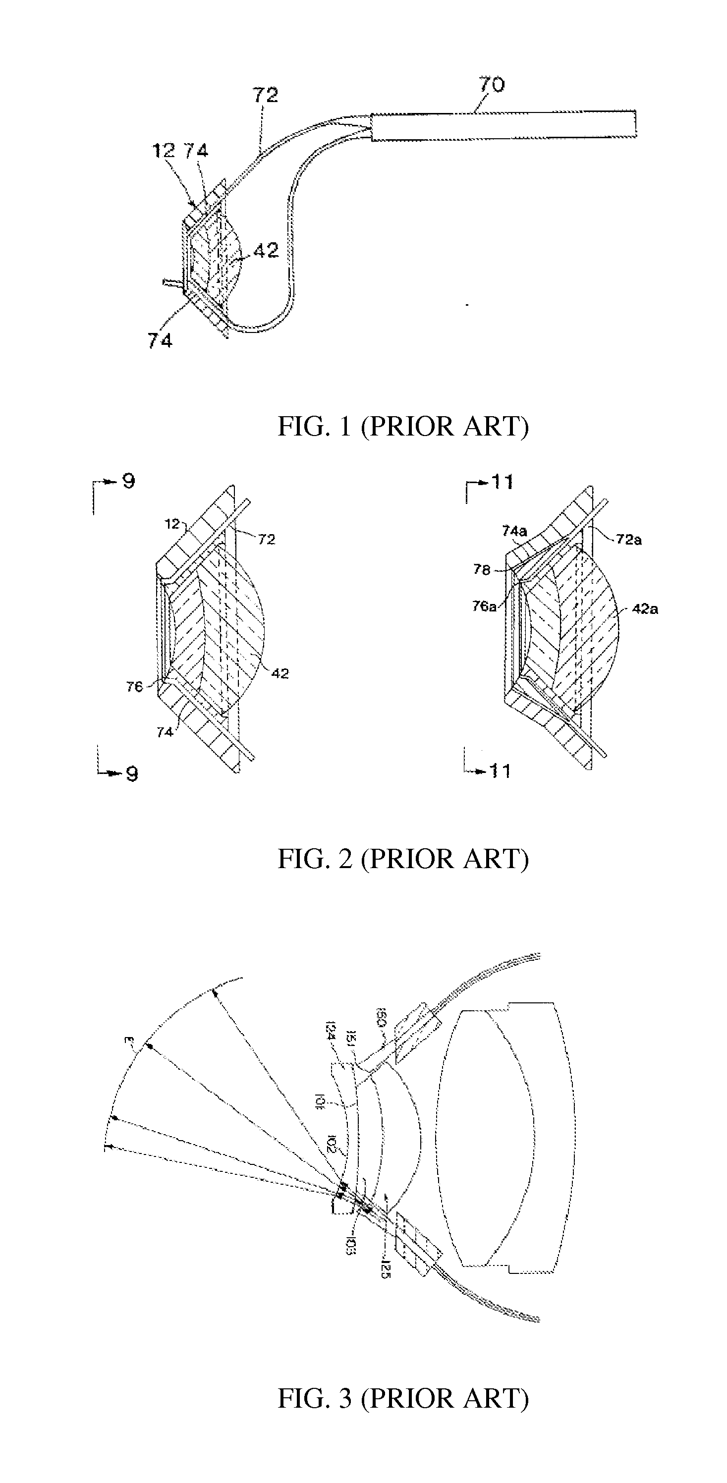
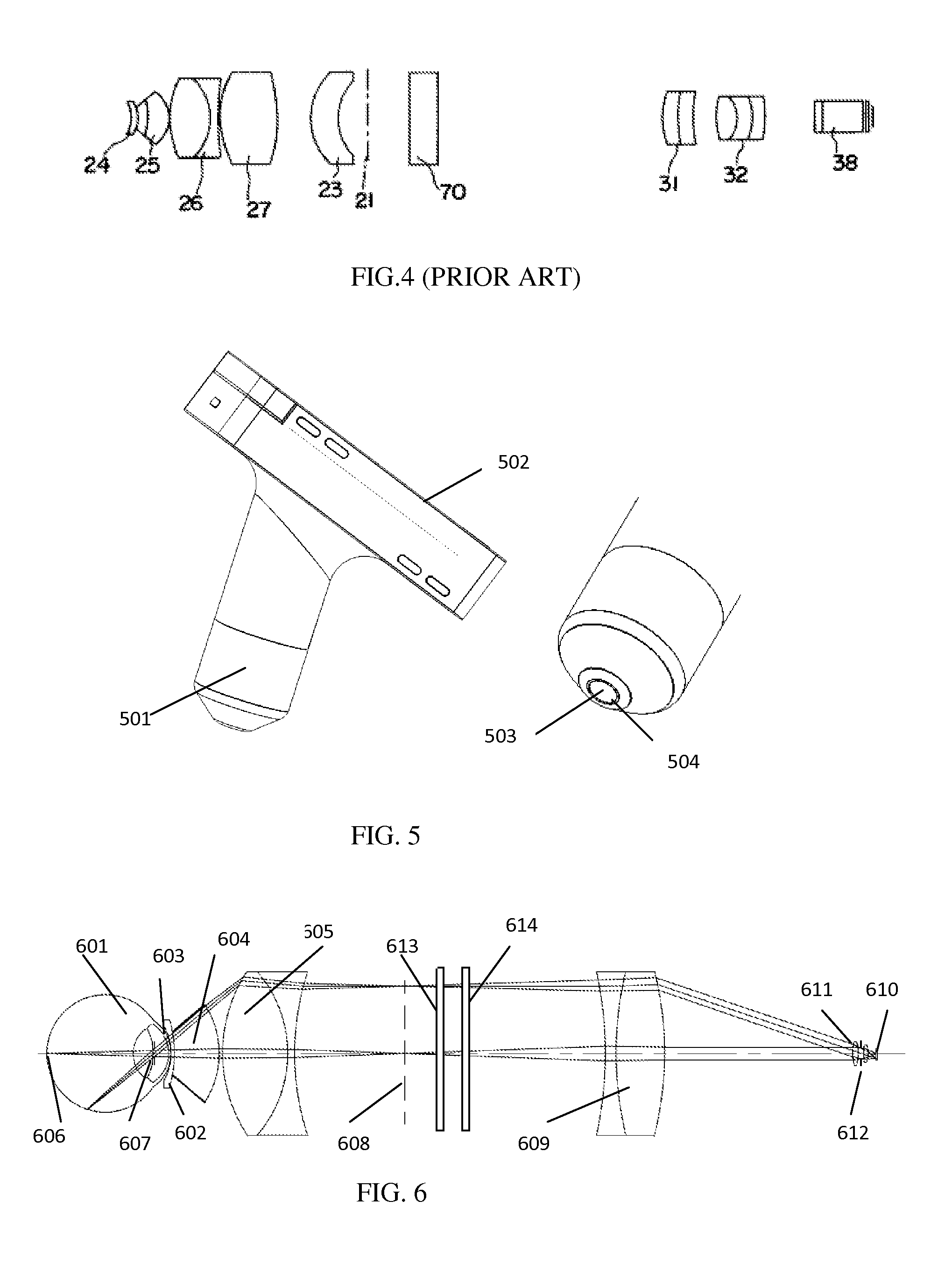
BACKGROUND OF THE INVENTION[0001]1. Field of the Invention[0002]The present invention is related to an ophthalmic examination apparatus, more particularly a contact eye imaging camera which uses the miniature digital image sensor for imaging and the solid state light emitting devices for lighting.[0003]2. Description of the Prior Art[0004]Several portable contact eye imaging systems have been proposed to photograph the posterior of human eyes (fundus camera) over the years. One of such, as disclosed in U.S. Pat. No. 5,608,472, is confined to use a fiber optical cable to guide the light from a remote light source to a hand held camera, which avoids the heat generated from the high brightness light source. The extension of the fiber cable also forms a circular light illumination device in order to distribute the light uniformly on to the posterior of an eye, as shown in FIG. 1. The details of the fiber optical ring structure are demonstrated in FIG. 2, where the fibers are tightly wra...

Claims


Application Information

Patent Timeline
Patent Type & Authority Applications(United States)
IPC IPC(8): A61B3/15
CPCA61B3/158A61B3/1208A61B3/132A61B3/0008A61B3/125
Inventor SU, WEI
Owner VISUNEX MEDICAL SYST
Learn More