Memory sensitive medical image browser

a medical image and memory technology, applied in image analysis, healthcare informatics, instruments, etc., can solve the problems of increasing cost and decreasing productivity

Inactive Publication Date: 2014-05-22
GENERAL ELECTRIC CO
View PDF4 Cites 11 Cited by
  • Summary
  • Abstract
  • Description
  • Claims
  • Application Information

AI Technical Summary

Benefits of technology

[0009]Certain examples provide a system including a medical image viewer. The example medical image viewer is configured to receive a request for a medical image study display at a browser on a client device. The example medical image viewer is configured to determine a size of the image study. The example medical image viewer is configured to determine available space on the client device for image data storage. The example medical image viewer is configured to determine a starting point for display of the image study via the browser. The example medical image viewer is configured to pre-load one or more images on the client device based on the starting point, image study size, and available space on the client device.
[0010]Certain e

Problems solved by technology

Often these are disparate, best-of breed systems that may or ma

Method used


Image

  • Memory sensitive medical image browser
  • Memory sensitive medical image browser
  • Memory sensitive medical image browser

Examples


Embodiment Construction

I. Overview

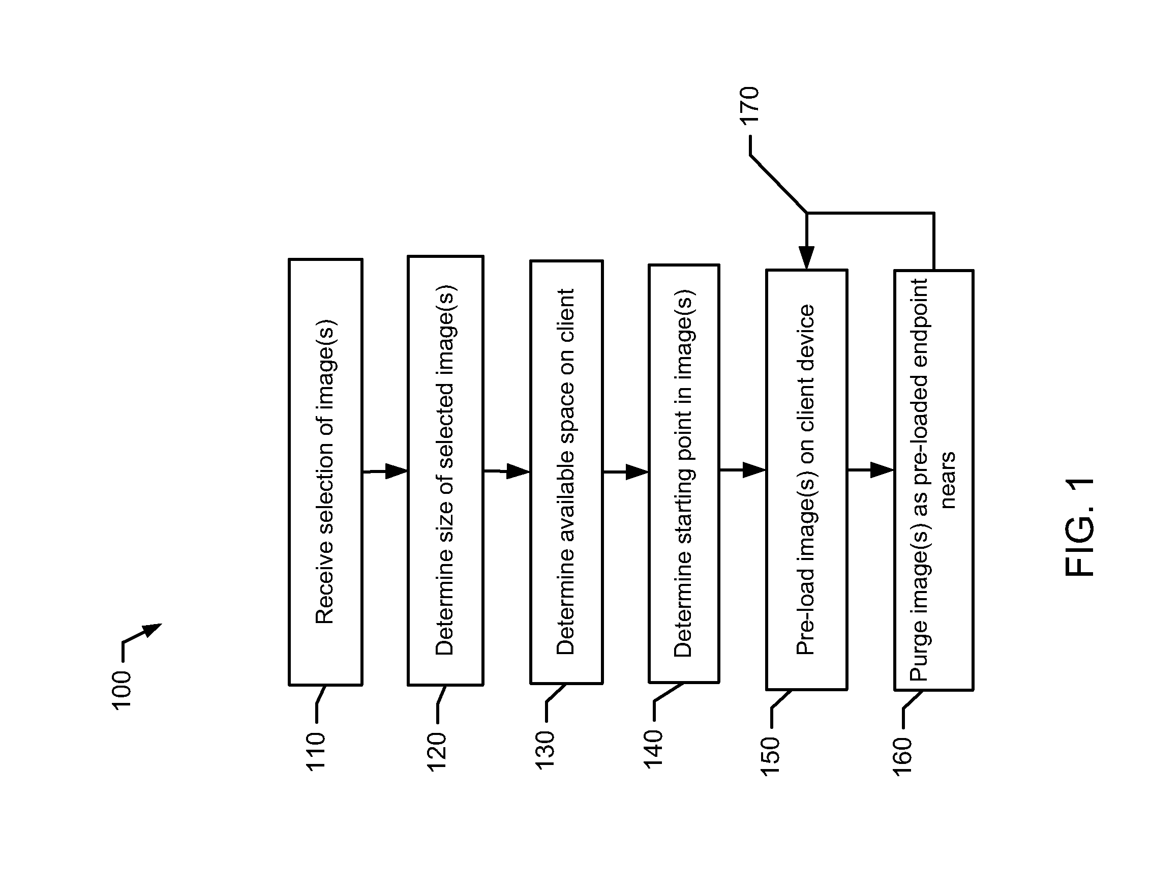
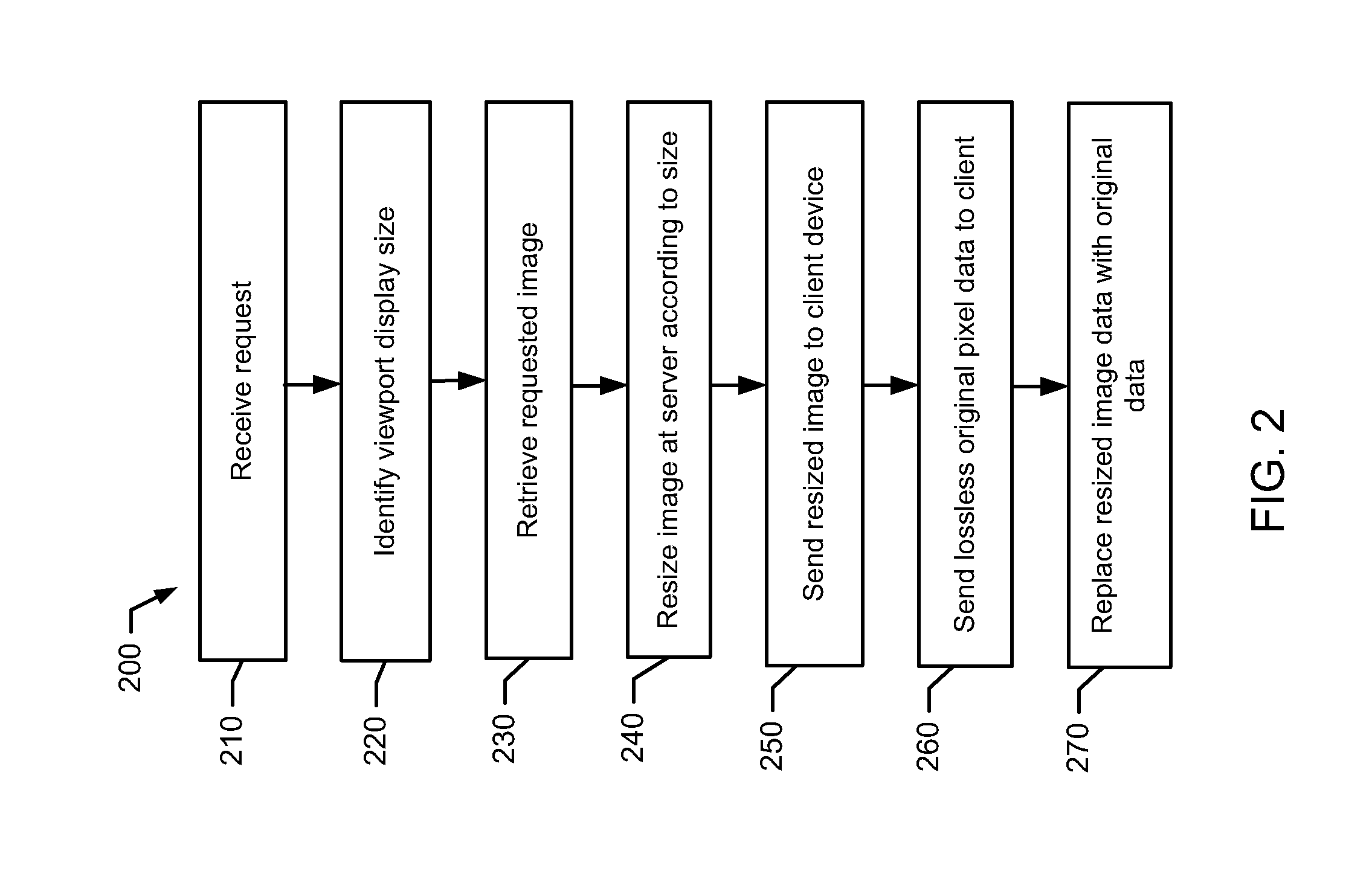
[0017]In certain examples, a unified viewer workspace for radiologists and clinicians brings together capabilities with innovative differentiators that drive optimal performance through connected, intelligent workflows. The unified viewer workspace enables radiologist performance and efficiency, improved communication between the radiologist and other clinicians, and image sharing between and across organizations, reducing cost and improving care.

[0018]The unified imaging viewer displays medical images, including mammograms and other x-ray, computed tomography (CT), magnetic resonance (MR), ultrasound, and / or other images, and non-image data from various sources in a common workspace. Additionally, the viewer can be used to create, update annotations, process and create imaging models, communicate, within a system and / or across computer networks at distributed locations.

[0019]In certain examples, the unified viewer implements smart hanging protocols, intelligent fetching ...


PUM


Abstract

Certain examples provide systems and methods for memory and/or device-sensitive image viewing. An example method includes receiving a request for a medical image study display at a browser on a client device. The example method includes determining a size of the image study. The example method includes determining available space on the client device for image data storage. The example method includes determining a starting point for display of the image study via the browser. The example method includes pre-loading one or more images on the client device based on the starting point, image study size, and available space on the client device.

Description

RELATED APPLICATIONS[0001][Not Applicable]FEDERALLY SPONSORED RESEARCH OR DEVELOPMENT[0002][Not Applicable]MICROFICHE / COPYRIGHT REFERENCE[0003][Not Applicable]BACKGROUND[0004]Prior to the rapid onset of digital imaging, patient images were “printed” to film. The film was “hung” and viewed by radiologists, who would then dictate a report. Reports were transcribed by individuals ranging for administrative staff to medical transcriptionists and sent to ordering physician via mail or fax. Critical results were delivered by phone or pager and business statistics were managed via paper reports and spreadsheets.[0005]As information systems for radiology came to market, the first commercially available solutions addressed the needs of the radiologist and the radiology department. These included Radiology Information Systems (RIS) and dictation transcription systems. RIS systems managed the ordering, scheduling, patient and management reporting processes while radiologists were still reading...

Claims


Application Information

Patent Timeline
IPC IPC(8): G06T7/00
CPCG06T7/0012G16H30/20G16H30/40
Inventor KLOTZER, JASON DIETER
Owner GENERAL ELECTRIC CO
Why Eureka
Try Eureka