
Mammography apparatus

a technology of mammography and x-ray image data, which is applied in the field of mammography equipment, can solve the problems of limited resolution of the plane from difficulty in detecting a malignant structure in the region of breast tissue, so as to improve the depth resolution of the 3d image data set, reduce scatter radiation during x-ray acquisition, and widen the selectable angle range

Active Publication Date: 2015-02-05
SIEMENS HEALTHCARE GMBH
View PDF3 Cites 19 Cited by
  • Summary
  • Abstract
  • Description
  • Claims
  • Application Information

AI Technical Summary

Benefits of technology

The present invention is a mammography apparatus that can improve image quality compared to conventional techniques. It reduces scatter radiation, improves depth resolution of a 3D x-ray image data set, and ensures that the distances traveled by the x-rays through the breast are approximately the same given different alignment angles of the x-ray source on the breast. It also provides an additional x-ray exposure in a second angle range to better depict malignant breast tissue and / or micro-calcification.

Problems solved by technology

A disadvantage of such imaging is that, due to overlapping tissue layers, it is often very difficult to detect a malignant structure in the region of a breast tissue.
Due to the incomplete scanning that results from this—i.e. due to the x-ray images being acquired only from a limited angle range of the breast—reconstruction of a plane from the 3D image data set has only a limited resolution.

Method used


Image

  • Mammography apparatus
  • Mammography apparatus
  • Mammography apparatus

Examples


Embodiment Construction

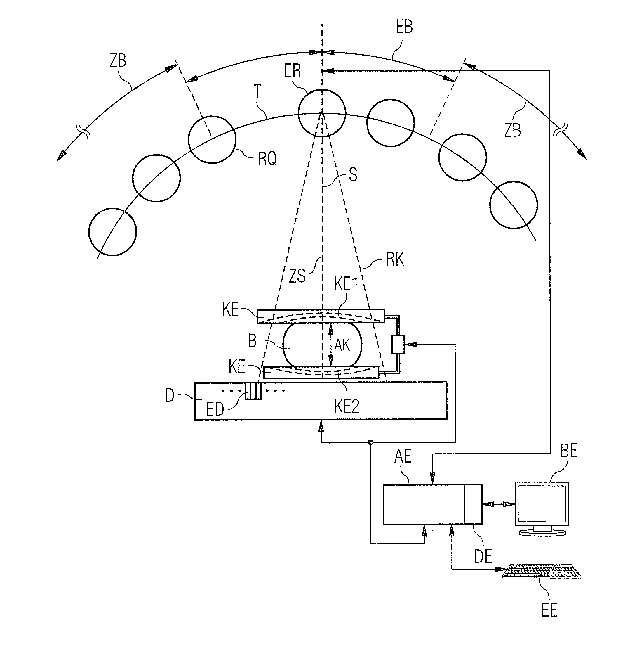
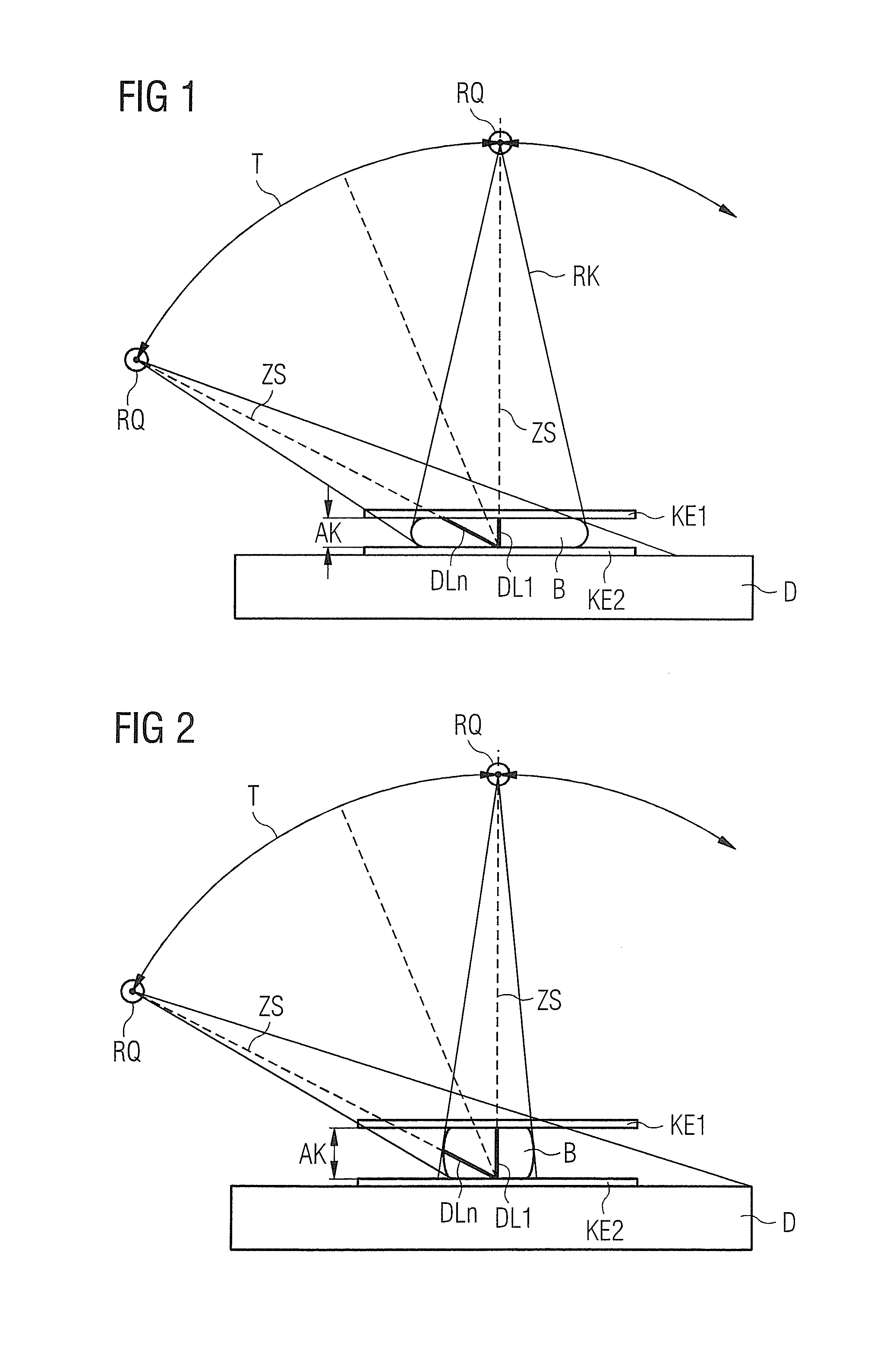
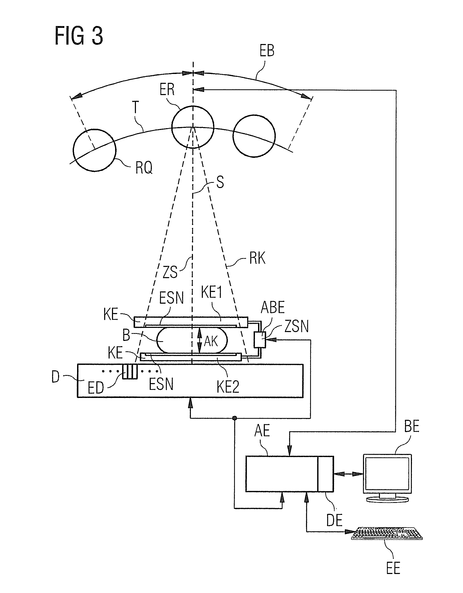
[0017]Among other things, with the mammography apparatus according to the invention the compression of the breast tissue is predetermined depending on the dimensions of the breast and / or the density of the breast tissue, such that, in the case of (for example) an arc-shaped trajectory of the x-ray source aligned on the detector, the path of the x-rays (for example of the central beam of an x-ray cone) through the breast remains within a predeterminable deviation at different positions of the x-ray source along the trajectory. The compression exerted on the breast by the compression unit is predetermined to achieve and is constantly checked. In the figures, it is described when a conventionally standardized compression exerted on the breast is adapted individually to the patient in the course of an x-ray examination. A first patient-specific adjustment of the compression pressure can take place directly after the overview acquisition, as described in FIGS. 1, 2 and 3. A variation of ...


PUM


Abstract

In a mammography apparatus, a deflection of an x-ray source aligned on a radiation detector is predetermined depending on the size of a breast compressed in the compression unit and / or the density of the breast tissue. Given an increasing deflection on an arc-shaped trajectory of the x-ray source, the compression of the breast is reduced at least once so that the path of the x-rays through the breast stays within a predetermined path length, starting from a first compression in a first x-ray acquisition.

Description

BACKGROUND OF THE INVENTION[0001]1. Field of the Invention[0002]The invention concerns a mammography apparatus with a compression unit, and a method for operating such a mammography apparatus.[0003]2. Description of the Prior Art[0004]Mammography is an x-ray examination of the breast with the goal of detecting tissue variations at an optimally early stage. By continuous improvement of the mammography method it is sought to generate x-ray images with high significance in order to differentiate benign tissue variations from malignant tissue variations. In conventional x-ray mammography, a single two-dimensional image of a breast is generated at a single projection direction. A disadvantage of such imaging is that, due to overlapping tissue layers, it is often very difficult to detect a malignant structure in the region of a breast tissue. In order to obtain a three-dimensional image of the breast tissue, a number of x-ray images of the breast are acquired from respectively from differ...

Claims


Application Information

Patent Timeline
Patent Type & Authority Applications(United States)
IPC IPC(8): A61B6/00A61B6/04
CPCA61B6/54A61B6/0414A61B6/502A61B6/025A61B6/4028A61B6/544
Inventor DORNBERGER, SUSANNEMERTELMEIER, THOMASNANKE, RALF
Owner SIEMENS HEALTHCARE GMBH
Learn More