Human monoclonal Anti-pd-l1 antibodies and methods of use

a monoclonal antibody and human antibody technology, applied in the field of antipdl1, can solve the problems of the progression physical deletion of the antigen-specific t cells themselves, and achieve the effect of increasing the activity of the antigen-specific t cell, enhancing the immune respons

Active Publication Date: 2015-10-01
DANA FARBER CANCER INST INC
View PDF3 Cites 60 Cited by
  • Summary
  • Abstract
  • Description
  • Claims
  • Application Information

AI Technical Summary

Benefits of technology

[0017]The present invention also provides methods of preventing or reversing T cell exhaustion comprising administering to a subject in need thereof a composition comprising any of the foregoing antibodies.
[0018]The present invention also provides methods of augmenting an immune response to an antigen comprising administering to a subject in need thereof a composition comprising any of the foregoing antibodies. In one aspect, the antigen is a viral antigen, a bacterial antigen or a tumor associated antigen. In another aspect, the viral antigen is HIV. In a further aspect, the tumor associated antigen is CAIX. In another aspect, the antibody is administered prior to or after exposure to the antigen. In another aspect, the administration of said antibody causes an increase in antigen specific T cell activity. In another aspect, the T-cell is an effector T cell.

Problems solved by technology

T cell exhaustion can also result in progressive physical deletion of the antigen-specific T cells themselves.

Method used


Image

  • Human monoclonal Anti-pd-l1 antibodies and methods of use
  • Human monoclonal Anti-pd-l1 antibodies and methods of use
  • Human monoclonal Anti-pd-l1 antibodies and methods of use

Examples


example 1

Generation of Human mAbs Against Pd-L1

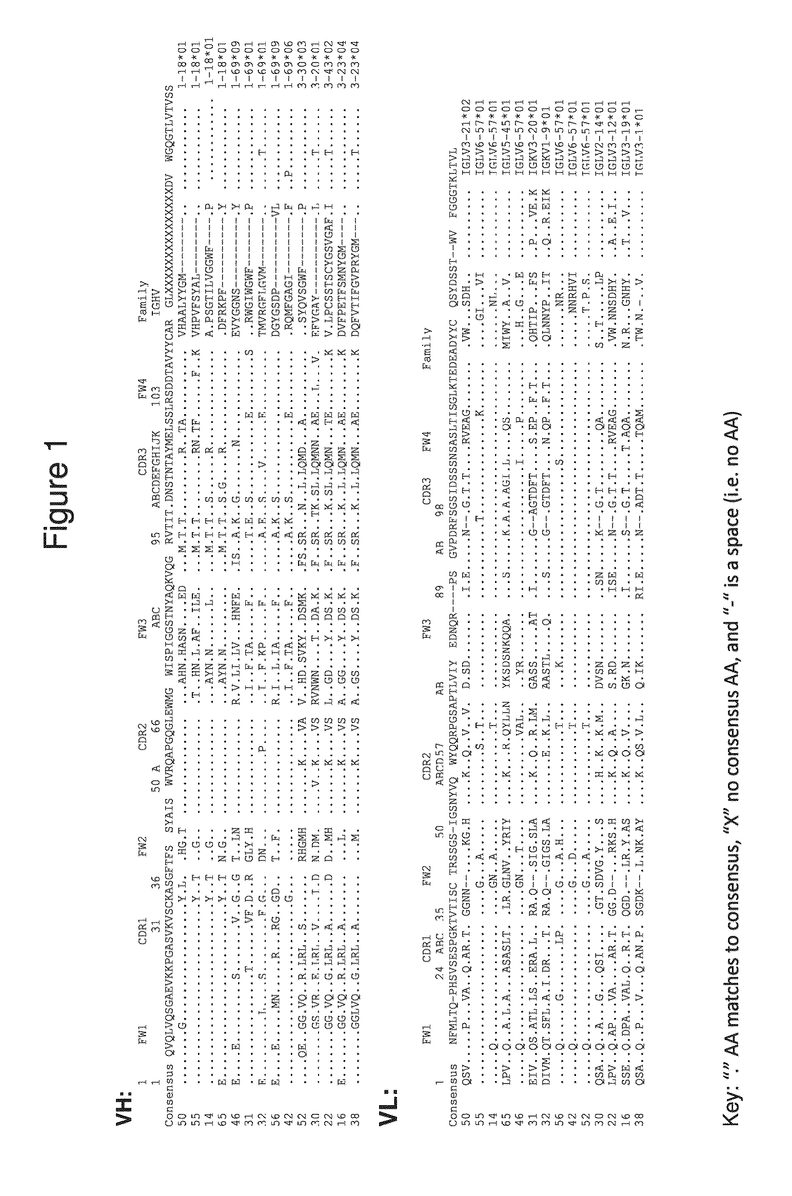
[0163]Human mAbs against human PD-L1 were generated by panning against a 27-billion member human scFv phage display library. Using full length PD-L1 in the form of paramagnetic proteoliposomes (PMPL), which assure proper orientation of the extracellular domain of PD-L1 for presentation to the library, 14 unique scFv-phage were identified that bind PD-L1. Human IgG constructs were constructed for these 14 unique scFv-phage: Ab-14, Ab-16, Ab-22, Ab-30, Ab-31, Ab-32, Ab-38, Ab-42, Ab-46, Ab-50, Ab-52, Ab-55, Ab-56 and Ab-65.

Example 2

Characterization of huPD-L1 mAbs Binding to Pd-L1

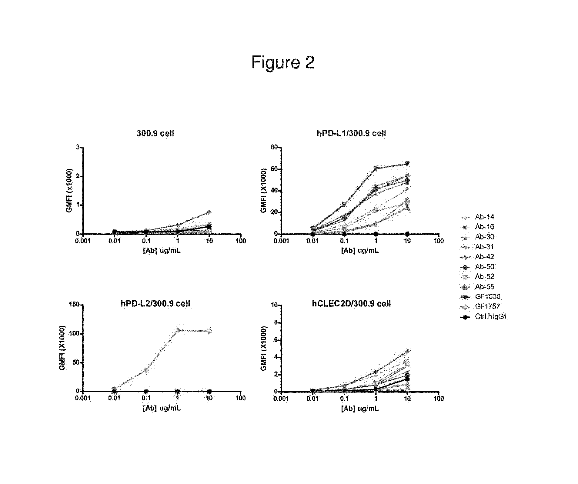
[0164]Binding analysis of huPD-L1 antibodies were performed using PD-L1-expressing cells. Four types of cells were tested, including parental cell line 300.19, and transfected cell lines expressing human PD-L1 (hPD-L1), human PD-L2 (HPD-L2), and human C-type lectin domain family 2 member (hCLEC2D). The binding assays were performed in duplicate, with the results summarize...

example 2

Characterization of Anti-PD-L1 Phage-Antibodies Blocking PD / PD-L1 Binding

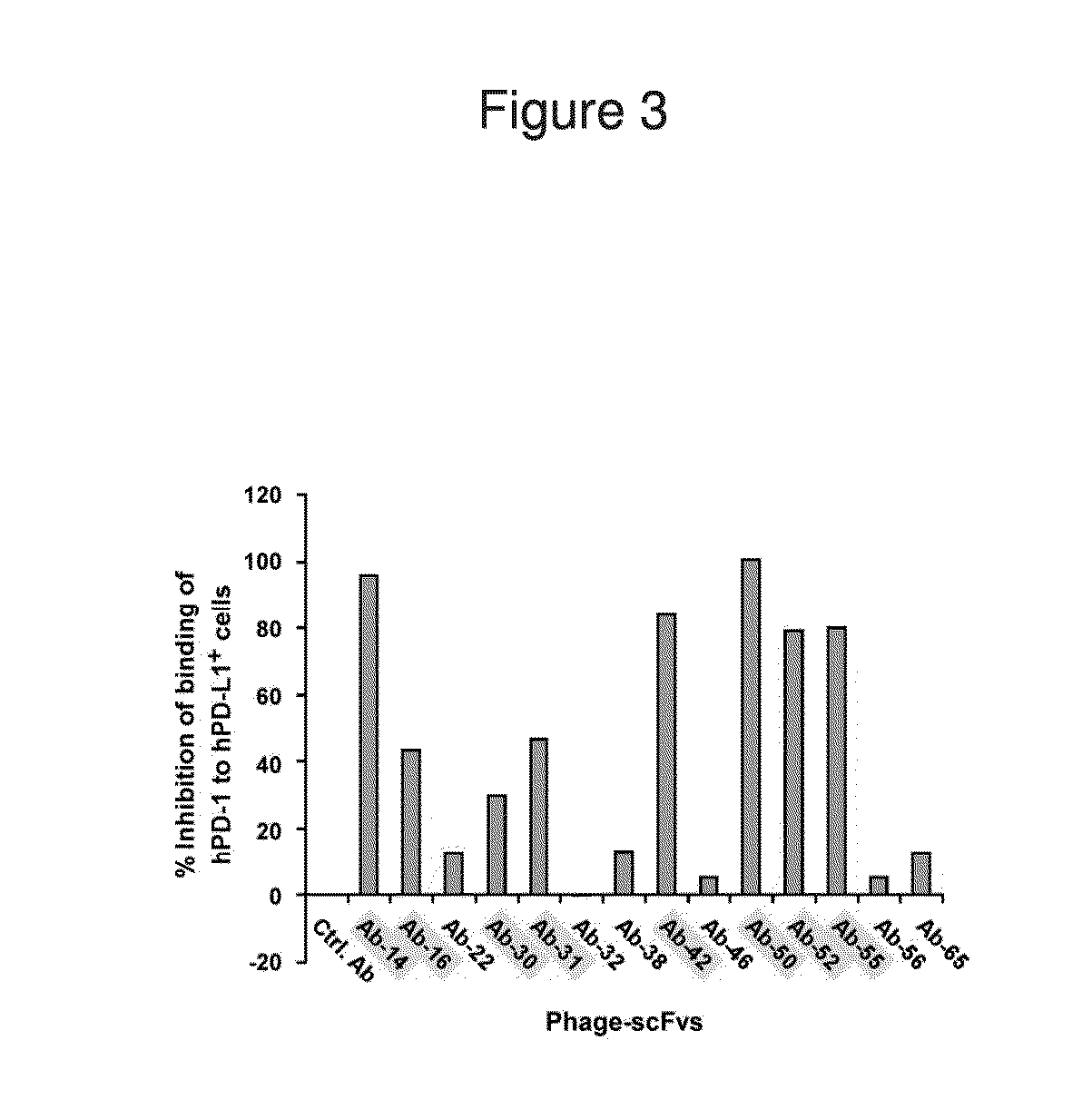
[0166]A competitive FACS analysis was performed to characterize the inhibition of hPD1 binding to hPD-L1 by anti-PD-L1 phage antibodies. All anti-hPD-L1 antibodies were in phage-scFv form. 293T cells were transfected with a vector encoding human PD-L1 fused to human Fc region for expression of hPD-L1-Fc. In this assay, 1012 plaque-forming units (pfu) of phage-scFvs were mixed with about 0.25 mg / ml of soluble hPD-1-hFc fusion protein and then added to the hPD-L1-expressing 293T cells. After washing, the cells were incubated with FITC-anti-human IgG antibody and analyzed by FACS to measure the binding of hPD1-hFc to hPD-L1 on the cell surface.

[0167]Fluorescence values were obtained by FACS analysis and used to generate a percentage of inhibition of binding of hPD-1 to hPD-L1+ cells. These values are displayed in FIG. 3. Almost all anti-PD-L1 phage scFvs demonstrated some ability to inhibit the binding of hPD-1 to...

example 3

Characterization of huPD-L1 Soluble Mabs Blocking PD / PD-L1 Binding

[0168]A competitive FACS analysis was performed to characterize the inhibition of hPD1 binding to hPD-L1 by the soluble huPD-L1 antibodies of the present invention. All huPD-L1 antibodies were tested for their ability to inhibit the binding of hPD1-IgG fusion protein with hPD-L1-expressing 300.19 cells. In this assay, 50,000 cells expressing hPD-L1 were pre-incubated with the huPD-L1 or control antibodies for 30 minutes at the following concentrations: 10 μg / ml, 1 μg / ml, 0.1 μg / ml and 0.01 μg / ml. After the pre-incubation, 0.125 μg of human PD1 fused to mouse IgG2α was added to the cells and incubated for another 30 minutes. The cells were washed twice, and then 0.125 μg of goat anti-mouse IgG2α-PE antibody was added to the cells. After 30 minutes of incubation, the cells were washed twice and then analyzed by FACS. The values obtained from FACS analysis are represented as mean fluorescence intensity units (MFI) and ar...


PUM

PropertyMeasurementUnit
timeaaaaaaaaaa
temperatureaaaaaaaaaa
concentrationsaaaaaaaaaa

Abstract

The present invention comprises human monoclonal antibodies that bind to PD-L1 (also known as programmed death ligand 1 or B7H1). Binding of the invented antibody to PD-L1 inhibits binding to its receptor, PD1 (programmed death 1), and ligand-mediated activities and can be used to treat cancer and chronic viral infections.

Description

RELATED APPLICATIONS[0001]This application claims priority to and benefit of U.S. Provisional Application No. 61 / 709,731, filed on Oct. 4, 2012, and U.S. Provisional Application No. 61 / 779,969 filed on Mar. 13, 2013; the contents of which are each hereby incorporated by reference in their entireties.FIELD OF THE INVENTION[0002]This invention relates generally to anti-PD-L1 (also known as programmed cell death 1 ligand 1 or B7H1) antibodies as well as to methods for use thereof.BACKGROUND OF THE INVENTION[0003]The immune system must achieve a balance between effective responses to eliminate pathogenic entities and maintaining tolerance to prevent autoimmune disease. T cells are central to preserving this balance, and their proper regulation is primarily coordinated by the B7-CD28 family of molecules. Interactions between B7 family members, which function as ligands, and CD28 family members, which function as receptors, provide critical positive signals that not only initiate, augment...

Claims


Application Information

Patent Timeline
Patent Type & Authority Applications(United States)
IPC IPC(8): C07K16/28C07K16/30C07K16/24C07K16/46
CPCC07K16/2896C07K16/468C07K16/3076C07K16/244C07K16/2803C07K16/30C07K2317/76C07K2317/622C07K2317/92C07K2317/31C07K2317/73C07K2317/732C07K2317/734C07K2317/565C07K16/2818C07K16/2827C07K16/2866C07K16/2878C07K2317/24C07K16/40C07K2317/21C07K2317/33C07K2317/526C07K2317/53A61P15/00A61P31/12A61P31/18A61P35/00
Inventor MARASCO, WAYNE A.SUI, JIANHUA
Owner DANA FARBER CANCER INST INC
Why Eureka
Try Eureka