Engineered three-dimensional breast tissue, adipose tissue, and tumor disease model
a three-dimensional breast tissue and tumor disease technology, applied in the field of three-dimensional breast tissue, adipose tissue, tumor disease model, can solve the problems of current two-dimensional cancer models not providing a physiologically relevant stromal milieu, current two-dimensional cancer models fail to accurately model disease initiation and progression, and current two-dimensional cancer models do not adequately demonstrate a native-like response to anti-cancer therapeutic agents
- Summary
- Abstract
- Description
- Claims
- Application Information
AI Technical Summary
Benefits of technology
Problems solved by technology
Method used
Image

Examples
example 1
Engineered Human Breast Tissue Model
Fabrication
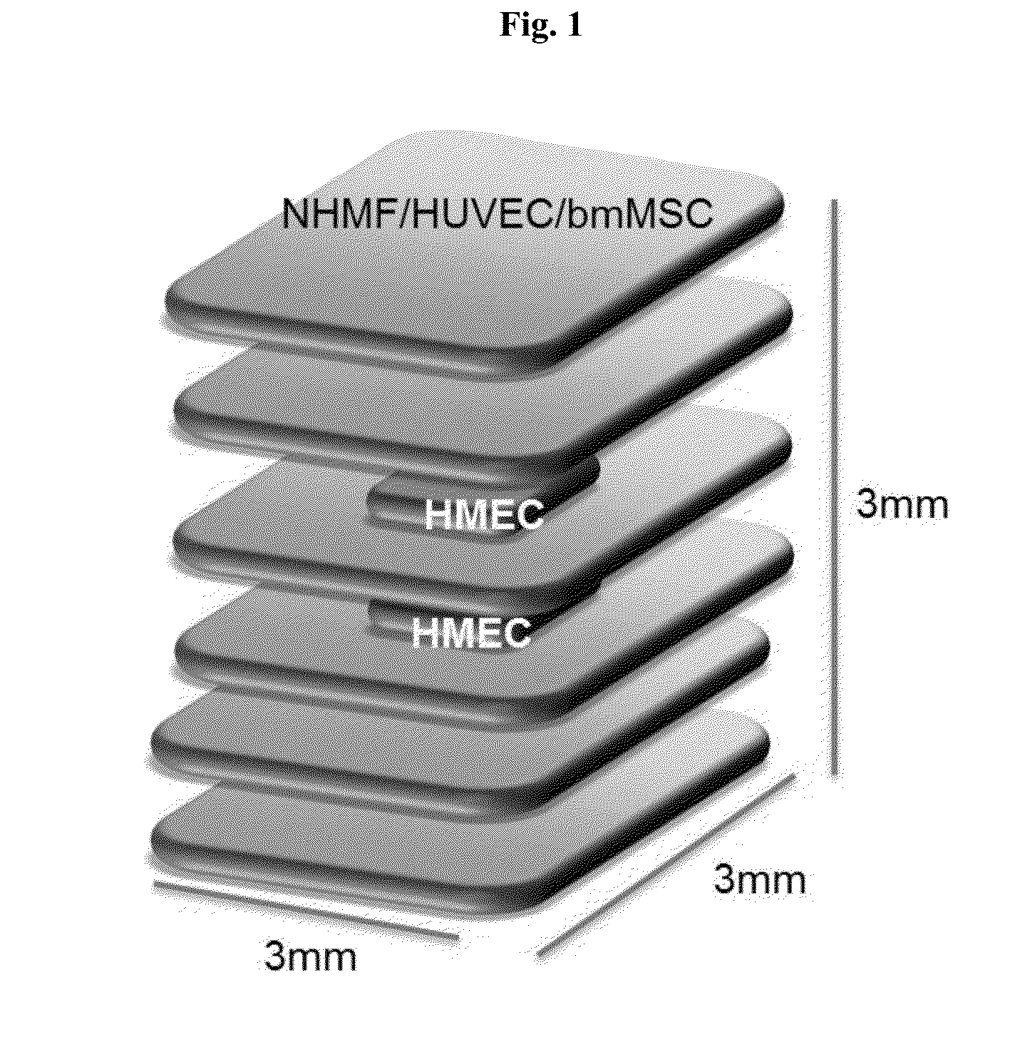
[0166]A 6-layer cube with dimensions 3 mm×3 mm×3 mm was bioprinted onto a Transwell membrane in a 6-well tissue culture plate according to the schematic diagram shown in FIG. 1 (Structure 1). The bottom two and top two layers were composed of 75% normal human mammary fibroblasts (NHMF) and 25% human umbilical vein endothelial cells (HUVEC). The middle two layers were comprised of a bioprinted square of 75% NHMF / 25% HUVEC surrounding a core of human mammary epithelial cells (HMEC) resuspended in an alginate and gelatin-containing hydrogel (Novogel® 2.0 System; Organovo, Calif.) to produce a bio-ink comprising 50-300 million cells / mL. Immediately following bioprinting, structures were incubated with 50 mM CaCl2 for 2 minutes, and cultured for 6 days. On day 6 of culture, the constructs were incubated with alginate lyase to degrade the hydrogel and incubated for a further 24 hours.
Results
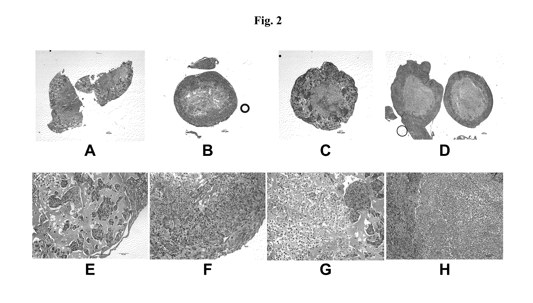
[0167]Assessment of Structure 1 was performed by his...
example 2
Engineered Human Breast Tissue Model with Adipose Tissue
Fabrication
[0172]A box with dimensions 5 mm×5 mm×500 μm was bioprinted onto a Transwell membrane in a 6-well tissue culture plate as shown in FIG. 7A (Structure 2). First, a 500 μm stromal bio-ink cylinder composed of 90% bone marrow-derived mesenchymal stem cells (bmMSC) and 10% HUVEC was bioprinted to form a box shape. HMEC cells were mixed with 10% gelatin hydrogel to form an epithelial bio-ink and bioprinted into the middle of the stromal bio-ink box, wherein the HMEC bio-ink did not touch the stromal bio-ink border. A third bio-ink composed of 75% NHMF and 25% HUVEC was used to fill the space between the stromal bio-ink border and the HMEC bio-ink in the middle. Constructs were incubated for 10 days. FIG. 7B depicts Structure 2 immediately post-fabrication and FIGS. 7C and 7D depict Structure 2 at day 10 post-fabrication. The bmMSC were provided with an adipocyte differentiation signal during incubation to generate viable,...
example 3
Engineered Human Breast Cancer Tumor Model
[0177]Bioprinted breast cancer constructs were generated in which a cancer cell node, composed of MCF7 breast cancer cells and human umbilical vein endothelial (HUVEC) cells, is surrounded on all sides by a stromal compartment composed of normal human mammary fibroblasts (NHMF), HUVEC cells, and subcutaneous preadipocytes (SPA). Bio-inks were produced by combining cells with a reversibly cross-linkable, alginate-containing hydrogel (Novogel® 3.0 System; Organovo, Calif.).
[0178]FIG. 10A shows a schematic diagram depicting the construction of the construct (Structure 3). FIG. 10B shows a photograph of the construct immediately after bioprinting and crosslinking of the hydrogel. FIG. 10C shows the construct 2 days after enzymatic treatment to remove the hydrogel, wherein the construct demonstrates condensation of tissue and generation of a smooth, solid nodule.
Cell Culture
[0179]Normal human mammary fibroblasts (NHMF) were acquired from ScienCel...
PUM
| Property | Measurement | Unit |
|---|---|---|
| height | aaaaa | aaaaa |
| height | aaaaa | aaaaa |
| temperature | aaaaa | aaaaa |
Abstract
Description
Claims
Application Information
 - R&D Engineer
- R&D Manager
- IP Professional
- Industry Leading Data Capabilities
- Powerful AI technology
- Patent DNA Extraction
Browse by: Latest US Patents, China's latest patents, Technical Efficacy Thesaurus, Application Domain, Technology Topic.
© 2024 PatSnap. All rights reserved.Legal|Privacy policy|Modern Slavery Act Transparency Statement|Sitemap