System and method for imaging with pinhole arrays

Active Publication Date: 2015-12-31
BAR ILAN UNIVERSITY +1
View PDF3 Cites 10 Cited by
  • Summary
  • Abstract
  • Description
  • Claims
  • Application Information

AI Technical Summary

Problems solved by technology

A larger pinhole transmits relatively higher radiation intensity, i.e. higher number of photons per time, but results in lower image resolution.
This may result in da

Method used


Image

  • System and method for imaging with pinhole arrays
  • System and method for imaging with pinhole arrays
  • System and method for imaging with pinhole arrays

Examples


Example

Example 1

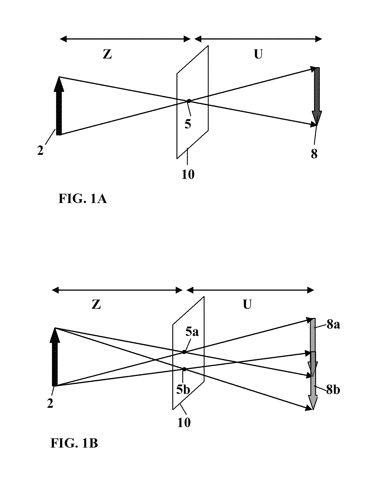
[0145]FIGS. 9A-9K show three aperture arrays, corresponding effective transmission functions and images of a test object. FIG. 9A-9C show aperture configuration of three aperture arrays having pinholes of diameter 170 μm; FIG. 9D illustrates the locations and numbering of the apertures. The set of aperture arrays is selected to provide 5.66 brighter imaging relative to a single pinhole of similar diameter. The locations of the apertures in the three arrays of FIGS. 9A-9C are shown in table 1 in relative units determined in accordance with pixel size of the detector and distances in the system (i.e. U and Z).

TABLE 1Location (x, y)[relative units]Location #Hole #(0, 0)11, 5, 9(1, 0)2 2(1, 1)3 3(0, 1)4 4(3, 0)56, 10(3, 3)67, 11(0, 3)78, 12(−6, 3) 813(−6, 0) 914(−6, −6)1015 (0, −6)1116 (3, −6)1217

[0146]As shown, apertures 1, 5 and 9 are located at the center of the mask and the rest of the apertures are arranged around to provide non-null effective transmission function. FIGS. ...

Example

Example 2

[0149]FIGS. 10A-10E show image data of simulation results using one-dimensional and two-dimensional aperture arrays configured as exemplified in FIGS. 9A-9C. The one-dimensional aperture arrays were configured with aperture locations including apertures 1, 2; 5, 6; and 14, 9, 10 as exemplified in FIG. 9D and provide image brightness increase of 2.33. FIG. 10A shows an image taken through a single pinhole of similar diameter; FIG. 10B shows a similar image taken through a pinhole diameter bigger by 150%; FIG. 10C shows reconstructed image taken in accordance with the above described technique utilizing one-dimensional aperture arrays; and FIGS. 10D and 10E show respectively raw image data and reconstructed image taken in accordance with the multi aperture technique utilizing two-dimensional aperture arrays.

[0150]As shown, increasing the aperture diameter increases image brightness at cost of resolution. This is while the reconstructed images according to the present inventio...

Example

Example 3

[0151]FIGS. 11A-11J illustrates simulation of Gamma imaging using the technique of the present invention. FIG. 11A shows the structure of arrays 1-3 and corresponding aperture dimensions; FIGS. 11B-11G show graphs of the arrays and corresponding effective transmission functions; FIG. 11H shows a single pinhole transmission as well the separate and total effective transmission functions of the arrays; and FIGS. 11I-11J show simulated images for imaging using a single pinhole (FIG. 11I) and reconstructed image (FIG. 11J) utilizing the technique of the invention with the pinhole arrays shown in FIG. 11A with conventional simulation techniques emulating bone scan for osteomyelitis transverse slice image (negative colors) using Gallium 67 radiation. Both FIGS. 11I and 11J illustrated a region of the figure in magnification, and as shown, the reconstructed image provides higher resolution and signal to noise ratio and thus provides information on features (marked with an arrow) t...


PUM


Abstract

An imaging system is provided, configured for providing three-dimensional data of a region of interest. The system comprising: an optical unit and a control unit. The optical unit comprises a radiation collection unit and a detection unit. The radiation collection unit comprises at least two mask arrangement defining at least two radiation collection regions respectively, the mask arrangements are configured to sequentially apply a plurality of a predetermined number of spatial filtering patterns formed by a predetermined arrangement of apertures applied on radiation collected thereby generating at least two elemental image data pieces corresponding to the collected radiation from said at least two collection regions. The control unit comprising is configured for receiving and processing said at least two elemental image data pieces and determining a plurality of at least two restored elemental images respectively being together indicative of a three dimensional arrangement of the region being imaged.

Description

RELATED APPLICATIONS[0001]This is a Continuation-in-Part of Application No. PCT / IL2015 / 050135 filed Feb. 5, 2015, which claims the benefit of U.S. Provisional Application No. 61 / 936,402 filed Feb. 6, 2014. The disclosure of the prior applications is hereby incorporated by reference herein in its entirety.GOVERNMENT LICENSE RIGHTS[0002]This invention was made with government support under W911NF-13-1-0485 awarded by US Army RDECOM. The US government has certain rights in the invention.TECHNOLOGICAL FIELD[0003]The invention is in the field of radiation imaging and relates to imaging techniques utilizing pinhole arrays.BACKGROUND ART[0004]References considered to be relevant as background to the presently disclosed subject matter are listed below:[0005][1] R. A. Vogel, D. Kirch, M. Lefree, and P. Steele, “A New Method of Multiplanar Emission Tomography Using a Seven Pinhole Collimator and an Auger Scintillation Camera,”J. Nucl. Med. 19 (6), 648-654 (1978).[0006][2] N. U. Schramm, G. Eb...

Claims


Application Information

Patent Timeline
IPC IPC(8): H04N13/00G02B27/22H04N13/02G06T5/00G21K1/04G02B5/20H04N13/133H04N13/122
CPCG02B26/06H04N13/211G02B5/201G21K1/04G06T5/006G06T5/001G02B27/2264G01T1/295G02B2207/129H04N13/133H04N2013/0081H04N2013/0092H04N13/218H04N13/122G02B27/46G02B30/24
Inventor ZALEVSKY, ZEEVSCHWARZ, MOSHE ARIE ARIELSHEMER, AMIRJAVIDI, BAHRAMWANG, JINGANG
Owner BAR ILAN UNIVERSITY
Why Eureka
Try Eureka