Methods and systems for computed tomography

a computed tomography and computed tomography technology, applied in the field of non-invasive diagnostic imaging, can solve problems such as the inability to govern a priori patient exposur

Active Publication Date: 2017-07-20
GENERAL ELECTRIC CO
View PDF3 Cites 19 Cited by
  • Summary
  • Abstract
  • Description
  • Claims
  • Application Information

AI Technical Summary

Benefits of technology

[0005]In one embodiment, a method comprises, in response to receiving an object-specific dose metric input by a user via an interface operably coupled to a scanner, predicting an image quality of a diagnostic scan, calculating an output source exposure level of the scanner for the predicted image quality, and performing the diagnostic scan at the specified output source exposure level responsive to the image quality above a threshold. In this way, the user may be able to input a dose specific to the patient, thereby governing the output level of the x-ray source that appropriately exposes the patient for a given clinical imaging task.

Problems solved by technology

Since the Deff is currently derived from reconstructed axial images of the patient, the SSDE is a post hoc dose metric and cannot govern the a priori patient exposure for a clinical imaging task.

Method used


Image

  • Methods and systems for computed tomography
  • Methods and systems for computed tomography
  • Methods and systems for computed tomography

Examples


Embodiment Construction

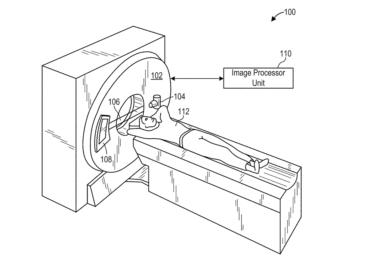
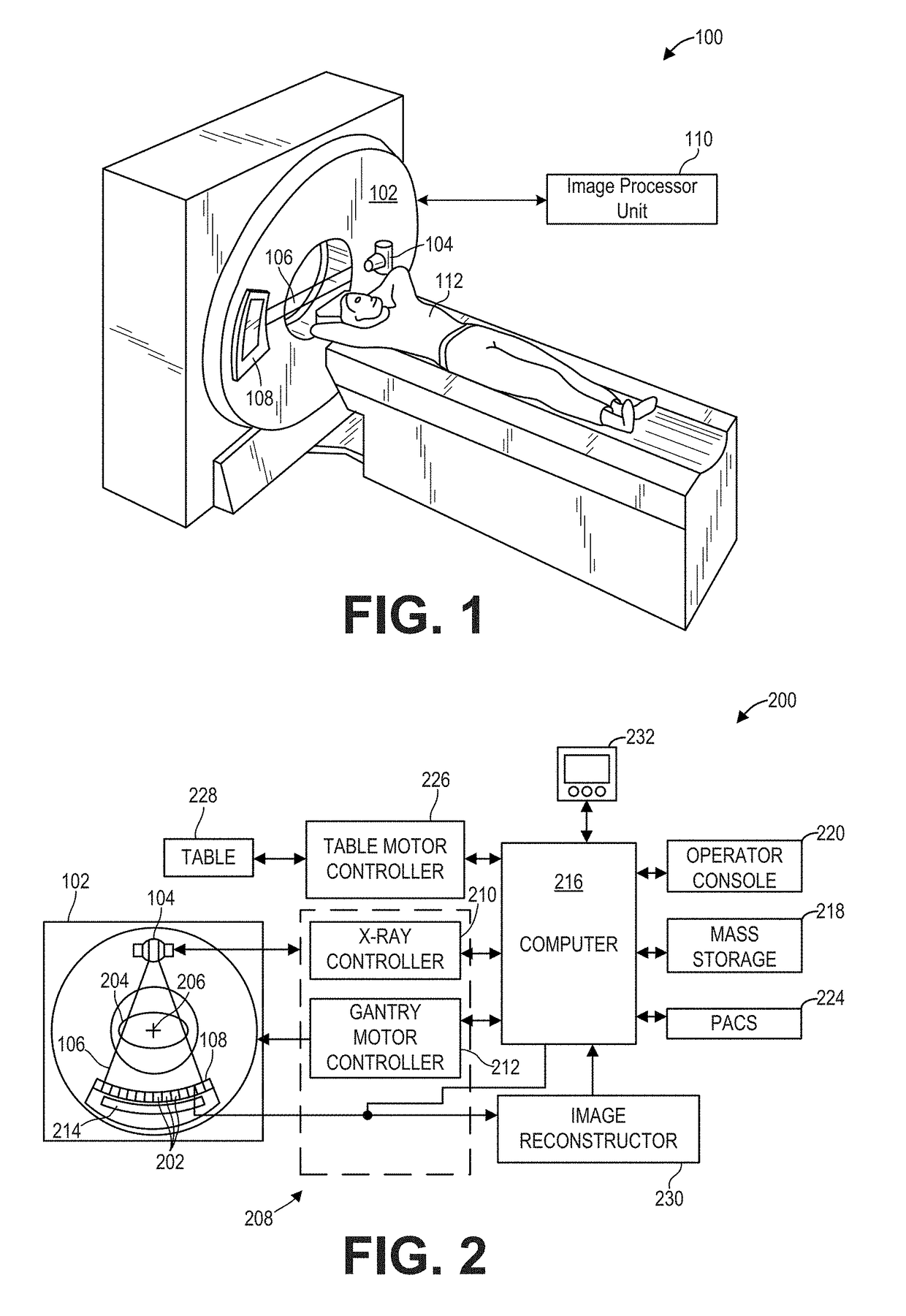
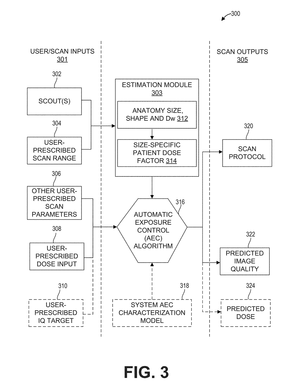
[0015]The following description relates to various embodiments of medical imaging systems. In particular, methods and systems are provided for automatic exposure control. An example of a computed tomography (CT) imaging system that may be used to acquire images processed in accordance with the present techniques is provided in FIGS. 1 and 2. CT systems may include an automatic exposure control (AEC) feature described in FIG. 3, wherein the output level of the source may be adjusted based on one of a user-prescribed dose input or image quality input (FIG. 5). Herein, a user may prescribe patient-specific dose metric input or an image quality input via a user interface as shown in FIG. 4. When a dose metric input is received, a method shown in FIG. 6, may include determining parameters of a diagnostic scan based on the dose metric input received, and further predicting an image quality of the diagnostic scan based on the dose metric input. Likewise, when an image quality input is rece...


PUM


Abstract

Methods and systems are provided for automatic exposure control in computed tomography imaging systems. In one embodiment, a method comprises in response to an object-specific dose metric input by a user via a user interface operably coupled to a scanner, predicting an image quality of a diagnostic scan, calculating an output level of a source of the scanner for the image quality, and performing the diagnostic scan at the output level when the image quality is higher than a threshold. In this way, patient-specific image quality settings may be determined.

Description

FIELD[0001]Embodiments of the subject matter disclosed herein relate to non-invasive diagnostic imaging, and more particularly, to automatic exposure control in computed tomography imaging systems.BACKGROUND[0002]Non-invasive imaging technologies allow images of the internal structures of a patient or object to be obtained without performing an invasive procedure on the patient or object. In particular, technologies such as computed tomography (CT) use various physical principals, such as the differential transmission of x-rays through the target volume, to acquire image data and to construct tomographic images (e.g., three-dimensional representations of the interior of the human body or of other imaged structures).[0003]Modern CT systems include automatic exposure control (AEC) configured to set the dose operating point to achieve optimized performance between image quality and radiation dose. Radiation exposure from CT scanners is measured in terms of the volumetric CT Dose Index ...

Claims


Application Information

Patent Timeline
Patent Type & Authority Applications(United States)
IPC IPC(8): A61B6/00A61B6/03
CPCA61B6/032A61B6/544A61B6/542A61B6/465A61B6/467A61B6/488A61B6/5205
Inventor CROTTY, DOMINIC JOSEPHNILSEN, ROY A.FAN, JIAHUAKULPINS, MARY SUERUPCICH, FRANCOSTEVENS, GRANT MOREY
Owner GENERAL ELECTRIC CO
Why Eureka
Try Eureka