Radiation imaging apparatus, and insertion state determination method

a radiation imaging and state determination technology, applied in the direction of radiation beam directing means, patient positioning for diagnostics, application, etc., can solve the problems of deteriorating the sensitivity and specificity of lesion detection, blind areas are likely to be generated, and subject's body tilt is a physical burden

Inactive Publication Date: 2017-09-21
CANON KK
View PDF2 Cites 12 Cited by
  • Summary
  • Abstract
  • Description
  • Claims
  • Application Information

AI Technical Summary

Problems solved by technology

However, it is known that in the case of taking a mammogram of a dense breast (a breast with many mammary glands), a lesion portion and a mammary gland structure overlap each other, which leads to deterioration in the sensitivity and specificity of lesion detection.
Particularly, it is difficult to insert a breast into an axilla-side area or an axilla area of the breast, so that the blind areas are likely to be generated in these areas.
However, tilting the body is a physical burden for the subject, and imaging of the area including the breast bone is not preferable in terms of imaging exposure.
In the upright breast CBCT, the body of the subject cannot be tilted using the weight of the subject to insert the breast into an insertion portion, unlike in the prone position type, so that the breast cannot be sufficiently inserted (i.e., the insertion state is unsuitable) in some cases and the blind area varies depending on the insertion state.
The variation in the blind area causes a problem that it is difficult to image an intended area.
Another problem is that the variation in the blind area in the upright breast CBCT is larger than the variation of the blind area in the mammogram that is taken by fixing the breast.

Method used


Image

  • Radiation imaging apparatus, and insertion state determination method
  • Radiation imaging apparatus, and insertion state determination method
  • Radiation imaging apparatus, and insertion state determination method

Examples


first embodiment

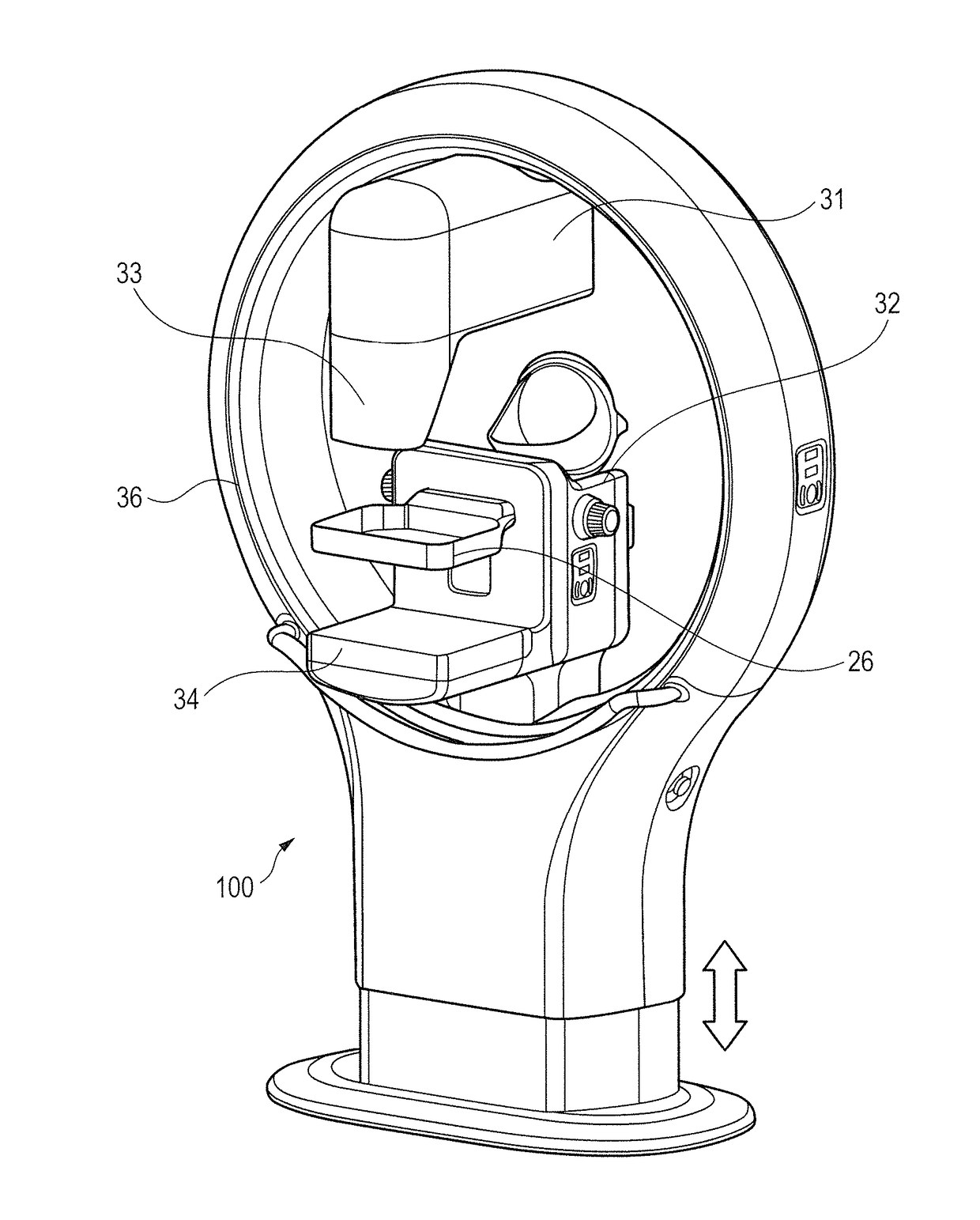
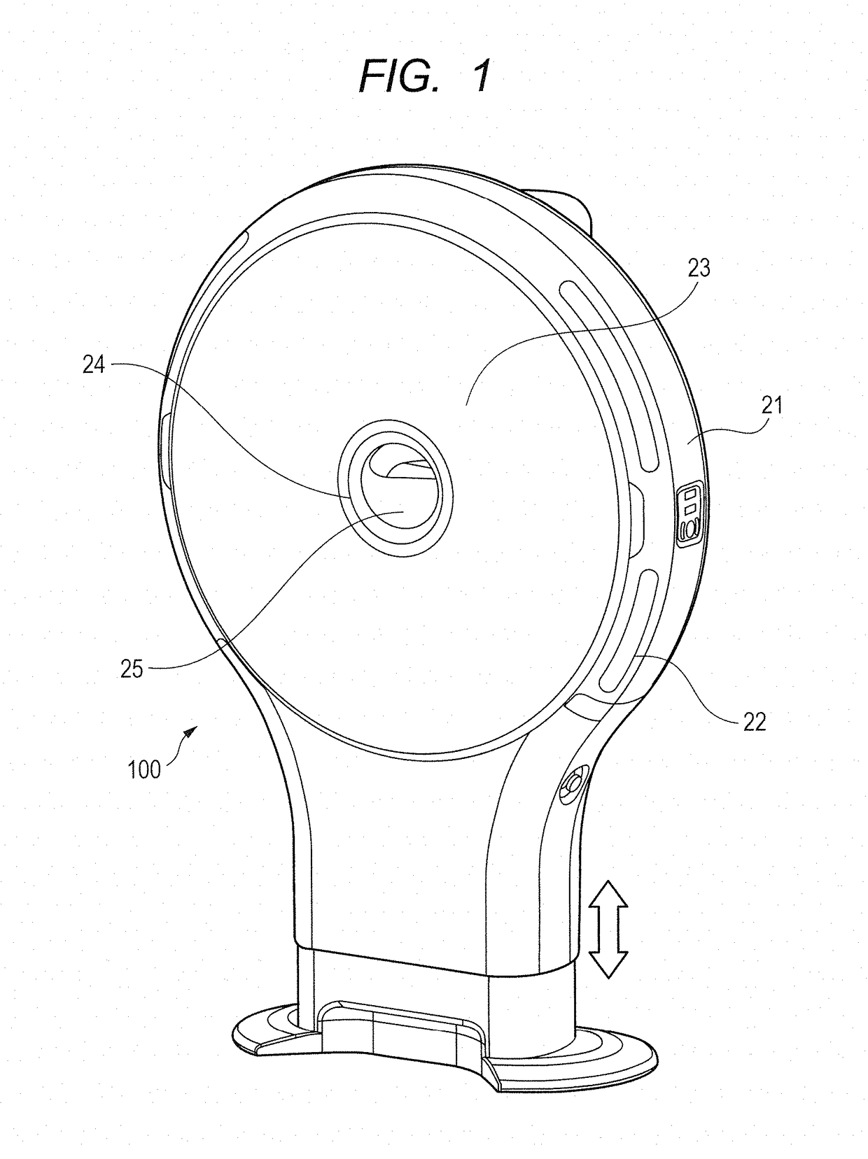
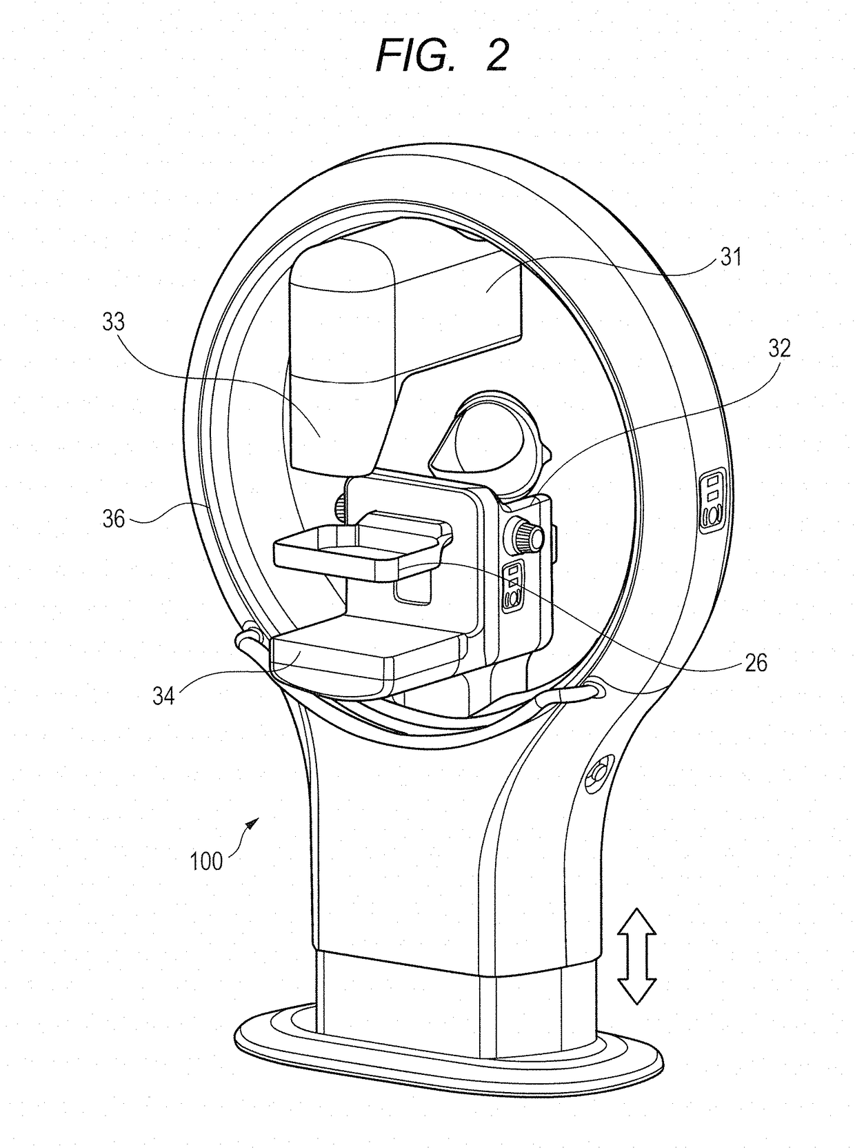
[0054]FIGS. 7Aa and 7Ab are diagrams each illustrating an example of the insertion state of the breast in the first embodiment. The radiation imaging system (medical image imaging system) 100 suitably manages blind areas in the C area and C′ area (outside upper area) which are predilection areas of breast cancer. The blind areas need to be suitably managed, except in a case where the radiological image of the whole body of the subject P is captured by a CT apparatus. In this case, to put it simply, the suitable management of blind areas indicates that the blind areas in the C area (outside upper area) and D area (outside lower area) of the breast is set to be smaller than the blind areas in the A area (inside upper area) and B area (inside lower area) of the breast.

[0055]FIG. 7Aa is a diagram illustrating a state where the left breast is inserted into the imaging area in the upright breast CBCT, as viewed from the leg portion side of the subject P. The rotatable frame 36 which is pr...

second embodiment

[0091]In the radiation imaging system (medical image imaging system) 100 of the first embodiment, the determination unit 104 determines the insertion state of the breast based on at least one of the length, the area, the volume, the shape and the pressure of the inserted breast inserted into the imaging area for the medical image from the insertion portion. On the other hand, in the radiation imaging system (medical image imaging system) 100 of a second embodiment, the determination unit 104 determines the insertion state of the breast based on at least one of the length, the area, the volume, the shape and the pressure of a non-inserted breast which is not inserted into the imaging area. Note that the descriptions of the components, functions, and operations of the second embodiment that are similar to those of the above embodiment are omitted, and differences between the second embodiment and the above embodiment are mainly described below.

[0092]FIG. 8 is a diagram illustrating an...


PUM


Abstract

Provided is a medical image imaging apparatus that images a medical image of a breast, including a determination unit configured to determine an insertion state of the breast based on a form of the breast inserted into an imaging area for the medical image from an insertion portion.

Description

BACKGROUND OF THE INVENTION[0001]Field of the Invention[0002]The present invention relates to a radiation imaging apparatus for imaging a radiological image of a breast, an insertion state determination method.[0003]Description of the Related Art[0004]A mammogram apparatus is normally used as an X-ray inspection apparatus for breast cancer. However, it is known that in the case of taking a mammogram of a dense breast (a breast with many mammary glands), a lesion portion and a mammary gland structure overlap each other, which leads to deterioration in the sensitivity and specificity of lesion detection. Tomosynthesis and CBCT apparatus exclusively for breast imaging have received attention as techniques for compensating for the shortcomings of the mammogram. These apparatuses have a feature that a 3D image of a breast is provided so that the lesion portion and the mammary gland structure can be observed separately.[0005]In the case of imaging a breast by the CBCT apparatus exclusivel...

Claims


Application Information

Patent Timeline
Patent Type & Authority Applications(United States)
IPC IPC(8): A61B6/00A61B6/04A61B5/00A61B6/03
CPCA61B6/502A61B6/032A61B6/0414A61B2562/0247A61B6/0492A61B5/0091A61B6/544A61B6/4085A61B6/44A61B5/1072A61B5/1073A61B5/1077A61B5/1079A61B6/037A61B6/08A61B6/46A61B6/462A61B6/463A61B6/589A61B8/0825A61B8/403
Inventor TSUJII, OSAMU
Owner CANON KK
Why Eureka
Try Eureka