Eureka AIR delivers breakthrough ideas for toughest innovation challenges, trusted by R&D personnel around the world.

Anatomical articulator for dental diagonostic method and prosthetic reconstruction

a technology for temporomandibular joints and anatomical articulators, which is applied in the field of apparatus for modeling the temporomandibular joints, can solve the problems of traditionally difficult modeling of the bone structure of the jaw, and achieve the effects of less cumbersome measurements and dials, accurate depiction of patients, and more anatomically accura

Inactive Publication Date: 2018-06-07
OHIO STATE INNOVATION FOUND
View PDF3 Cites 5 Cited by
  • Summary
  • Abstract
  • Description
  • Claims
  • Application Information

AI Technical Summary

Benefits of technology

The patent describes an anatomical articulator for modeling the temporomandibular joint (TMJ). It consists of a lower base portion with a upright support and a set of replica condyles made from digital data representing a patient's jaw structure. There is also an upper frame portion with receptacles for attaching a set of replica mandibular fossae, which are also made from digital data. These parts meet to form a hinge for the articulator. The technical effect of this invention is to provide a useful tool for research and treatment of TMJ disorders.

Problems solved by technology

Modeling the bone structure of the jaw, of course, has traditionally been much more difficult, given that no internal impression is available for making a mold and associated cast of an individual's internal skull, bones, and joints.
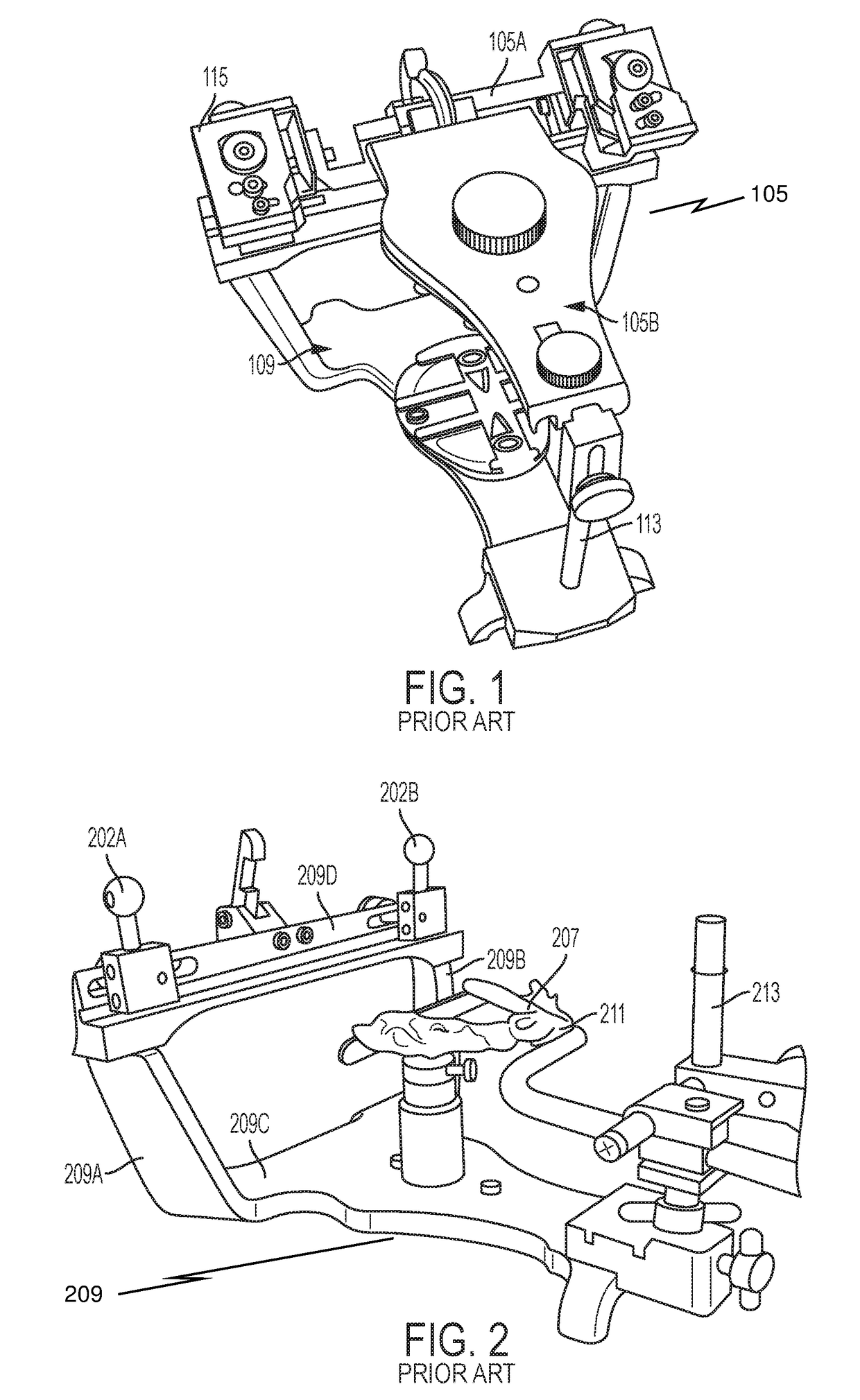
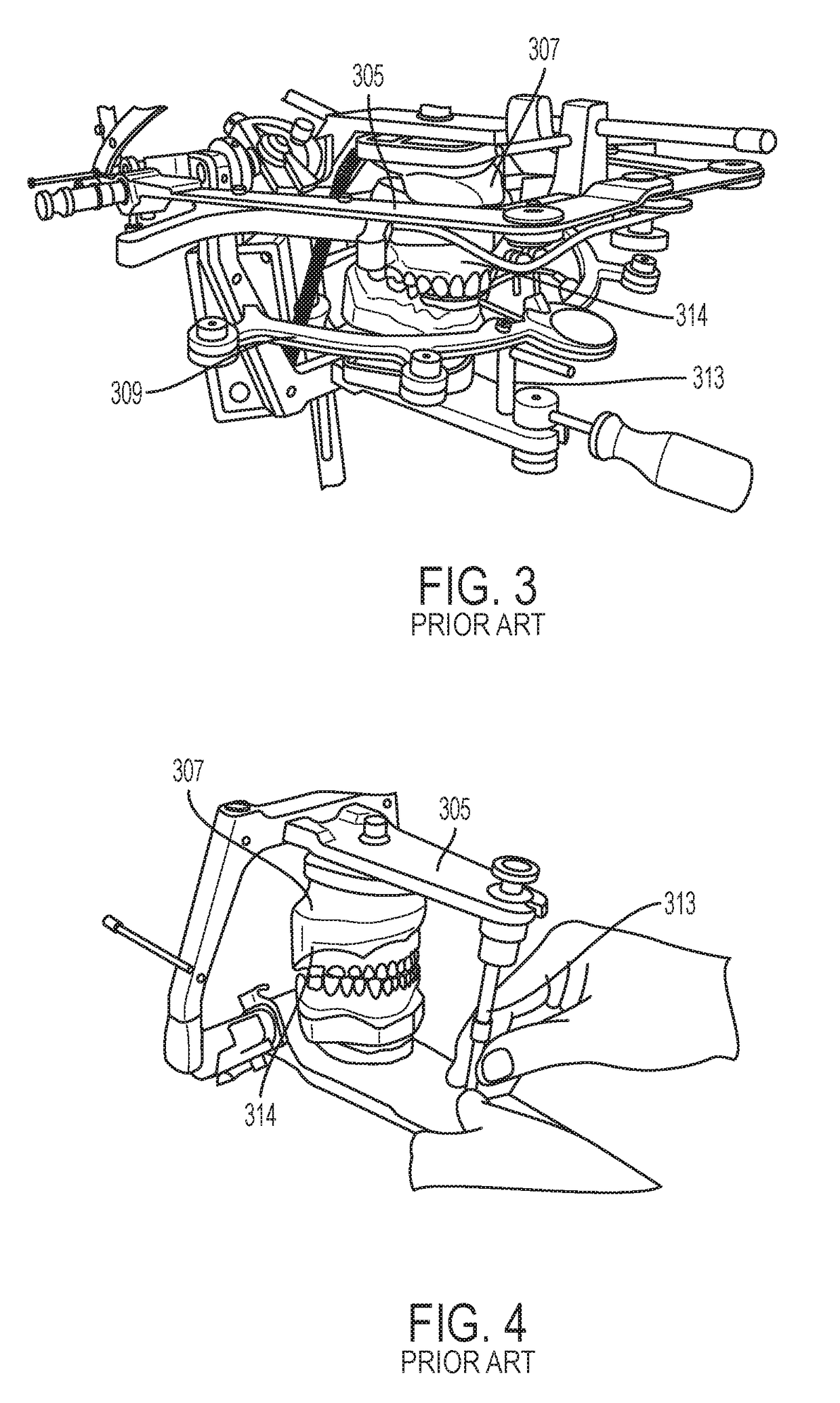
The biggest limitation of this set up lies in the generic temporomandibular joint (TMJ) replicas shown in FIG. 6.

Method used


Image

  • Anatomical articulator for dental diagonostic method and prosthetic reconstruction
  • Anatomical articulator for dental diagonostic method and prosthetic reconstruction
  • Anatomical articulator for dental diagonostic method and prosthetic reconstruction

Examples


Embodiment Construction

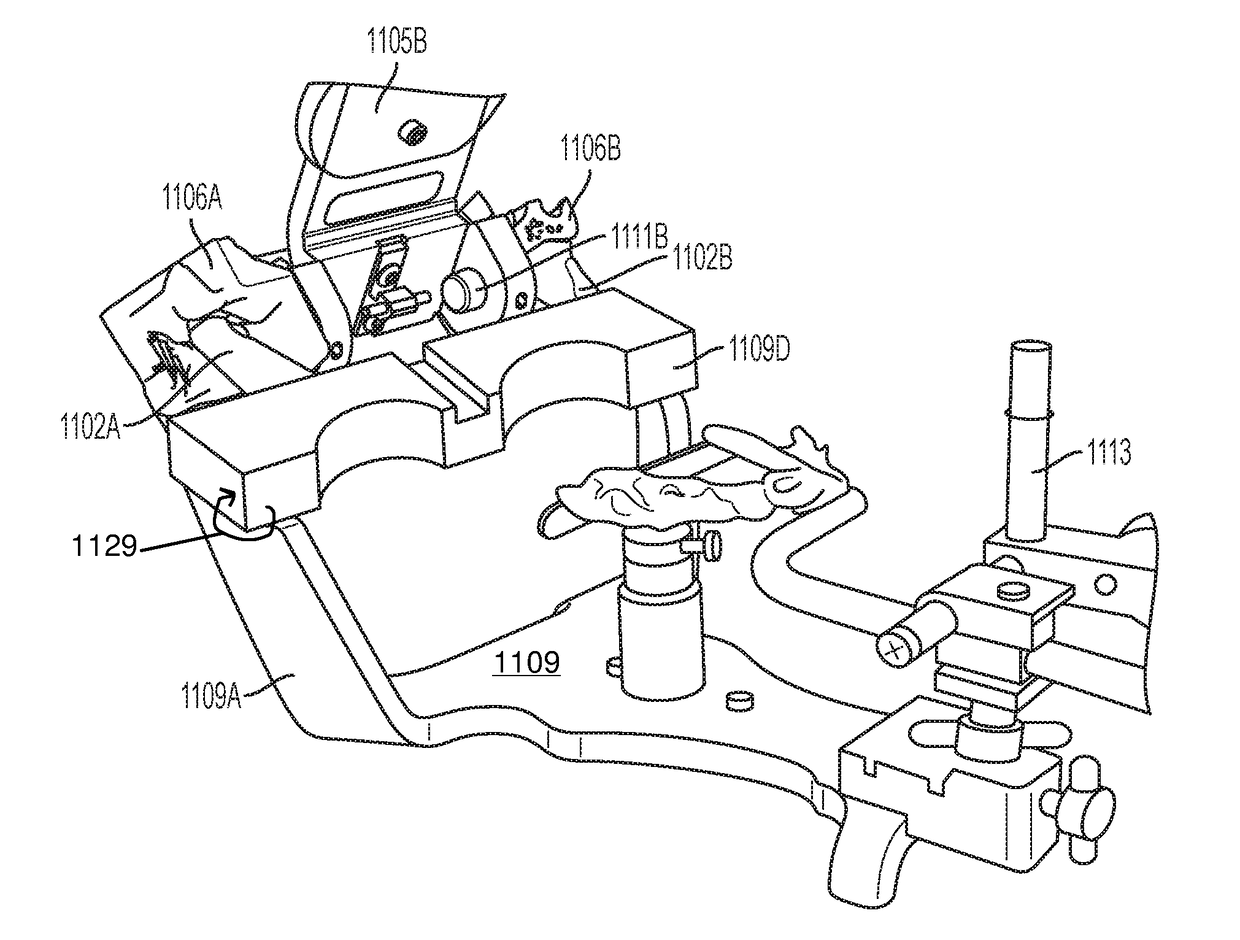
[0036]In one embodiment, an articulator, according to the disclosure herein, includes common portions of standard articulators with significant changes in the replicated temporomandibular joint (TMJ). Instead of the above noted prior art replicas with generic and ineffective condyle termini in the form of simple spheres (202) and right angled or at least sharply angled fossae (606), one embodiment of an articular (900) includes a lower base (909) on which a lower jaw (not shown) would be mounted on the above-noted elevated platform. Instead of the mechanical replicas described above, however, the articulator of FIG. 9 includes an exact replica of a patient's condyles (902) and fossae (906) that have been printed via stereolithographic three-dimensional printing techniques. Accordingly references to anatomically modeled or printed condyles and fossae may be considered “replica condyles” and “replica fossae.” Other additive layer three-dimensional printing options are also available t...


PUM


Abstract

An anatomical articulator for modeling a temporomandibular joint includes a lower base portion comprising at least one upright support. A set of replica condyles produced from digital data representing a patient jaw structure is connected to the at least one upright support. An upper frame portion defines receptacles on either side. A set of replica mandibular fossae produced from the digital data representing a patient jaw structure is configured to connect to the upper frame portion via the receptacles. The replica condyles and the replica mandibular fossae meet to form a hinge of the articulator.

Description

CROSS REFERENCE TO RELATED APPLICATIONS[0001]This application claims priority to and incorporates entirely by reference U.S. Provisional Patent Application Ser. No. 62 / 430,058 filed on Dec. 5, 2016, and entitled “Anatomical Articulator for Dental Diagnostic Methods and Prosthetic Reconstruction.”STATEMENT REGARDING FEDERALLY SPONSORED RESEARCH OR DEVELOPMENT[0002]No federally sponsored research or development has been used for this disclosure.FIELD OF THE INVENTION[0003]This disclosure relates to the field of apparatuses for modeling the temporomandibular joint of a living being, particularly humans, and diagnosing dental procedures to alleviate disorders of the temporomandibular joint, as well as for prosthetic reconstruction of the masticatory system.BACKGROUND[0004]The temporomandibular joint is a bone structure in the jaw that provides a hinge that connects the jaw to the skull. This joint is the structure by which a jaw moves up and down and side to side for chewing, speaking, ...

Claims


Application Information

Patent Timeline
IPC IPC(8): A61C11/02A61C19/045A61B6/03A61B6/00
CPCA61C11/02A61C19/045A61B6/032A61B6/4085A61C11/022A61C11/06B33Y80/00
Inventor AZER, SHEREEN S.
Owner OHIO STATE INNOVATION FOUND
Why Eureka
Eureka Blog
Learn More