Fixation of intraluminal device

a technology of intraluminal devices and fixation methods, applied in medical science, surgical staples, non-surgical orthopedic devices, etc., to achieve the effect of facilitating such explantation

Active Publication Date: 2018-09-06
BFKW
View PDF1 Cites 0 Cited by
  • Summary
  • Abstract
  • Description
  • Claims
  • Application Information

AI Technical Summary

Benefits of technology

[0004]While the use of tissue ingrowth patented in the above-identified patents has been found to provide a satisfactory solution for fixation to resist distal migration, aspects of the present invention includes providing short-term fixation of the device until the tissue ingrowth providing long-term fixation is in place. Such short-term fixation is easy to carry out and capable of complete fixation over the days or weeks that it takes for the long-term fixation to occur.
[0005]Aspects of the present invention provide techniques for explantation of an intraluminal device having a wall defining first and second wall portions configured to be positioned in a lumen. The first wall portion may be an esophageal portion that is configured to the size and shape of a portion of the esophagus. The second wall portion may be a cardiac portion that is configured to the size and shape of the cardiac portion of the stomach. A connector connecting the esophageal and cardiac portions is positioned against lumen tissue, such as in the gastroesophageal (GE) junctions wherein lumen tissue bridges or encases the connector during deployment of the device. Explantation of the device should not substantially damage the GE junction of the recipient. Aspects of the present invention facilitate such explantation and provide techniques that may beneficially utilize such tissue bridging the connector for long-term fixation of an intraluminal device.
[0006]An intraluminal device adapted to be positioned in a lumen, according to an aspect of the invention, includes a wall having a first wall portion configured to the size and shape of a first portion of the lumen and a second wall portion configured to the size and shape of a second portion of the lumen. A connector connects the first wall portion with the second wall portion. The connector is configured to be positioned against the lumen and wherein the connector is separably connected with one or both of the first and second wall portions. This allows the device to be explanted by disconnecting the connector and axially withdrawing the connector from tissue that encases the connector without substantial damage to the tissue.
[0018]An intraluminal device adapted to be deployed at the gastroesophageal (GE) region of a recipient, according to an aspect of the invention, includes a wall defining a cardiac portion that is configured to the size and shape of the cardiac portion of the stomach. A fastener temporarily fixes the wall to the recipient to resist distal migration. A wall characteristic fixes the wall to the GE region through growth of tissue causing long-term fixing of the wall to the GE region to resist distal migrations. The wall characteristic facilitates tissue ingrowth. The temporarily fixing fixes the device at the GE region while the tissue grows at the characteristics to provide the long-term fixation.
[0020]The wall characteristic may facilitate tissue ingrowth openings in the wall. The device may include an esophageal portion that is configured to the size and shape of a portion of the esophagus and a connector connecting the esophageal portion and the cardiac portion wherein the tissue ingrowth openings are at the esophageal portion. The wall characteristic may include the connector being an elongated member that is configured to be positioned at the GE junction wherein tissue at least partially envelopes the connector to provide long-term fixation.
[0024]A method of fixation of an intraluminal device at the gastroesophageal (GE) region of the recipient to resist distal migration, according to an aspect of the invention, includes the device having a wall defining a cardiac portion that is configured to the size and shape of the cardiac portion of the stomach, an esophageal portion that is configured to the size and shape of a portion of the esophagus and a connector connecting the esophageal portion and the cardiac portion. The intraluminal device is positioned at the GE region wherein tissue will grow to a wall characteristic of the connector at the GE junction to provide long-term fixation to resist distal migration. The device is temporarily fixed at the GE region while tissue grows to the wall characteristic of the connector.

Problems solved by technology

A challenge with such devices and methods is fixation of a portion of the device against a surface of the GI tract, such as the cardiac portion of the stomach in the presence of peristalsis tending to cause distal migration of the device.

Method used


Image

  • Fixation of intraluminal device
  • Fixation of intraluminal device
  • Fixation of intraluminal device

Examples


Embodiment Construction

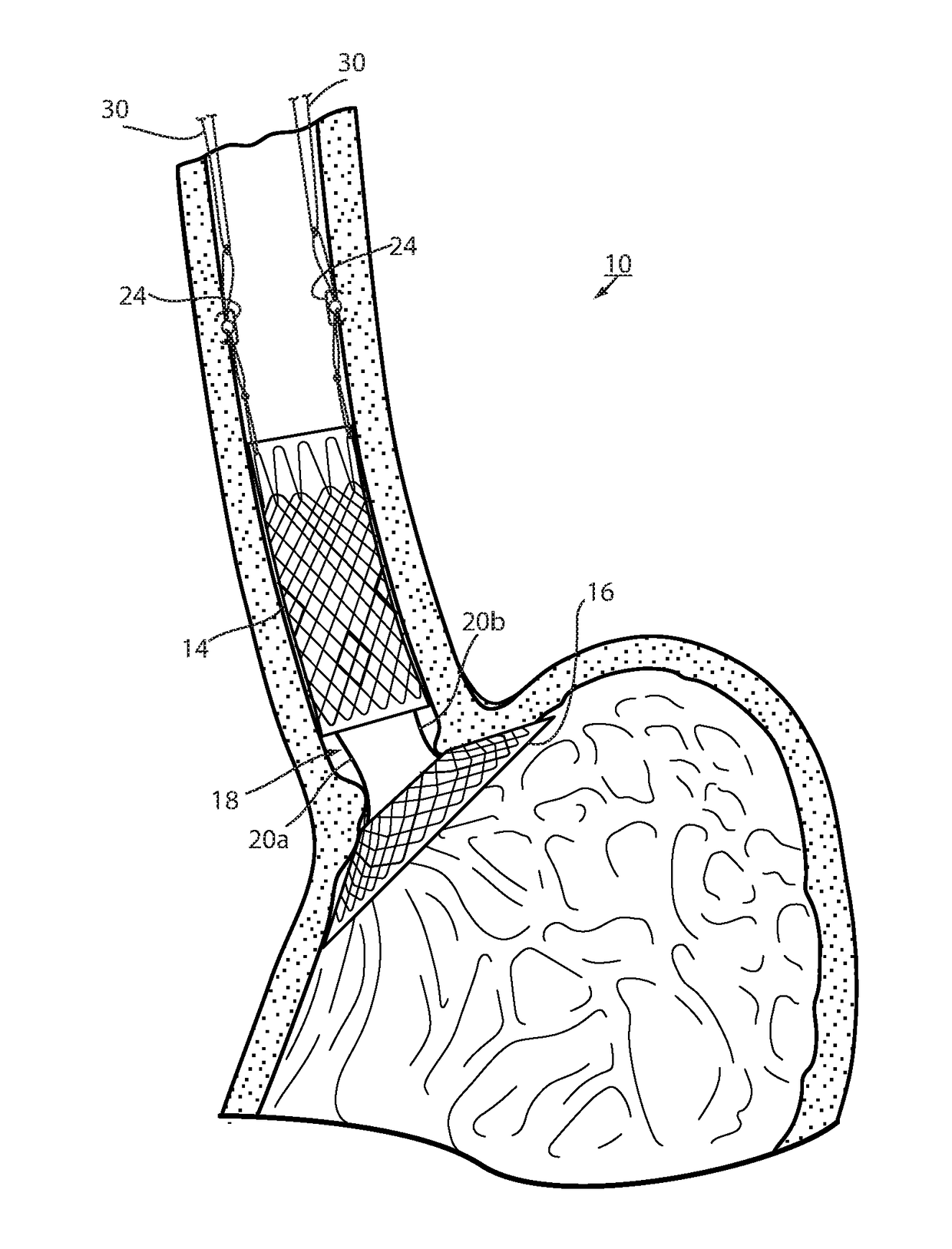
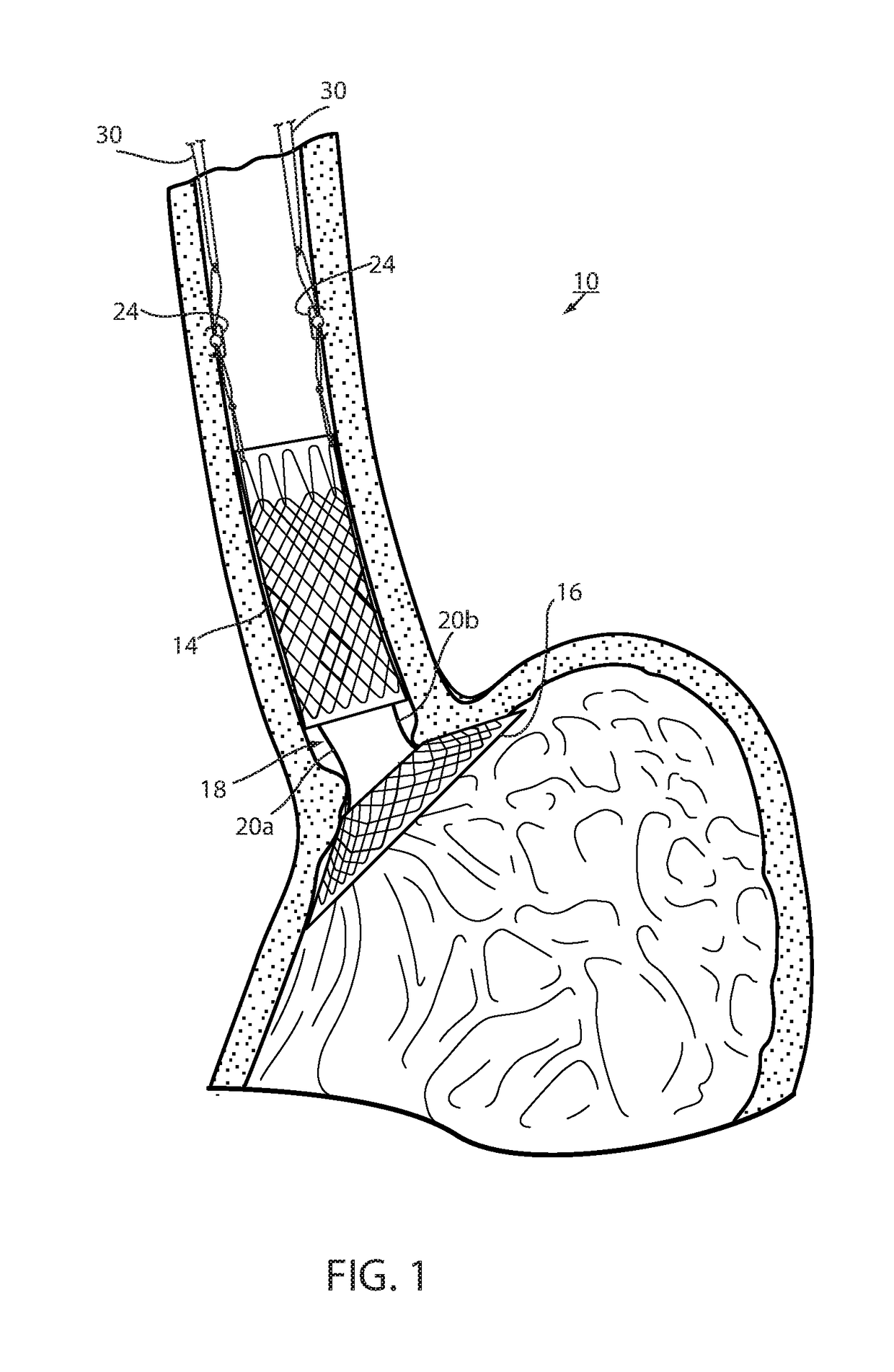
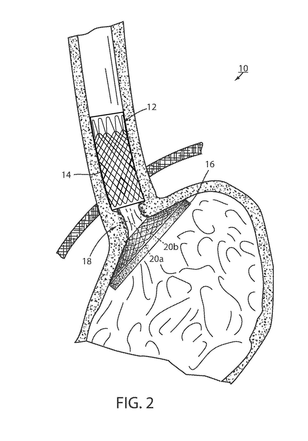
[0053]Referring now to the drawings and the illustrative embodiment depicted therein, an intraluminal device, such as a bariatric device 10, has a wall 12 defining an esophageal portion 14 that is configured to the size and shape of a portion of a mammalian lumen or hollow organ, namely, the esophagus, a cardiac portion 16 that is configured to the size and shape of a separated portion of mammalian lumen or hollow organ, namely, the cardiac portion of the stomach and a connector 18 connecting esophageal portion 14 and cardiac portion 16 (FIGS. 1-5). While illustrated as a bariatric device, it should be understood that that principles of the invention are applicable to other intraluminal devices that are positioned in a lumen or hollow organ that experiences peristalsis, such as an esophageal stent, an anti-reflux device, a nasal gastric tube, an intestinal sleeve, and the like. Also, the invention may be applied to a metabolic disease treatment device and method as disclosed in comm...


PUM


Abstract

An intraluminal device and method of fixation of an intraluminal device to resist distal migration in a mammalian lumen or hollow organ that is subject to peristalsis, according to an aspect of the invention, includes spaced apart wall portions connected with a connector. The wall portions are configured to the size and shape of a portion of the lumen or hollow organ and the connector is configured to be positioned against a wall of the lumen or hollow organ. The intraluminal device is positioned in a mammalian lumen or hollow organ that is subject to peristalsis. The device is fixed in the lumen or hollow organ against distal migration, wherein tissue lining the lumen or hollow organ bridges over the connector. The device is explanted after tissue bridges over the connector including separating the connector from one or both of the wall portions and withdrawing the connector axially from the tissue bridging over the connector.

Description

CROSS REFERENCE TO RELATED APPLICATIONS[0001]The present application is a continuation of U.S. patent application Ser. No. 15 / 534,891, filed Jun. 9, 2017, which claims the priority benefits of International Patent Application No. PCT / US2015 / 067407, filed Dec. 22, 2015, which claims priority from U.S. patent application Ser. No. 62 / 234,335, filed on Sep. 29, 2015, and U.S. patent application Ser. No. 62 / 151,150, filed on Apr. 22, 2015, and U.S. patent application Ser. No. 62 / 115,689, filed on Feb. 13, 2015, and U.S. patent application Ser. No. 62 / 097,295, filed on Dec. 29, 2014, which are all hereby incorporated by reference herein in their entireties.BACKGROUND OF THE INVENTION[0002]The present invention is directed to an intraluminal device and method of fixation of an intraluminal device and, in particular, a technique that enhances both fixation and removeability of the device. While the invention is illustrated for use with a bariatric device and / or a metabolic device, it may be...

Claims


Application Information

Patent Timeline
Patent Type & Authority Applications(United States)
IPC IPC(8): A61F2/04A61F5/00
CPCA61F2/04A61F5/0089A61F2002/045A61F5/0036A61F2002/044A61F5/0069A61B17/064A61B2017/0641A61F2220/0016A61F2220/0033A61F2230/0093A61F2250/006
Inventor FOOTE, JAMES A.WALBURN, FREDERICK J.BAKER, RANDAL S.
Owner BFKW
Why Eureka
Try Eureka