Fixation of intraluminal device

a technology applied in the field of intraluminal devices and fixation methods of intraluminal devices, to achieve the effect of facilitating such explantation

Active Publication Date: 2021-06-01
BFKW
View PDF228 Cites 0 Cited by
  • Summary
  • Abstract
  • Description
  • Claims
  • Application Information

AI Technical Summary

Benefits of technology

[0004]While the use of tissue ingrowth patented in the above-identified patents has been found to provide a satisfactory solution for fixation to resist distal migration, aspects of the present invention includes providing short-term fixation of the device until the tissue ingrowth providing long-term fixation is in place. Such short-term fixation is easy to carry out and capable of complete fixation over the days or weeks that it takes for the long-term fixation to occur.
[0005]Aspects of the present invention provide techniques for explantation of an intraluminal device having a wall that is configured to be positioned in a lumen. At least one core is removably connected with a portion of the wall and is positioned against lumen tissue when the wall is positioned in the lumen such that tissue envelopes of or encases the core during deployment or implantation of the device. Explantation of the device should not substantially damage the lumen of the recipient or require that the lumen be incised in order to separate the core from the lumen. Aspects of the present invention facilitate such explantation and provide techniques that may beneficially utilize such tissue encasing the connector for immediate and / or long-term fixation of an intraluminal device against distal migration caused by peristalsis.

Problems solved by technology

A challenge with such devices and methods is fixation of a portion of the device against a surface of the GI tract, such as the cardiac portion of the stomach in the presence of peristalsis tending to cause distal migration of the device.

Method used


Image

  • Fixation of intraluminal device
  • Fixation of intraluminal device
  • Fixation of intraluminal device

Examples


Embodiment Construction

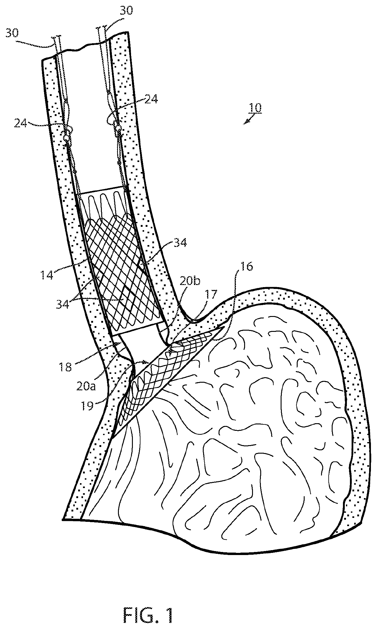
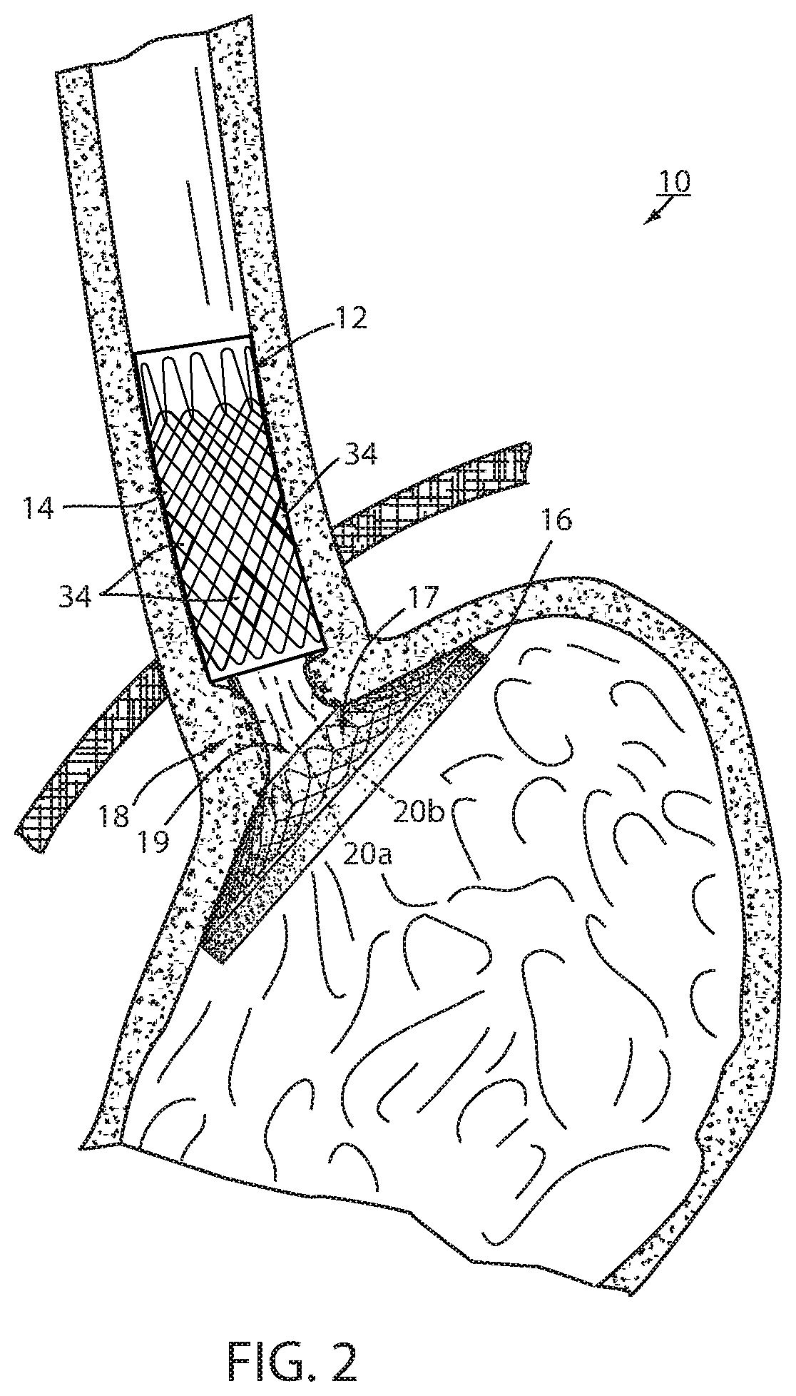
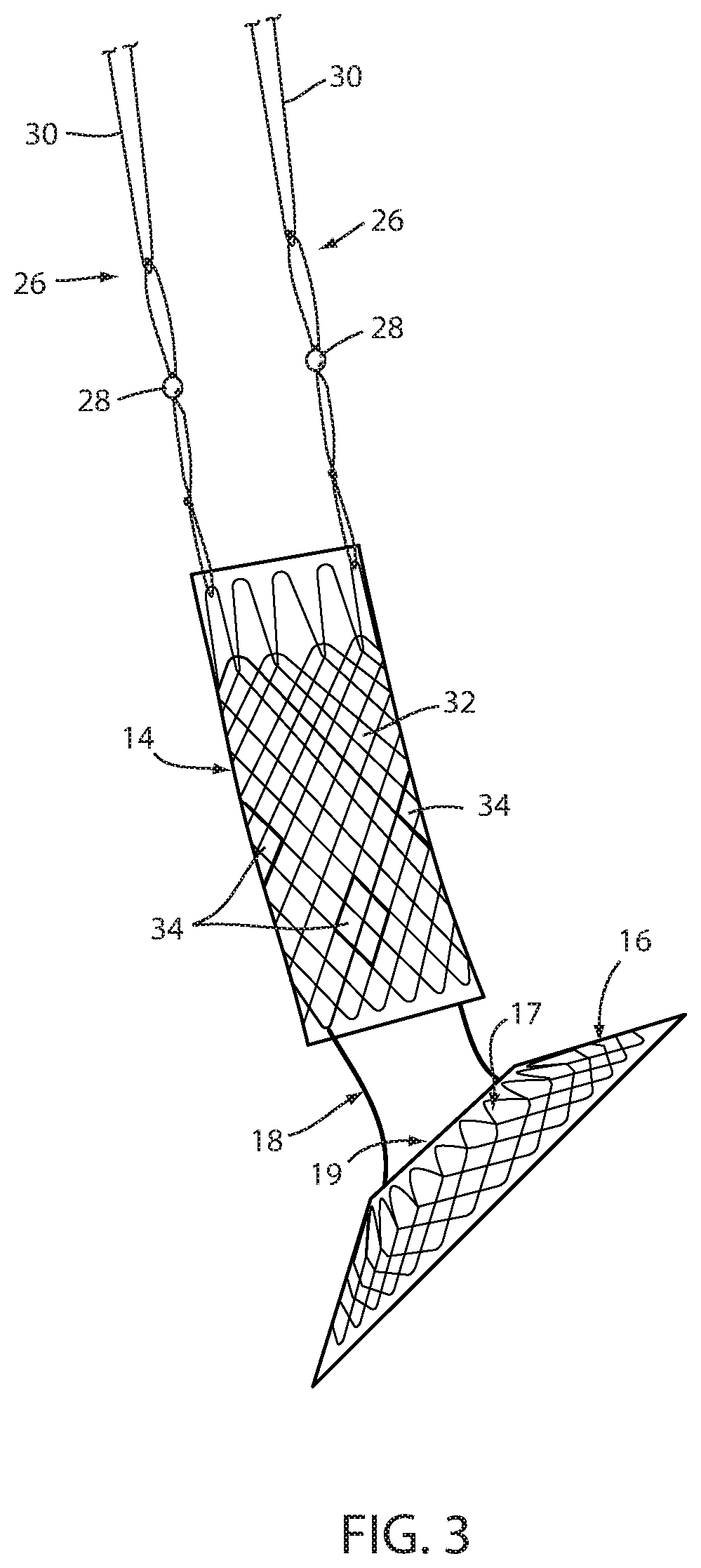
[0057]Referring now to the drawings and the illustrative embodiment depicted therein, an intraluminal device, such as a bariatric device or a metabolic disease treatment 10, has a wall 12 defining an esophageal portion 14 that is configured to the size and shape of a portion of a mammalian lumen or hollow organ, namely, the esophagus, a cardiac portion 16 that is configured to the size and shape of a separated portion of mammalian lumen or hollow organ, namely, the cardiac portion of the stomach and a connector 18 connecting esophageal portion 14 and cardiac portion 16 (FIGS. 1-5). While illustrated as a bariatric device, it should be understood that that principles of the invention are applicable to other intraluminal devices that are positioned in a lumen or hollow organ that experiences peristalsis, such as an esophageal stent, an anti-reflux device, a nasal gastric tube, an intestinal sleeve, and the like. Also, the invention may be applied to a metabolic disease treatment devic...


PUM


Abstract

An intraluminal device adapted to be positioned in a lumen that experiences peristalsis and method of fixation of an intraluminal device in the lumen, includes a wall configured to the size and shape of a portion of the lumen and at least one core. The at least one core is removably connected with a portion of the device wall. The at least one core is configured to be positioned against the lumen when the wall is positioned in the lumen. In this manner tissue envelopes the core during implantation of the intraluminal device. The at least one core is configured to be axially removable in situ from the tissue enveloping the core after the device has been deployed by disconnecting the core from the portion of the wall in order to explant the intraluminal device from lumen.

Description

CROSS REFERENCE TO RELATED APPLICATIONS[0001]The present application is a continuation-in-part of U.S. patent application Ser. No. 15 / 534,891, filed Jun. 9, 2017, which claims the priority benefits of International Patent Application No. PCT / US2015 / 067407, filed Dec. 22, 2015, which claims priority from U.S. patent application Ser. No. 62 / 234,335, filed on Sep. 29, 2015, and U.S. patent application Ser. No. 62 / 151,150, filed on Apr. 22, 2015, and U.S. patent application Ser. No. 62 / 115,689, filed on Feb. 13, 2015, and U.S. patent application Ser. No. 62 / 097,295, filed on Dec. 29, 2014, which are all hereby incorporated by reference herein in their entireties.BACKGROUND OF THE INVENTION[0002]The present invention is directed to an intraluminal device and method of fixation of an intraluminal device and, in particular, a technique that enhances both fixation and removability of the device. While the invention is illustrated for use with a bariatric device and / or a metabolic device, it...

Claims


Application Information

Patent Timeline
Patent Type & Authority Patents(United States)
IPC IPC(8): A61F2/04A61F5/00A61B17/064
CPCA61F2/04A61B17/064A61F5/0036A61F5/0069A61F5/0089A61B2017/0641A61B2017/0645A61F2002/044A61F2002/045A61F2220/0016A61F2220/0033A61F2230/0093A61F2250/006
Inventor FOOTE, JAMES A.WALBURN, FREDERICK J.BAKER, RANDAL S.
Owner BFKW
Why Eureka
Try Eureka