Positioning support and fetal heart rate registration support for ctg ultrasound transducers

a technology of ultrasound transducers and support posts, which is applied in the field of positioning support and fetal heart rate registration support of ctg ultrasound transducers, can solve the problems of difficult finding the optimal transducer position, long period of signal loss reported during fhr monitoring, and strong limitations in clinical practice to interpret fhr traces, etc., to achieve better assessment of fetal health status, facilitate the initial placement of the transducer, and facilitate the effect of fetal heart ra

Active Publication Date: 2019-05-09
KONINKLJIJKE PHILIPS NV
View PDF0 Cites 4 Cited by
  • Summary
  • Abstract
  • Description
  • Claims
  • Application Information

AI Technical Summary

Benefits of technology

[0008]The present disclosure describes a system that overcome common problems related to fetal heart rate monitoring as part of a CTG (Cardio Toco Graphy) system based on ultrasound Doppler heart beat registration. The system uses an array of independent piezo acoustic transducers distributed on a horizontal surface, such that 3D spatial information on the location of the fetal heart can be obtained. Presenting the spatial location of the fetal heart within the volume of observation covered by the transducer can facilitate the initial placement of the transducer. Furthermore, by being able to exclude maternal arteries from the measurement volume, this will help to avoid mistakenly measuring the maternal heart rate instead of the fetal heart rate. This results in more robust recordings of fetal heart rate, allowing better assessment of fetal health status, which improves safety of the fetus.

Problems solved by technology

However, for Doppler ultrasound (US) as well as for abdominal electrodes, long periods of signal loss have been reported during fHR monitoring.
For the assessment of the health state of the fetus, these long signal loss periods strongly limit the interpretation of the fHR traces in clinical practice.
However, finding the optimal transducer position can be a challenging task.
Sometimes obtaining a good signal is not possible, such that real time imaging techniques are required for further assistance to find the location of the fetal heart.
Finding the location of the fetal heart can be especially challenging in early weeks of gestation.
A common problem of today's ultrasound CTG transducers is that the signal containing fetal heartbeat information represents only a portion of the entire received signal.
Signal contributions from other moving structures, like maternal arteries lying behind the fetal heart, are superimposed but actually unwanted.
Any signal contribution, other than the reflection from the fetal heart, reduce the accuracy of the subsequent heart beat calculation or can, at the worst, lead to a registration of the maternal signal.
However, this does not allow to get any depth information on the fetal heart location.

Method used


Image

  • Positioning support and fetal heart rate registration support for ctg ultrasound transducers
  • Positioning support and fetal heart rate registration support for ctg ultrasound transducers
  • Positioning support and fetal heart rate registration support for ctg ultrasound transducers

Examples


Embodiment Construction

[0030]The following are definitions of terms as used in the various embodiments of the present disclosure.

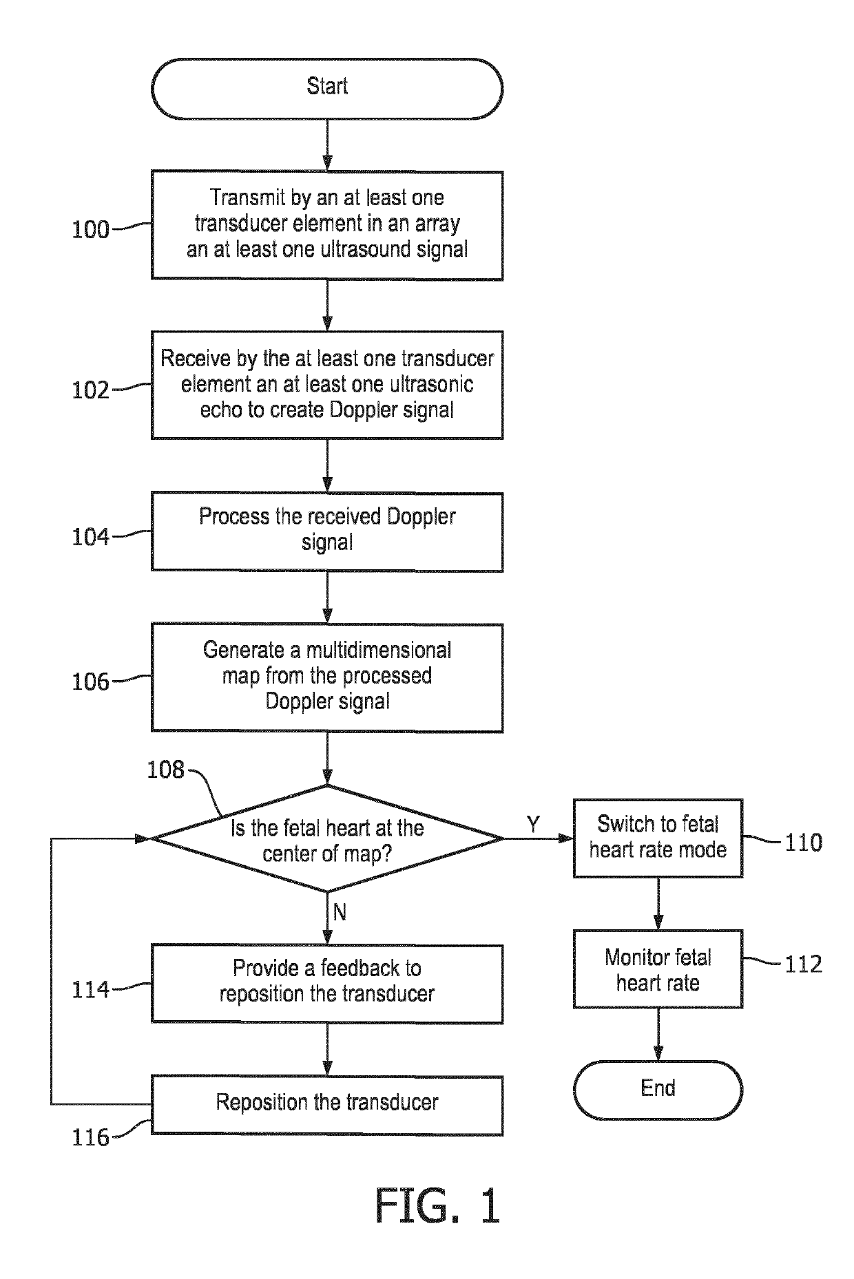
[0031]The term “depth” as used herein refers to a distance measured from the transducer array plane to a point within the maternal abdomen volume that is being scanned. Depth is set to have a value of “0” at the inner wall of the maternal abdomen and a maximum value at the farthest point from which a Doppler signal can be obtained. The depth may be, in some embodiments, the middle of the sample volume from which the Doppler signal is calculated.

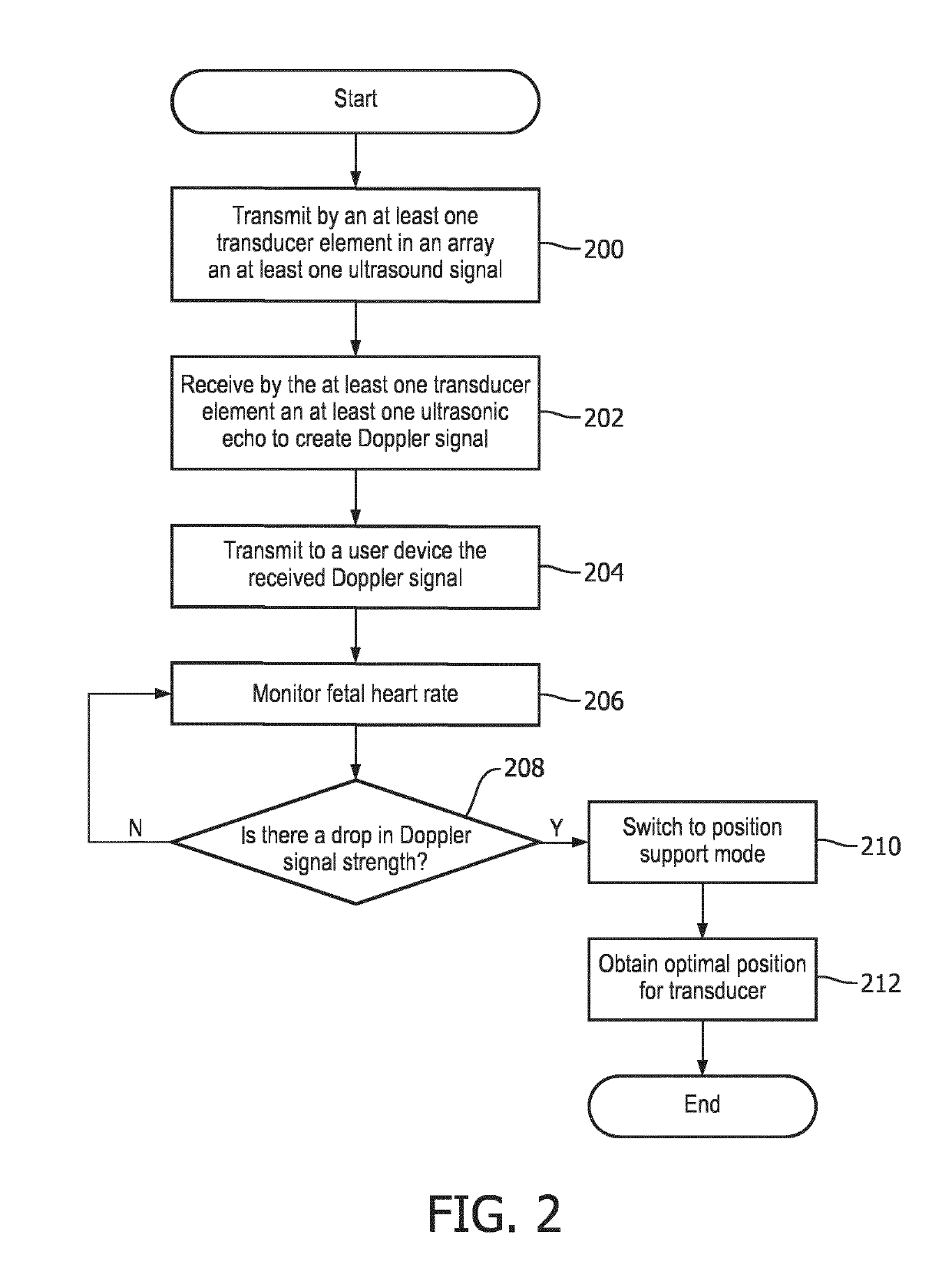
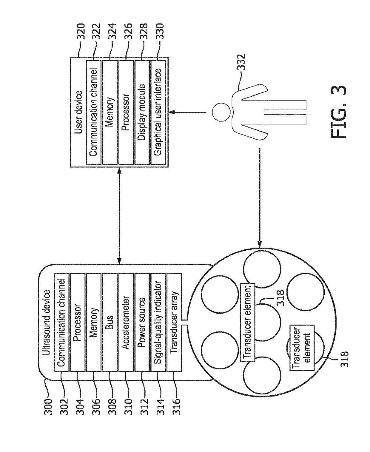
[0032]The term “channel” as used herein refers to a wired or wireless data pathway allocated to each transducer element in a transducer array disclosed in the present disclosure. One channel is set aside for processing the Doppler signal received from all transducer elements when the ultrasound device is used in the fetal heart rate mode (fUR-mode).

[0033]The term “feedback” as used herein refers to an audio, visual, or combined audio and ...


PUM


Abstract

A method for monitoring fetal health. The method of the present disclosure comprises transmitting (100) an ultrasound signal from an at least one transducer element in an array towards a maternal abdomen. An at least one ultrasonic echo signal corresponding to an at least one depth of the maternal abdomen is then received (102) by the at least one transducer element. The at least one Doppler signal is determined after the echo is received by the at least one transducer element is then processed (104) in an at least one channel. A multidimensional map is then generated (106) from the processed Doppler signal. A feedback (114) to reposition the at least one transducer element is then provided when the fetal heart is determined to lie outside an ultrasound beam volume.

Description

BACKGROUND OF THE INVENTION[0001]Measuring the fetal heart rate (fHR) during labor and delivery is the most commonly applied obstetric procedure to assess the health state of the fetus and to determine if early intervention is required. However, for Doppler ultrasound (US) as well as for abdominal electrodes, long periods of signal loss have been reported during fHR monitoring. For the assessment of the health state of the fetus, these long signal loss periods strongly limit the interpretation of the fHR traces in clinical practice.[0002]The ultrasound Doppler recording quality highly depends on the correct placement of the transducer on the maternal abdomen, since the fetal heart needs to be located within the ultrasound beam. However, finding the optimal transducer position can be a challenging task. In clinical practice, skilled nurses palpate the maternal abdomen and determine the fetal presentation from the outside. By relying on a signal-quality indicator and by listening to t...

Claims


Application Information

Patent Timeline
Patent Type & Authority Applications(United States)
IPC IPC(8): A61B8/08
CPCA61B8/0866A61B8/0883A61B8/488A61B8/4245A61B8/5269A61B8/42
Inventor HAMELMANN, PAULSCHMITT, LARSKOLEN, ALEXANDER FRANCISCUSWOHLSCHLAGER, MARKUS
Owner KONINKLJIJKE PHILIPS NV
Why Eureka
Try Eureka