Selection and Cloning of T Lymphocytes in a Microfluidic Device

a microfluidic device and t lymphocyte technology, applied in the field of selection and expansion of t lymphocytes, can solve the problems of lack of ex vivo expansion methods and therapies that still require further refinement, and achieve the effect of facilitating linkage of peptide-mhc complexes

Inactive Publication Date: 2020-04-16
BRUKER CELLULAR ANALYSIS INC
View PDF2 Cites 6 Cited by
  • Summary
  • Abstract
  • Description
  • Claims
  • Application Information

AI Technical Summary

Benefits of technology

[0017]In certain embodiments, the activating agent can include CD3 and CD28 agonists. The CD3 and / or CD28 agonist can be an antibody. In some embodiments, the anti-CD3 agonist (e.g., antibody) is conjugated to a solid support. In some embodiments, the anti-CD28 agonist (e.g., antibody) is conjugated to a solid support. In some embodiments, the anti-CD28 agonist (e.g., antibody) is a solute in an aqueous solution. Thus, for example, the activating agent can comprise one or more beads conjugated to anti-CD3 and anti-CD28 agonist antibodies. Alternatively, the activating agent can comprise one or more beads conjugated to anti-CD3 agonist antibodies and a solution of soluble anti-CD28 agonist antibody. In still other alternatives, the CD3 and / or CD28 agonists can be linked (covalently or non-covalently) to one or more surfaces of a sequestration pen. The surface(s) of the sequestration pen can be conditioned in a manner that facilitates linkage of the CD3 and / or CD28 agonists to the surface(s).
[0019]In certain embodiments, the activating agent can comprise a complex between a peptide antigen and an MHC molecule. Such complexes can be linked, as in the case of peptide-MHC tetramer complexes. The peptide-MHC complexes can be conjugated to a solid support. In some embodiments, the solid support can be one or more beads. In other embodiments, the solid support can be one or more surfaces of a sequestration pen. Thus, the surface(s) of the sequestration pen can be conditioned in a manner that facilitates linkage of the peptide-MHC complexes to the surface(s).

Problems solved by technology

However, the therapies still require further refinement.
One of the key problems in both autologous T cell therapies and CAR-T therapies is the lack of methods for expanding T cells ex vivo in a manner that selectively expands T cells having the highest tumor killing potential.

Method used


Image

  • Selection and Cloning of T Lymphocytes in a Microfluidic Device
  • Selection and Cloning of T Lymphocytes in a Microfluidic Device
  • Selection and Cloning of T Lymphocytes in a Microfluidic Device

Examples


example 1

ell Expansion in an OptoSelect™ Chip

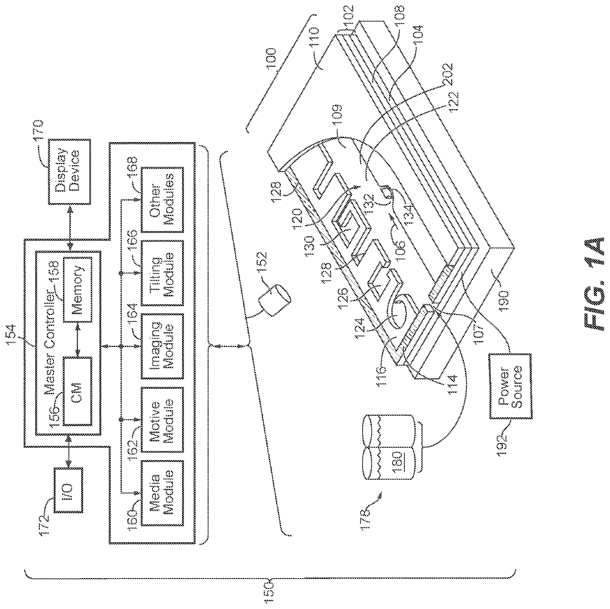
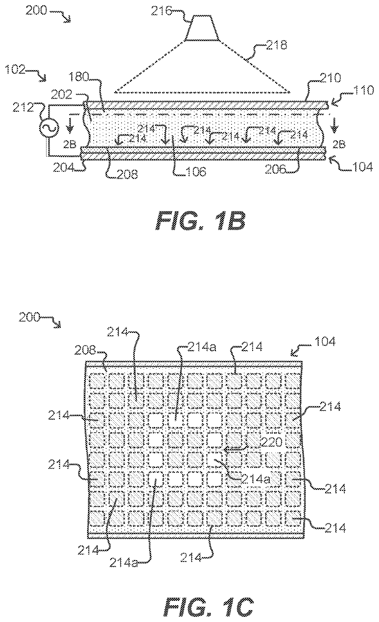
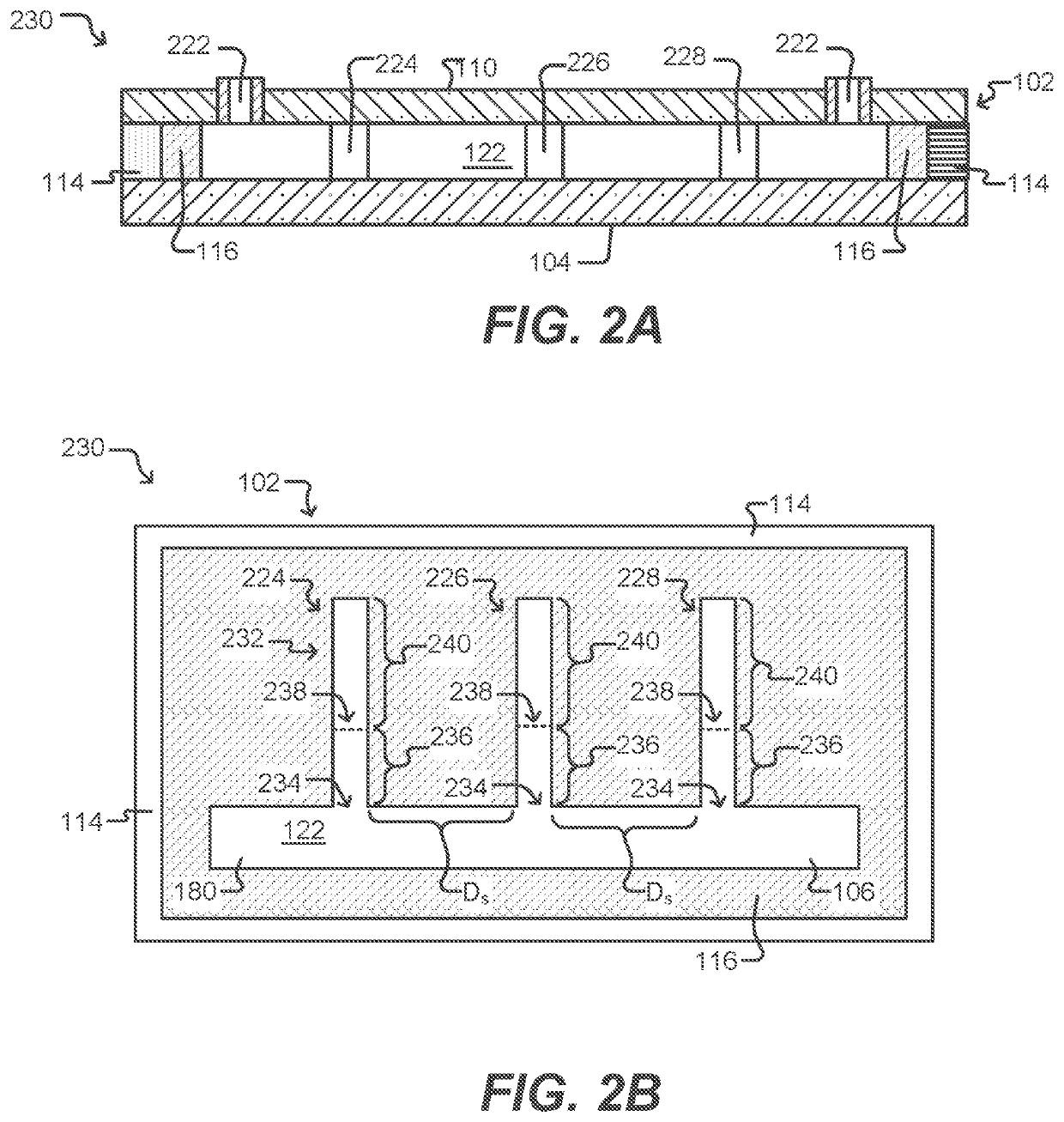
[0258]T cell expansion was achieved within an OptoSelect chip, a nanofluidic device manufactured by Berkeley Lights, Inc. and controlled by an optical instrument which was also manufactured by Berkeley Lights, Inc. The instrument included: a mounting stage for the chip coupled to a temperature controller; a pump and fluid medium conditioning component; and an optical train including a camera and a structured light source suitable for activating phototransistors within the chip. The OptoSelect™ chip included a substrate configured with OptoElectroPositioning (OEP™) technology, which provides a phototransistor-activated OET force. The chip also included a plurality of microfluidic channels, each having a plurality of NanoPen™ chambers (or sequestration pens) fluidically connected thereto. The volume of each sequestration pen was around 1×10⁶ cubic microns.

[0259]CD3+ human T lymphocytes isolated from peripheral blood were mixed with anti-CD3 / anti-CD2...

example 2

Expansion of Human T Cells in an OptoSelect™ Chip

[0264]T cell expansion was achieved within an OptoSelect chip (Berkeley Lights, Inc.), which was controlled by an optical instrument also manufactured by Berkeley Lights, Inc., as described in Example 1.

[0265]Initially, human CD14+ monocytes isolated from peripheral blood were cultured for 7 days in DC culture medium (RPMI, 10% FBS, 2% Human AB serum, 100 ng / ml GM-CSF, 50 ng / ml IL-4; R&D Systems) to promote differentiation of dendritic cells (DCs). 250 μg / ml LPS (R&D Systems) was added to the culture medium during the last 2 days of culture to promote DC activation.

[0266]Allogeneic donor T lymphocytes were mixed with DCs from the foregoing culture at a ratio of ˜10 T cells / 1 DC and incubated for 5 hours in a 5% CO2 incubator at 37° C. Following the incubation, the T cells / DCs mixture was resuspended, then flowed through a fluidic inlet and into the microfluidic channels within the chip. The flow was stopped and T cells / DCs were random...

example 3

pecific Expansion of Human T Cells in an OptoSelect™ Chip

[0271]T cell expansion was achieved within an OptoSelect chip (Berkeley Lights, Inc.), which was controlled by an optical instrument also manufactured by Berkeley Lights, Inc., as described in Example 1.

[0272]Initially, human CD14+ monocytes isolated from peripheral blood were cultured for 7 days in DC culture medium (RPMI, 10% FBS, 2% Human AB serum, 100 ng / ml GM-CSF, 50 ng / ml IL-4; R&D Systems) to promote differentiation of dendritic cells (DCs). 250 μg / ml LPS (R&D Systems) was added to the culture medium during the last 2 days of culture to promote DC activation. At the same time as the addition of the LPS, the DCs were also pulsed with 10 μM Tetanus toxin (TT) antigen (Sigma-Aldrich Co.) and 10 μM Epstein Barr Virus (EBV) antigen (EastCoast Bio, Inc.).

[0273]Autologous donor T lymphocytes were mixed with TT- and EBV-pulsed DCs from the foregoing culture at a ratio of ˜10 T cells / 1 DC and incubated for 5 hours in a 5% CO2 in...


PUM

PropertyMeasurementUnit
volumeaaaaaaaaaa
width Wconaaaaaaaaaa
volumeaaaaaaaaaa

Abstract

Methods of expanding T lymphocytes in a microfluidic device are provided. The methods can include introducing one or more T lymphocytes into a microfluidic device; contacting the one or more T lymphocytes with an activating agent; and perfusing culture medium through the microfluidic device for a period of time sufficient to allow the one or more T lymphocytes to undergo at least one round of mitotic cell division. The expansion can be non-specific or antigen-specific. T lymphocytes produced according to the disclosed methods are also provided, along with methods of treating cancer in a subject. The methods of treating cancer can include isolating T lymphocytes from a tissue sample obtained from the subject; expanding the isolated T lymphocytes in a microfluidic device; exporting the expanded T lymphocytes from the microfluidic device; and reintroducing the expanded T lymphocytes into the subject.

Description

CROSS REFERENCE TO RELATED APPLICATIONS[0001]This application is a continuation application of U.S. patent application Ser. No. 15 / 802,100, filed on Nov. 2, 2017, which is a continuation application of International Patent Application No. PCT / US2017 / 022846, filed on Mar. 16, 2017, which claims the benefit under 35 U.S.C. § 119 of U.S. Patent Application No. 62 / 309,454, filed on Mar. 17, 2016, U.S. Patent Application No. 62 / 326,667, filed on Apr. 22, 2016, U.S. Patent Application No. 62 / 412,212, filed on Oct. 24, 2016, and U.S. Patent Application No. 62 / 470,744, filed on Mar. 13, 2017, the entire disclosure of each of which is incorporated herein by reference.FIELD[0002]The field generally relates to methods, systems and devices for selecting and expanding T lymphocytes within a microfluidic environment.BACKGROUND OF THE INVENTION[0003]Immunotherapy is the burgeoning field of using a patient's immune system to help fight disease. A variety of immunotherapy strategies have been evalua...

Claims


Application Information

Patent Timeline
Patent Type & Authority Applications(United States)
IPC IPC(8): C12N5/0783A61K39/00C12M1/00C12M3/06A61P35/00
CPCC12N2501/2302A61K39/0011A61K2035/124C12N2501/515C12M23/16C12M29/10C12N2502/1121C12N2501/51C12N2533/50C12N5/0636A61P35/00A61K2039/5158A61K35/17
Inventor BRONEVETSKY, YELENAWANG, XIAOHUABEEMILLER, PETER J.BEAUMONT, KRISTIN G.LOWE, JR., RANDALL D.MASTROIANNI, ALEXANDER J.CHAPMAN, KEVIN T.MARKS, NATALIE C.
Owner BRUKER CELLULAR ANALYSIS INC
Why Eureka
Try Eureka