Medical image processing apparatus and endoscope apparatus

a technology of endoscope and medical image, which is applied in the field of medical image processing apparatus and endoscope apparatus, can solve the problems of heavy burden on the patient, discrimination of the type of the degree of progress of the lesion or the lik

Active Publication Date: 2020-07-30
FUJIFILM CORP
View PDF4 Cites 2 Cited by
  • Summary
  • Abstract
  • Description
  • Claims
  • Application Information

AI Technical Summary

Benefits of technology

[0007]An object of the present invention is to provide a medical image processing apparatus and an endoscope apparatus that can present more detailed information regarding a lesion or the like than before.
[0024]The medical image processing apparatus and the endoscope apparatus of the present invention can provide more detailed information regarding a lesion or the like than before.

Problems solved by technology

A doctor or the like discriminates a type and a degree of progress of a lesion or the like presented by the medical apparatus, but discrimination of the type and the degree of progress of a lesion or the like is still a heavy burden.

Method used


Image

  • Medical image processing apparatus and endoscope apparatus
  • Medical image processing apparatus and endoscope apparatus
  • Medical image processing apparatus and endoscope apparatus

Examples


first embodiment

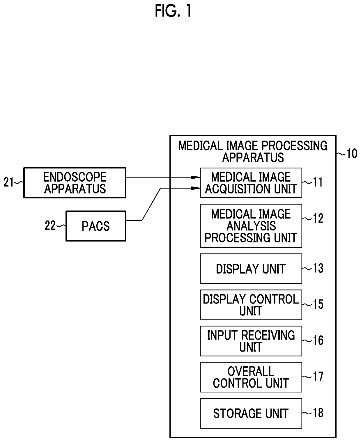
[0038]As shown in FIG. 1, a medical image processing apparatus 10 comprises a medical image acquisition unit 11, a medical image analysis processing unit 12, a display unit 13, a display control unit 15, an input receiving unit 16, an overall control unit 17, and a storage unit 18.

[0039]The medical image acquisition unit 11 acquires a medical image including a subject image, directly from an endoscope apparatus 21 or the like that is a medical apparatus, or through a management system such as a picture archiving and communication system (PACS) 22, or other information systems. The medical image is a still image or a motion picture (a so-called examination motion picture). In a case where the medical image is a motion picture, the medical image acquisition unit 11 can acquire a frame image forming a motion picture after examination as a still image. In addition, in a case where the medical image is a motion picture, display of the medical image includes not only displaying a still im...

second embodiment

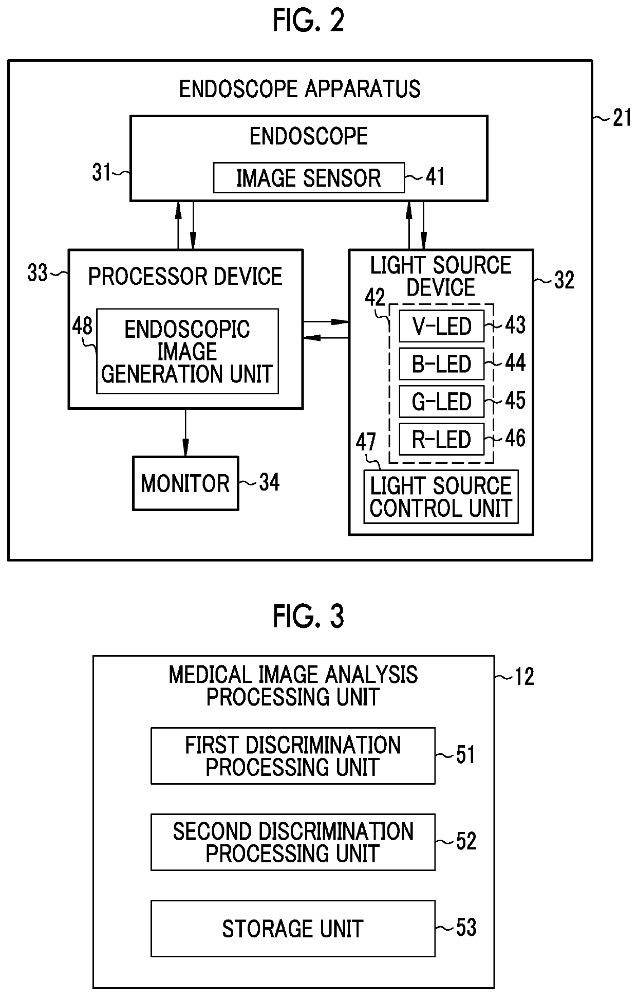
[0079]In the first embodiment described above, the medical image processing apparatus 10 and the endoscope apparatus 21 are separate apparatuses. However, the endoscope apparatus 21 can include the medical image processing apparatus 10. In this case, as an endoscope apparatus 510 shown in FIG. 10, each unit 520 forming the medical image processing apparatus 10 is provided in the processor device 33. Here, the display unit 13 can share the monitor 34 of the endoscope apparatus 21. In addition, the medical image acquisition unit 11 corresponds to an “endoscopic image acquisition unit” formed by the image sensor 41 and the endoscopic image generation unit 48. For this reason, it is sufficient to provide the processor device 33 with each unit other than the medical image acquisition unit 11 and the display unit 13. The configuration of each of other units is the same as in the first embodiment. In addition, a new endoscope apparatus can be configured by all of the medical image processi...


PUM


Abstract

A medical image processing apparatus includes a medical image acquisition unit that acquires a medical image including a subject image, a first discrimination processing unit that performs a first discrimination process of detecting a lesion and discriminating a degree of progress of the detected lesion by using a first medical image captured using first illumination light having a specific spectrum, among the medical images, and a second discrimination processing unit that performs a second discrimination process of discriminating the degree of progress of the lesion by using a second medical image captured using second illumination light having a spectrum different from the first illumination light, among the medical images.

Description

CROSS-REFERENCE TO RELATED APPLICATIONS[0001]This application is a Continuation of PCT International Application No. PCT / JP2018 / 038496 filed on 16 Oct. 2018, which claims priority under 35 U.S.C § 119(a) to Japanese Patent Application No. 2017-201333 filed on 17 Oct. 2017. The above application is hereby expressly incorporated by reference, in its entirety, into the present application.BACKGROUND OF THE INVENTION1. Field of the Invention[0002]The present invention relates to a medical image processing apparatus that uses analysis results of medical images, and an endoscope apparatus.2. Description of the Related Art[0003]In the related art, an apparatus that acquires an image including a subject image (hereinafter, referred to as a medical image) among apparatuses relevant to medical care (hereinafter, referred to as a medical apparatus) presents the acquired medical image to a doctor. Then, the doctor performs diagnosis or the like using the medical image obtained from the medical ...

Claims


Application Information

Patent Timeline
Patent Type & Authority Applications(United States)
IPC IPC(8): G06T7/00A61B1/06A61B5/00
CPCA61B1/0638G06T7/0012G06T2207/10068G06T2207/30096A61B5/7275G16H30/40G16H50/20G16H50/30G06T2207/10016G06T2207/10024G06T2207/10152A61B1/000094
Inventor AOYAMA, TATSUYA
Owner FUJIFILM CORP
Why Eureka
Try Eureka