Reusable specimen imaging device holder system with replaceable membranes

a technology of imaging device and membrane, which is applied in the field of reusable specimen imaging device holder system with replaceable membrane, can solve the problems of not utilizing the full design and methodology of the rotational component, dismantling the rotational component, and increasing the risk of poorer cosmetics, so as to reduce the cost, simplify and inexpensive, and minimize the excised tissue

Inactive Publication Date: 2021-03-18
CURETON JANICE ANN +1
View PDF0 Cites 0 Cited by
  • Summary
  • Abstract
  • Description
  • Claims
  • Application Information

AI Technical Summary

Benefits of technology

[0009]There is a limited availability of surgical specimen image holders that minimally compress the excised tissue and offer reusability. Our system provides a simplified and inexpensive alternative to current one time use device holders. It can provide reduced cost and reduced medical waste associated with these procedures. Because our invention features a plastic framed device that is easily sanitized with medical grade sanitizers, and allows for replacement of the radiolucent membrane that prevents cross contamination of tissue particles it is a useful tool in radiographic specimen imaging.

Problems solved by technology

We continue using the Wedge as it provides adequate retention to hold the specimen in proper orientation with minimal compression of the surgical specimen; however, we are dismantling the rotational component and not utilizing its full design and methodology.
However, both were criticized in prior art referenced Devlin et al., U.S. Pat. No. 5,383,472, due to their cleaning process and potential for cross contamination of tissue particles, and concern for excess compression of the surgical specimen.
The need for more surgeries can create emotional stress and financial hardship, as well as increased risk for a poorer cosmetic result, and could lead to mastectomy in some cases.
There is a limited availability of surgical specimen image holders that minimally compress the excised tissue and offer reusability.

Method used


Image

  • Reusable specimen imaging device holder system with replaceable membranes
  • Reusable specimen imaging device holder system with replaceable membranes
  • Reusable specimen imaging device holder system with replaceable membranes

Examples


Embodiment Construction

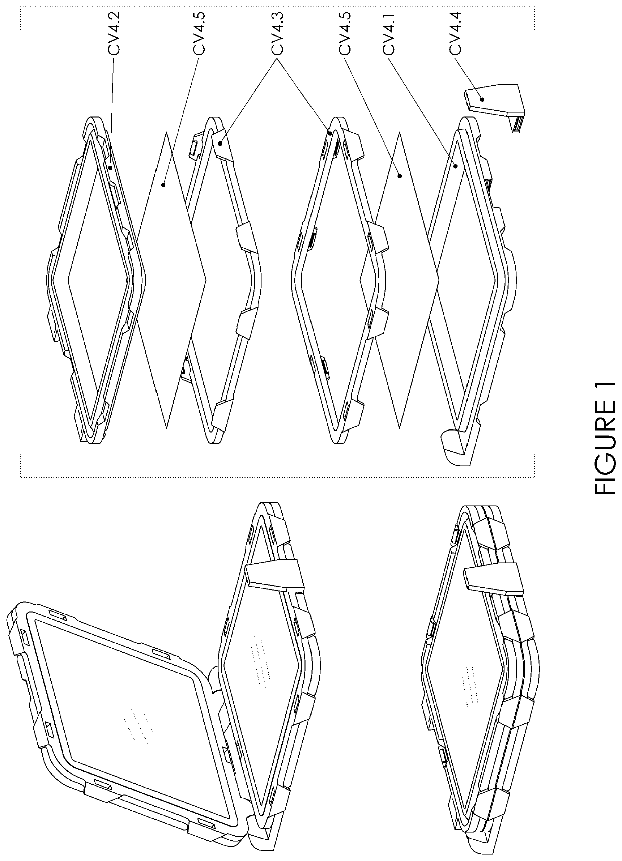
[0017]FIG. 1 shows a fully assembled perspective view of the embodiment. It is composed of CV 4.1, CV 4.2, two pieces of CV4.3, CV.4, and CV 4.5.

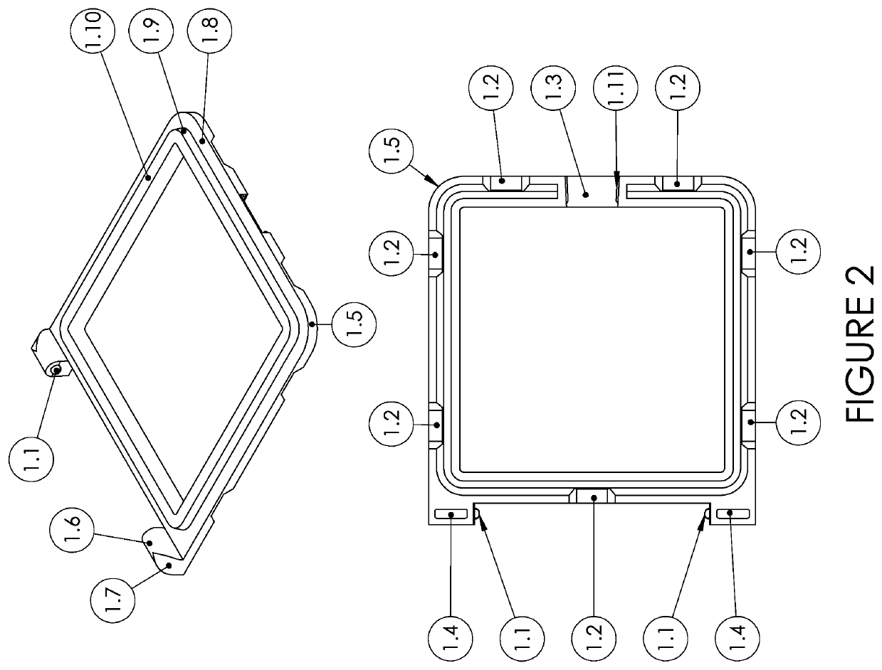
[0018]FIG. 2 shows a perspective view and a bottom view of CV4.1. It identifies the individual working components.[0019]1.1 is a convex sphere that makes up half of the working hinge.[0020]1.2 is the shelf that allows the clip on component 3.1.[0021]1.3 is a cut out to allow room for CV 4.4 to be inserted.[0022]1.4 is a cavity to lower thickness of material to optimize even shrinkage of the medium.[0023]1.5 is a curved edge to aid in ease of use.[0024]1.6 is the structure to maximize support for the hinge features.[0025]1.7 is curved and reduces material.[0026]1.8 is one of four surfaces that trap the working membrane on CV 4.1.[0027]1.9 is the second of four surfaces that trap the working membrane on CV 4.1.[0028]1.10 is one of two surfaces that push the two membranes together to form the outer limits where the specimen will rest.[0029]1.1...


PUM

PropertyMeasurementUnit
thicknessaaaaaaaaaa
sizesaaaaaaaaaa
radiographic imagingaaaaaaaaaa

Abstract

The present invention is for the retention of surgically removed tissue for radiographic imaging. In breast conservation surgery specimen radiographic imaging is used to evaluate the success of removed diseased tissue with a negative margin. Accurate positioning of tissue orientation with minimal compression of the removed pathologic neoplasm is employed. The radiolucent device holder's purpose is to secure the tissue in proper orientation for radiographic imaging and margin evaluation. The present invention is a reusable system. It features a snap together component that allows for the replacing of the radiolucent membranes. Components are properly disinfected and membranes replaced making the device holder ready for reuse. This reusability is innovative, more cost effective and reduces medical waste. While this present invention, reusable specimen imaging holder device system with replaceable membranes has particular use in breast conservation surgery specimen imaging for optimal margin evaluation it is not exclusive to that purpose.

Description

BACKGROUND1. Field of the Invention[0001]This invention relates to an apparatus for holding surgically excised breast tissue for radiographic imaging for the purpose of optimal tissue margin evaluation.2. Brief Description of the Related Art[0002]My passion for this invention began after our hospital bought four new intraoperative radiographic units for imaging our breast specimens at the various hospital surgical sites. We began using a new specimen device holder called the Wedge by Faxitron, Bioptics, prior art referenced Akif, B., U.S. Pat. No. 8,741,232. Our breast radiologists prefer a true 90 degree orthogonal view for breast specimen imaging for margin evaluation. Although, the Wedge was very unique in its new design and methodology, our radiologist did not feel it resulted in a true 90 degree orthogonal view with its rotational component, but rather felt it resulted in more of a 45 degree view. We continue using the Wedge as it provides adequate retention to hold the specime...

Claims


Application Information

Patent Timeline
Patent Type & Authority Applications(United States)
IPC IPC(8): A61B10/00B01L9/00G01N33/483
CPCA61B10/0096B01L9/52G01N2223/6126B01L2300/0609B01L2300/0809G01N33/4833B01L3/5055B01L2300/0681
Inventor CURETON, JANICE ANNCHATMAN, MATTHEW LUCAS
Owner CURETON JANICE ANN
Why Eureka
Try Eureka