Image display apparatus and method, and storage medium

Inactive Publication Date: 2005-02-08
CANON KK
View PDF4 Cites 22 Cited by
  • Summary
  • Abstract
  • Description
  • Claims
  • Application Information

AI Technical Summary

Benefits of technology

It is an object of the present invention to provide an image display apparatus and an image display method, which are capable of clearly indicating to an observer subtle differences between

Problems solved by technology

A specific problem being researched is how to cope with changes in the radiographic conditions between the past image and the current image.
Regardless of how this projected image which gives only 2-dimensional information is chan

Method used


Image

  • Image display apparatus and method, and storage medium
  • Image display apparatus and method, and storage medium
  • Image display apparatus and method, and storage medium

Examples


first embodiment

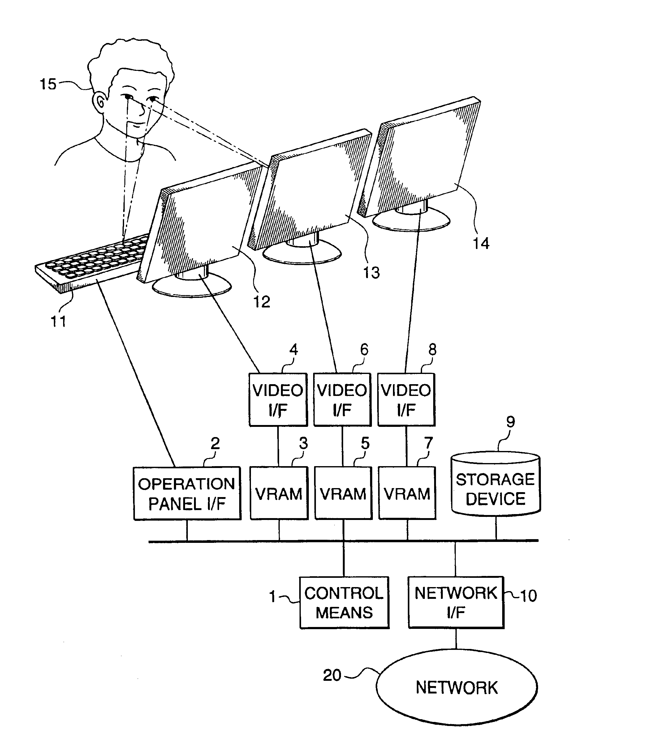
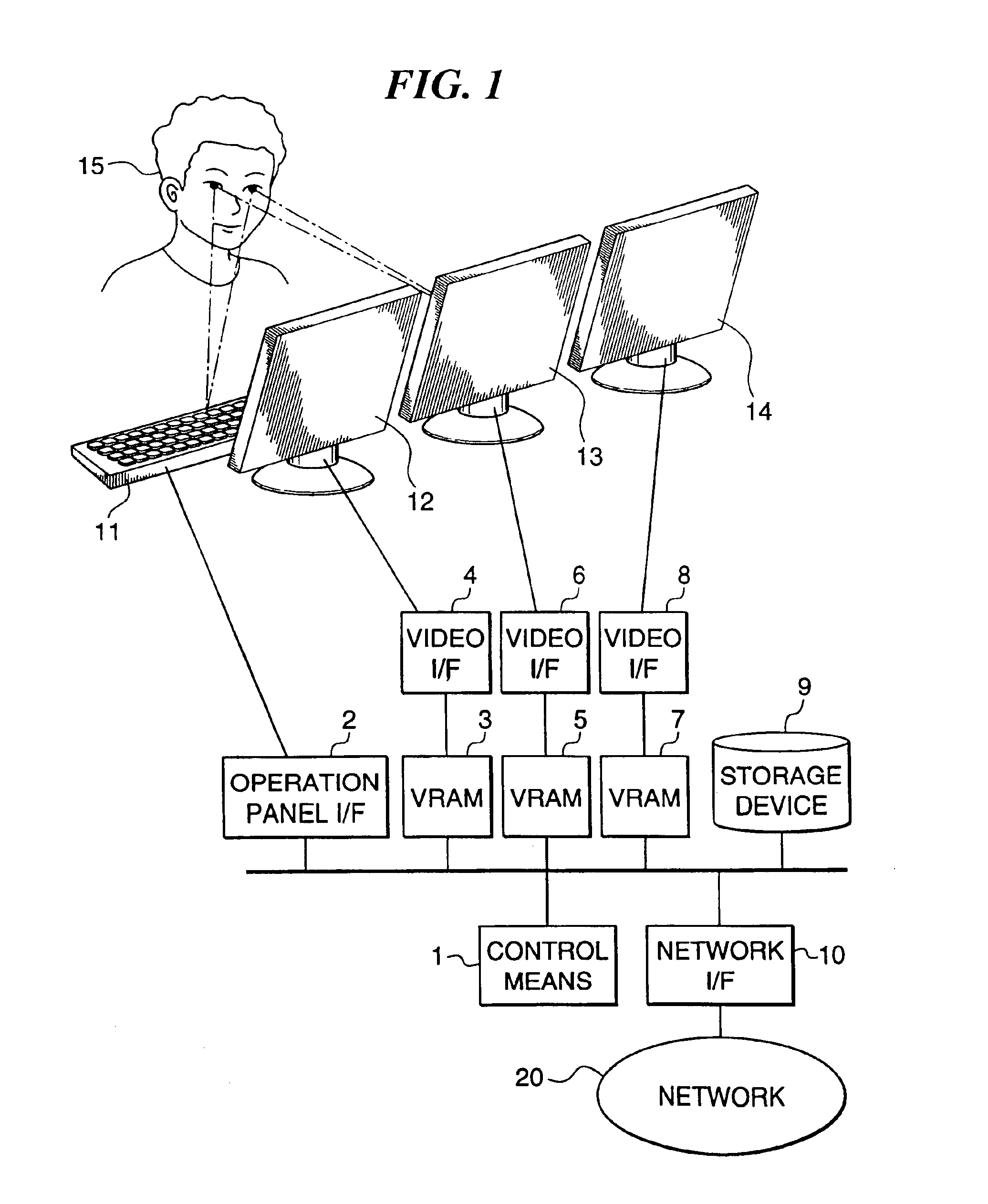
A description will now be given of a first embodiment of the present invention with reference to the drawings. FIG. 1 is a block diagram showing the constitution of an image display apparatus according to the first embodiment. In this embodiment, a description will be given of an image display apparatus for displaying medical images such as chest X-ray images, CT images, MRI images or ultrasound diagnosis images.

The image display apparatus displays at least two medical images such as chest X-ray images taken of the same subject at different times. As shown in FIG. 1, the image display apparatus has a network interface (network I / F) 10 for inputting medical images (including a past image and a current image) and information relating to the patient in question from a network 20.

A chest X-ray radiography device or the like is connected to the network I / F 10 via the network 20. Chest X-ray images taken by the chest X-ray radiography device are transferred along with patient information ...

second embodiment

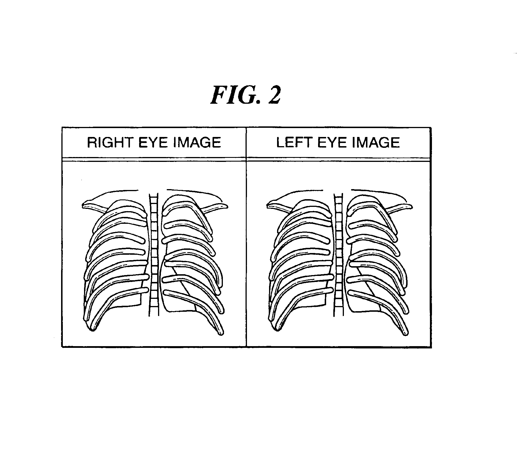
A description will now be given of a second embodiment of the present invention with reference to FIG. 4. FIG. 4 shows an example of images displayed on a stereo display device of an image display apparatus according to the second embodiment. In FIG. 4, the image captured by the right eye is shown on the left side and the image captured by the left eye on the right side. The image display apparatus has the same constitution in the present embodiment as in the first embodiment described above, and hence the description of this constitution will be omitted here.

The present embodiment differs from the first embodiment in that there is a function for displaying the left and right eye images in different colors.

For example, the right eye image may be displayed in red on the stereo display device 12 and the left eye image in blue, as shown in FIG. 4. The colors used can be freely changed in accordance with the preference of the observer 15. The chosen colors are displayed, for example, us...

third embodiment

A description will now be given of a third embodiment of the present invention with reference to FIG. 5. FIG. 5 shows an example of images displayed on a stereo display device of an image display apparatus according to the third embodiment. In FIG. 5, the image captured by the right eye is shown on the left side and the image captured by the left eye on the right side. The image display apparatus has the same constitution in the present embodiment as in the first embodiment described above, and hence the description of this constitution is omitted here.

The present embodiment differs from the first or second embodiment in that there is a function for making either the left eye image or the right eye image flash on and off. For example, in FIG. 5, the left eye image can be made to flash. The rate of flashing can be freely changed by the observer 15.

This flashing is effective particularly when the colors of the two images are changed as in the second embodiment. If one of the images is...


PUM


Abstract

There are provided an image display apparatus and an image display method, which are capable of clearly indicating to an observer subtle differences between two images of the same subject obtained at different times, without the need for complex image processing, and a storage medium storing a program for implementing the image display method. Two images of the same subject obtained at different times are inputted. A stereo display device displays the two inputted images in a manner enabling an observer to fuse the two inputted images together for stereoscopic viewing. A control means controls the stereo display device to display the two inputted images such that the two images are projected separately into the left and right eyes of the observer.

Description

BACKGROUND OF THE INVENTION1. Field of the InventionThe present invention relates to an image display apparatus and image display method for displaying two images of the same subject obtained, for example, at different times, and a storage medium storing a program for implementing the image display method.2. Prior ArtMedical X-ray images, in particular X-ray images of the human chest, are indispensable to medical diagnosis, and are very widely used even today.Chest X-ray images are used for examining the extent of progress of a disease or for examining for changes indicative of outbreak of a disease in a patient who is the subject of the X-ray radiography. Such examinations are often carried out by comparing current and past X-ray images of the same patient. In such a comparison, the past image and the current image are generally displayed simultaneously or films thereof are presented simultaneously, and an observer looks for differences between the two.Moreover, as described in “Ap...

Claims


Application Information

Patent Timeline
IPC IPC(8): G06F3/14G09G3/00H04N13/00A61B6/02A61B6/00G09G5/00G09G5/36H04N7/18H04N13/122
CPCA61B6/464G09G3/003H04N13/0011H04N13/0018H04N13/0025H04N13/004G06F3/1438H04N13/044H04N13/0037H04N13/0055H04N13/0059H04N13/0207H04N13/0253H04N13/0285H04N13/0404H04N13/0431H04N13/0434H04N13/0438H04N13/337H04N13/341H04N13/305H04N13/344H04N13/194H04N13/254H04N13/189H04N13/156H04N13/111H04N13/207H04N13/122H04N13/334H04N13/286H04N13/15H04N13/133
Inventor INOUE, HITOSHISANADA, SHIGERU
Owner CANON KK
Why Eureka
Try Eureka