
Nasolacrimal duct tube used for lacrimal duct reformation operation, and nasolacrimal duct tube instrument

a technology of lacrimal duct reformation and duct tube instrument, which is applied in the field of nasolacrimal stent, can solve the problems of difficult work for the insertion of silicon tube device in the lacrimal passage, large hemorrhage, and insufficient stent siz

Inactive Publication Date: 2005-04-12
TORAY IND INC +1
View PDF10 Cites 30 Cited by
  • Summary
  • Abstract
  • Description
  • Claims
  • Application Information

AI Technical Summary

Benefits of technology

According to the present invention, a nasolacrimal stent device for lacrimal passage plastic surgery includes a flexible tube capable of being inserted and detained in a lacrimal passage and provided with at least an opening formed in a part thereof at a predetermined distance from one of the opposite ends thereof, and an illuminating device capable of being inserted in and extracted from the flexible tube through the opening formed in the flexible tube. The illuminating device may be an optical fiber or a light-emitting device.

Problems solved by technology

Therefore the silicon tube device must be operated by feel in the nasal duct when inserting the silicon tube device into the lacrimal passage, and hence it often occurs that the silicon tube device deviates from a correct passage, a false passage is formed in tissues, and nasal mucous membranes are damaged causing massive hemorrhage.
Thus the work for the insertion of the silicon tube device in the lacrimal passage is very difficult.
However, the former method requires difficult work to use fingers for surgical operations, observing pictures taken by an endoscope and displayed on a monitor.
The use of a surgical lamp is not perfectly effective because it is difficult to illuminate the interior of the nasal duct directly with the surgical lamp.

Method used


Image

  • Nasolacrimal duct tube used for lacrimal duct reformation operation, and nasolacrimal duct tube instrument
  • Nasolacrimal duct tube used for lacrimal duct reformation operation, and nasolacrimal duct tube instrument
  • Nasolacrimal duct tube used for lacrimal duct reformation operation, and nasolacrimal duct tube instrument

Examples


Embodiment Construction

A preferred embodiment of the present invention will be described hereinafter.

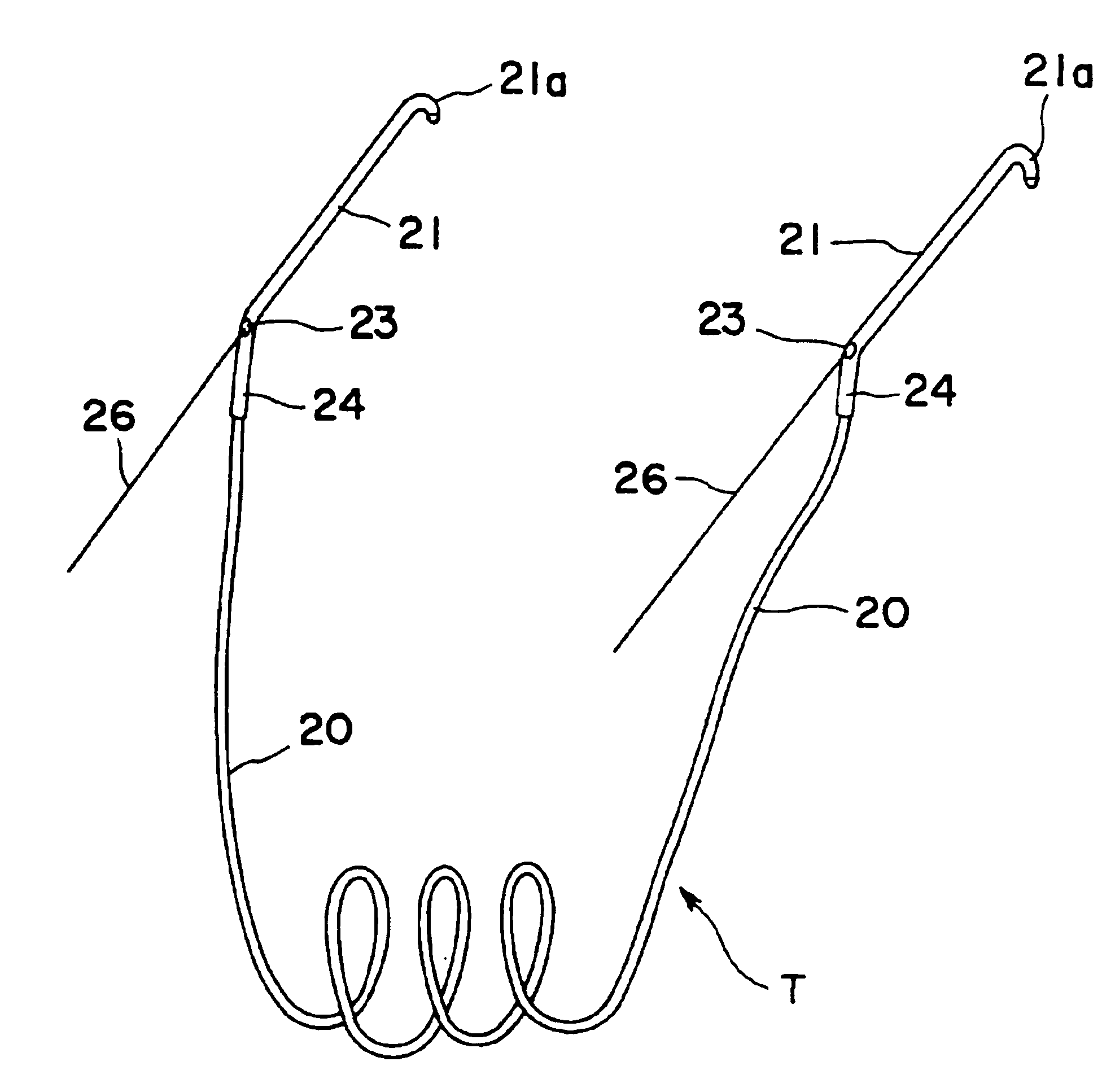
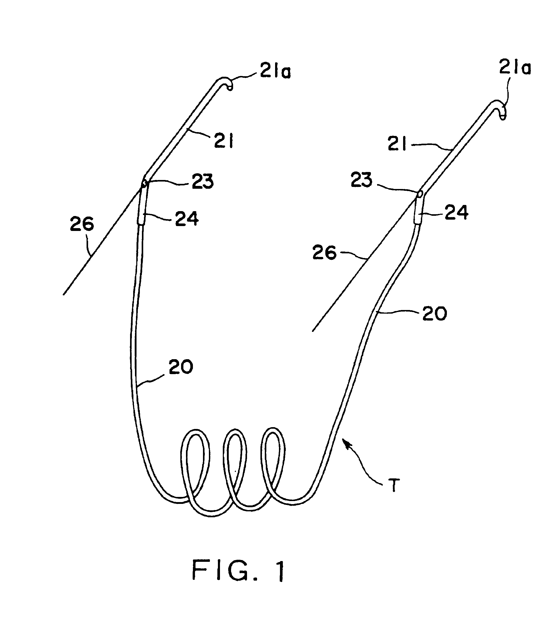
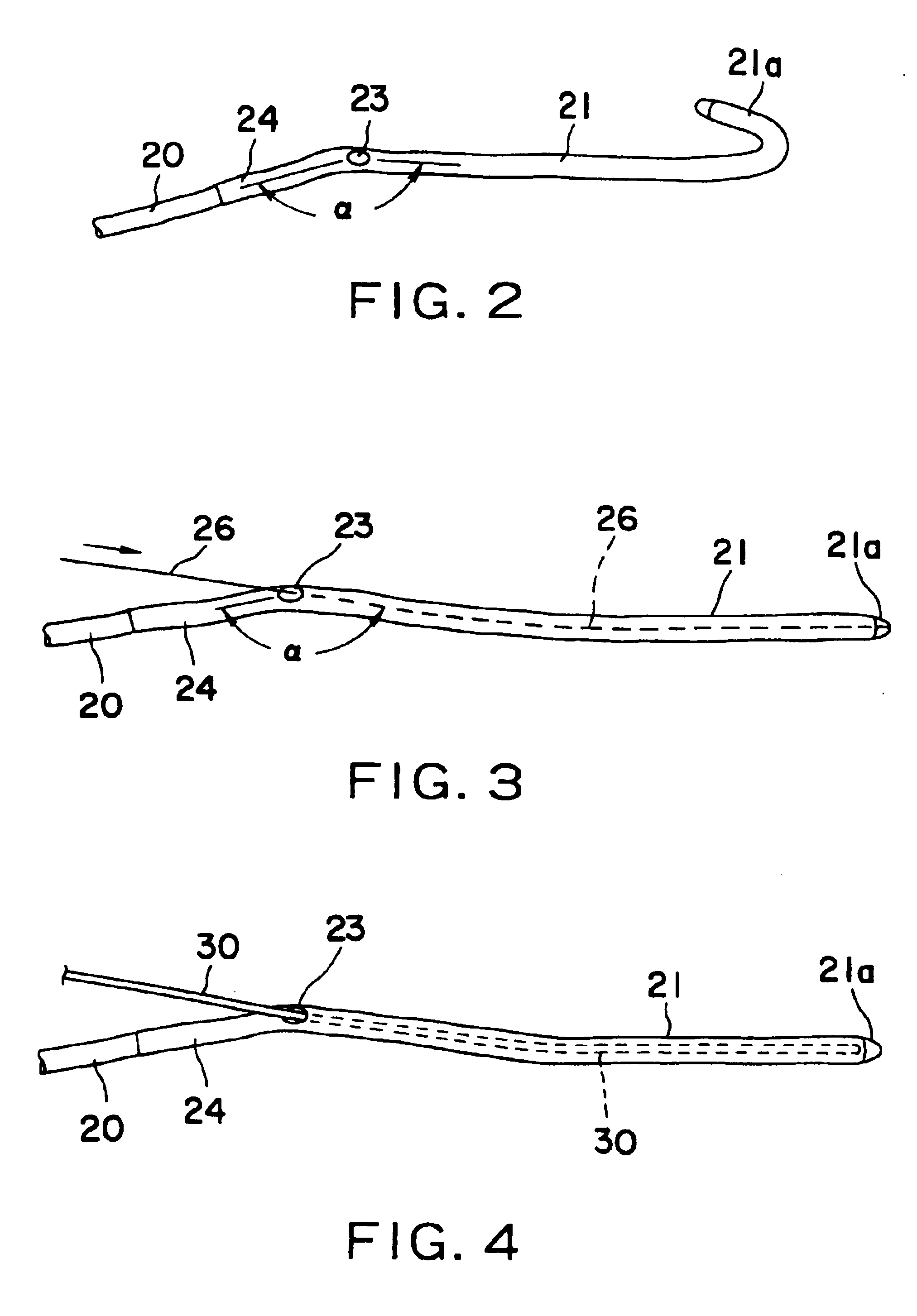
Referring to FIG. 1, a nasolacrimal stent and a nasolacrimal stent device in a preferred embodiment according to the present invention for lacrimal passage plastic surgery includes a tube T having a detention tube segment 20 and flexible, light-transmitting (transparent or translucent) first and second probe tube segments 21 connected to the opposite ends of the flexible detention tube segment 20, respectively. The flexible detention tube segment 20, similarly to a conventional one, may be formed of silicone. The probe tube segments 21 may be formed of, for example, a polyolefin resin, a polyamide resin, a polyurethane resin or a mixture of some of those resins. Tubes of those resins including polyolefin resins are harder and more excellent in shape retention than silicone tubes, and can be readily inserted in the lacrimal passage. Preferably, the inside diameter of the detention tube segment 20 is, for ex...


PUM


Abstract

A nasolacrimal stent device illuminates a dark nasal cavity so that the interior of the nasal cavity can be directly observed to facilitate and ensure the correct insertion of a nasolacrimal stent into the nasal duct. Flexible, transparent probe tube segments (21) are connected to the opposite ends of a flexible detention tube segment (2) having a diameter that permits the flexible detention tube segment (2) to be inserted in a lacrimal passage, respectively. The respective extremities of distal end parts (21a, 21b) of the probe tube segments (21) are closed. The probe tube segments (21) are provided in their base end parts with openings (23), respectively. When inserting each probe tube segment (21) into the nasal cavity, an illuminating device, such as an optical fiber (30) is inserted through the opening (23) into the probe tube segment (21) to illuminate the interior of the nasal cavity through the transparent probe tube segment (21). The probe tube segment (21) can be surely caught with a hook and can be pulled out from the nasal cavity. An ultrasonic probe or an endoscope may be used instead of the illuminating device to find the position of the nasolacrimal stent in the nasal cavity.

Description

TECHNICAL FIELDThe present invention relates to a nasolacrimal stent to be employed in operations for the plastic surgery of a lacrimal punctum, a canaliculus, a lacrimal sac or a lacrimal passage of a nasolacrimal duct for the treatment of lacrymatory diseases.BACKGROUND ARTReferring to FIG. 15 showing the nose and the eye E of a man, the interior of the eye E communicates with a superior canaliculus 2a and an inferior canaliculus 2b by means of small pores opening in a medial part of the eye E on the side of the nose, i.e., a superior lacrimal punctum 1a and an inferior lacrimal punctum 1b. The superior canaliculus 2a and the inferior canaliculus 2b communicate with a lacrimal sac 4 by means of a collective canaliculus 3. The lacrimal sac 4 connects with a nasolacrimal duct 5 extending downward and opening into an inferior nasal meatus 6. In FIG. 15, indicated at 7 is a middle nasal meatus and at 8 is a middle turbinate bone.A lacrimal silicone tube is a nasolacrimal stent for the...

Claims


Application Information

Patent Timeline
IPC IPC(8): A61B1/267A61B19/00
CPCA61F9/00772A61B2019/5206A61B2090/306
Inventor MAKINO, HIROYUKI
Owner TORAY IND INC
Learn More