Endoscopic instrument for forming an artificial valve

a technology of endoscopic instruments and valves, applied in the field of apparatus and a method for forming an artificial valve, can solve the problems of inflexible treatment, insufficient flexibility of treatment, and difficulty in rotatable graspers to hold and suspend, so as to prevent gastroesophageal reflux effectively

Inactive Publication Date: 2005-07-26
APOLLO ENDOSURGERY INC +1
View PDF12 Cites 189 Cited by
  • Summary
  • Abstract
  • Description
  • Claims
  • Application Information

AI Technical Summary

Benefits of technology

[0013]The first object of the present invention is to provide the apparatus and method for forming a valve including the proper muscularis below the mucous membrane to prevent gastroesophageal reflux effectively.
[0014]The second object of the present invention is to provide the apparatus and method for holding and suspending the junction between the stomach and the esophagus using a holding device extending out of the distal end of an endoscope to improve the ease of the operation for forming the protrusion.

Problems solved by technology

Thus, the valve formed in this application is not large and thick enough to prevent reflux satisfactorily.
The field of view of the endoscope t is blocked by the rotatable fastener head p, which makes difficult for the rotatable grasper s to hold and suspend the junction v between the stomach and the esophagus.
With this apparatus, treatment is not flexible enough to allow a small protrusion to facilitate food flow in the case of moderate GERD.

Method used


Image

  • Endoscopic instrument for forming an artificial valve
  • Endoscopic instrument for forming an artificial valve
  • Endoscopic instrument for forming an artificial valve

Examples


first embodiment

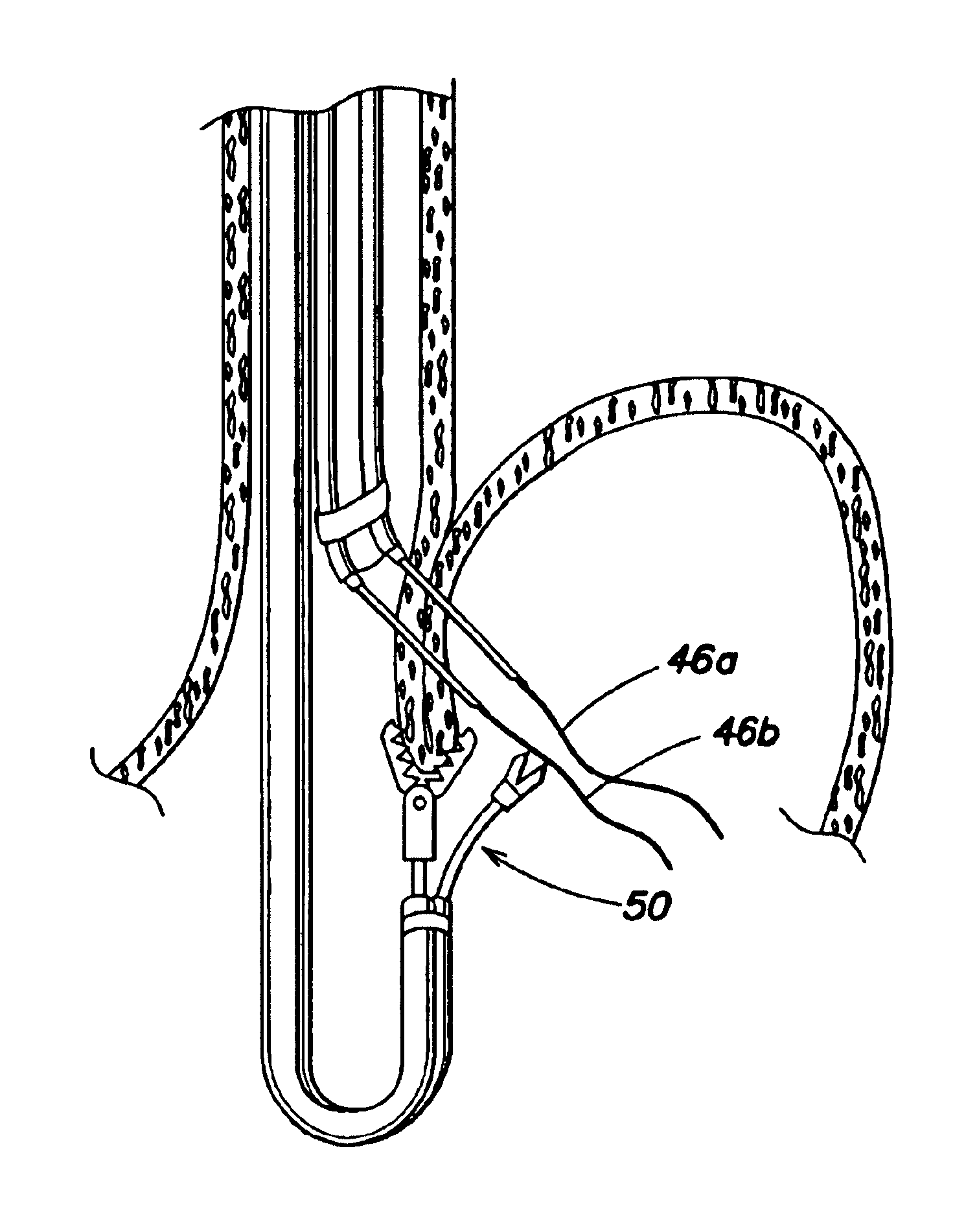
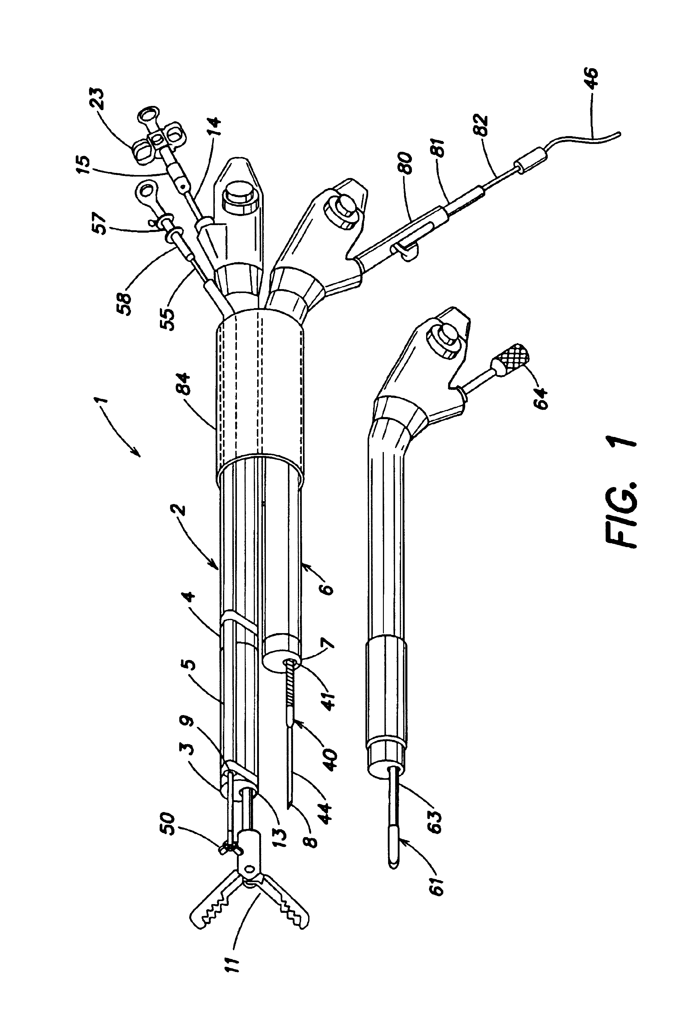
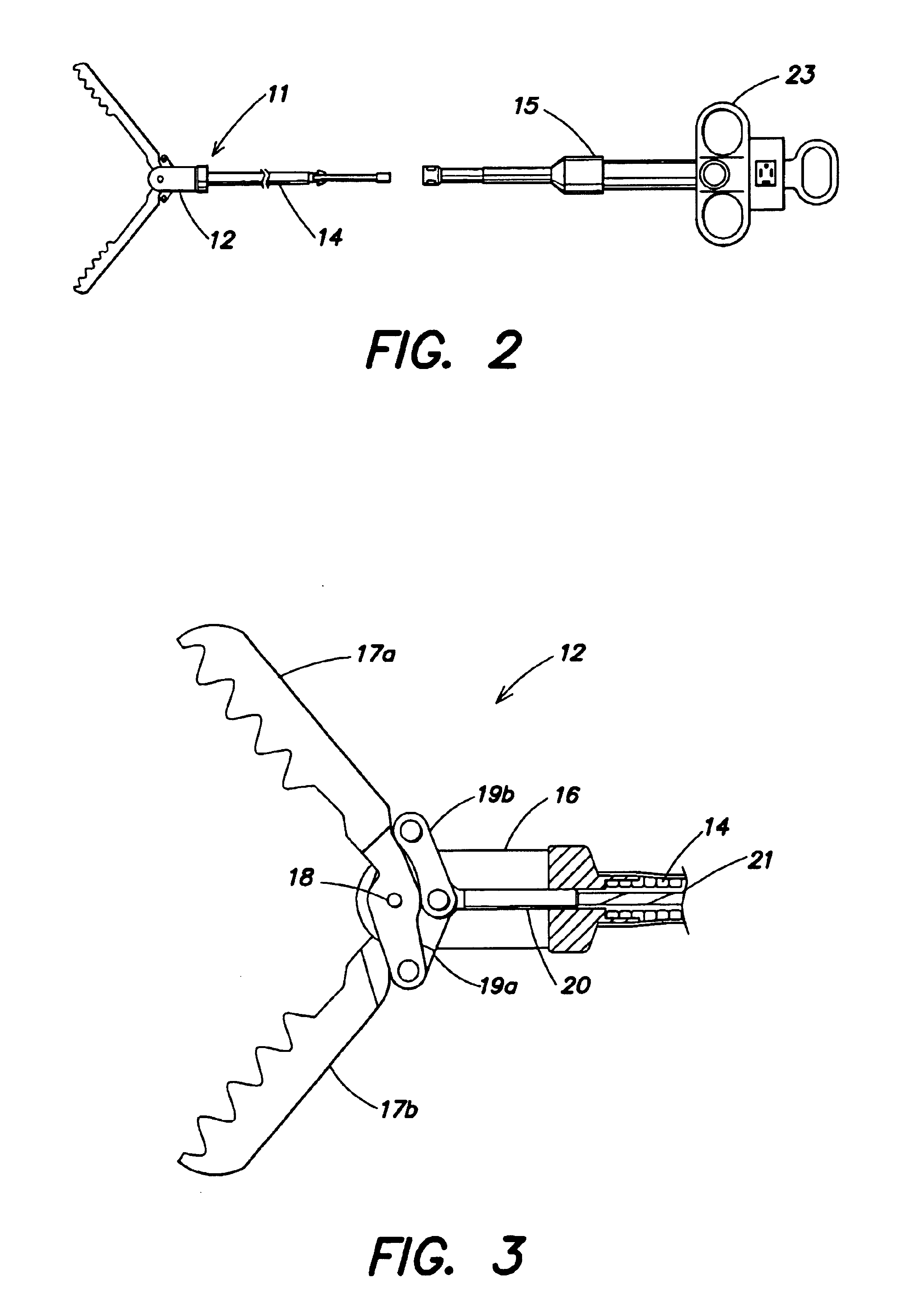
[0040]FIGS. 2 to 6 depict the first endoscope 2 and holding device 11 of the The holding device 11 comprise a distal portion 12, a sheath 14 fixed at the proximal end of the distal portion 12 and inserted in a channel 13 of the endoscope 2, and an manipulation section 15 fixed detachably at the proximal end of the sheath 14 for operating the distal portion 12. In the distal portion 12, a pair of jaws 17a and 17b, which may be forcep jaws, are fixed pivotally with a pin 18 to a cover 16 and via links 19a and 19b to a cable anchor 20. A cable 21 is fixed to the cable anchor 20, and inserted in the sheath 14. The cable 21 is fixed detachably to a sliding part 22 of the manipulation section 15. A slider handle 23 is fixed to the sliding part 22, and is translated lengthwise in relation to the manipulation section 15.

[0041]The distal portion 12 has a larger outside diameter than the channel 13 of the first endoscope 2. The jaws 17a and 17b have a plurality of teeth 24a and 24b for grasp...

second embodiment

[0069]In the second embodiment, a needle tool 110 is composed as follows. Two sheaths 111a and 111b are fixed at the distal ends 112a and 112b parallel to the distal end 113 of the second endoscope 6. The sheaths 111a and 111b are fixed at the proximal portion on the outer periphery of the second endoscope 6 using medical tape at several points. The two sheaths 111a and 111b are fixed to the manipulation section 114 and accommodate needles 115a and 115b, which slide inside. Grips 116a and 116b are fixed to the proximal ends of the needles 115a and 115b and connected to connecting section 117 which is detachable.

[0070]After the first endoscope 2 or the holding device 11 holds and suspends the tissue 91, the endoscope 6 and the needle tool 110 fixed to the endoscope 6 are inserted into the body cavity of a patient. The endoscope 6 is operated to bring the distal ends of the sheaths 111a and 111b to contact with the entering points 93a and 93b and press the grips 116a and 116b to pierc...

third embodiment

[0074]In the third embodiment, a needle 120 does not have a lumen. Instead, a suture 121 is inserted and fixed in a through hole 122 of the needle 120.

[0075]The needle 120 pierces the tissue with the suture 121 inserted and fixed in the through hole 122. The suture grasping forceps 50 pulls the suture 121 out of the through hole 122 and removed through the guide 5 out of the body.

[0076]In addition to the effect of the first embodiment, because it is not necessary to insert the suture 121 in the needle, the needle 120 is thinner than the hollow needle 44 in the first embodiment. The needle 120 can be tapered to have a sharp tip, which pierces the tissue with smaller force to improve operability.

[0077]FIGS. 32 to 36 depict the fourth embodiment of the present invention. The same components as the first embodiment are indicated by the same numbers, and their description will be omitted.

[0078]In the forth embodiment, an endoscope 140 has an optical system capable of viewing in the side ...


PUM


Abstract

The holding device of the endoscope allows suspension of the tissue while it is held and fixed securely. Use of holding device with large distal ends will not damage tissue and allows holding and suspension of a large area. A needle pierces at least the proper muscularis, thereby forming a large protrusion including the proper muscularis of the stomach and the esophagus as artificial valve to prevent reflux effectively. The holding device, formed extending out of the distal end of the endoscope, can touch the tissue easily under observation of the endoscope. Treatment is simple, and takes a shorter time.

Description

[0001]This application claims the priority of U.S. Provisional Patent Application Ser. No. 60 / 220,204, filed Jul. 24, 2000.FIELD OF INVENTION[0002]The present invention relates to an apparatus and a method for forming an artificial valve to treat gastroesophageal reflux disease (GERD).[0003]1. Background of the Invention[0004]The incidence of GERD has increased recently. The main symptoms of GERD are heartburn and mucosal breaks in the esophagus. Although it is a benign disease, GERD is accompanied by serious pain, and often requires treatment. The main cause of GERD is decreased function of the lower esophageal sphincter (LES) at the bottom of the esophagus followed by reflux of acid into the esophagus.[0005]GERD is usually treated by administration of acid secretion controlling agents such as proton-pump inhibitors. Moderate GERD will improve and may be treated completely by medication. If, however, the LES function is damaged seriously or if anatomic problems such as hiatal herni...

Claims


Application Information

Patent Timeline
Patent Type & Authority Patents(United States)
IPC IPC(8): A61B17/04A61B17/28A61B1/012A61B1/018A61B1/273A61B17/06
CPCA61B1/00098A61B1/00181A61B17/0469A61B17/0483A61B1/018A61B1/2736A61B17/0482A61B17/29A61B2017/003A61B2017/00827A61B2017/0472A61B2017/0474A61B2017/06042A61B2017/06052A61B2017/061A61B2017/2926
Inventor SUZUKI, TAKAYUKIPASRICHA, PANKAJ P.
Owner APOLLO ENDOSURGERY INC
Why Eureka
Try Eureka