
Systems and methods for evaluating objects with an ultrasound image

an object evaluation and ultrasound technology, applied in the field of automatic characterization and identification of objects, can solve the problems of limited approaches, computational and time-consuming algorithms in cross-sectional area computation, and few automated approaches in the field of in-vivo ultrasonic object definition and identification

Inactive Publication Date: 2005-09-20
BOSTON SCI SCIMED INC
View PDF31 Cites 153 Cited by
  • Summary
  • Abstract
  • Description
  • Claims
  • Application Information

AI Technical Summary

Benefits of technology

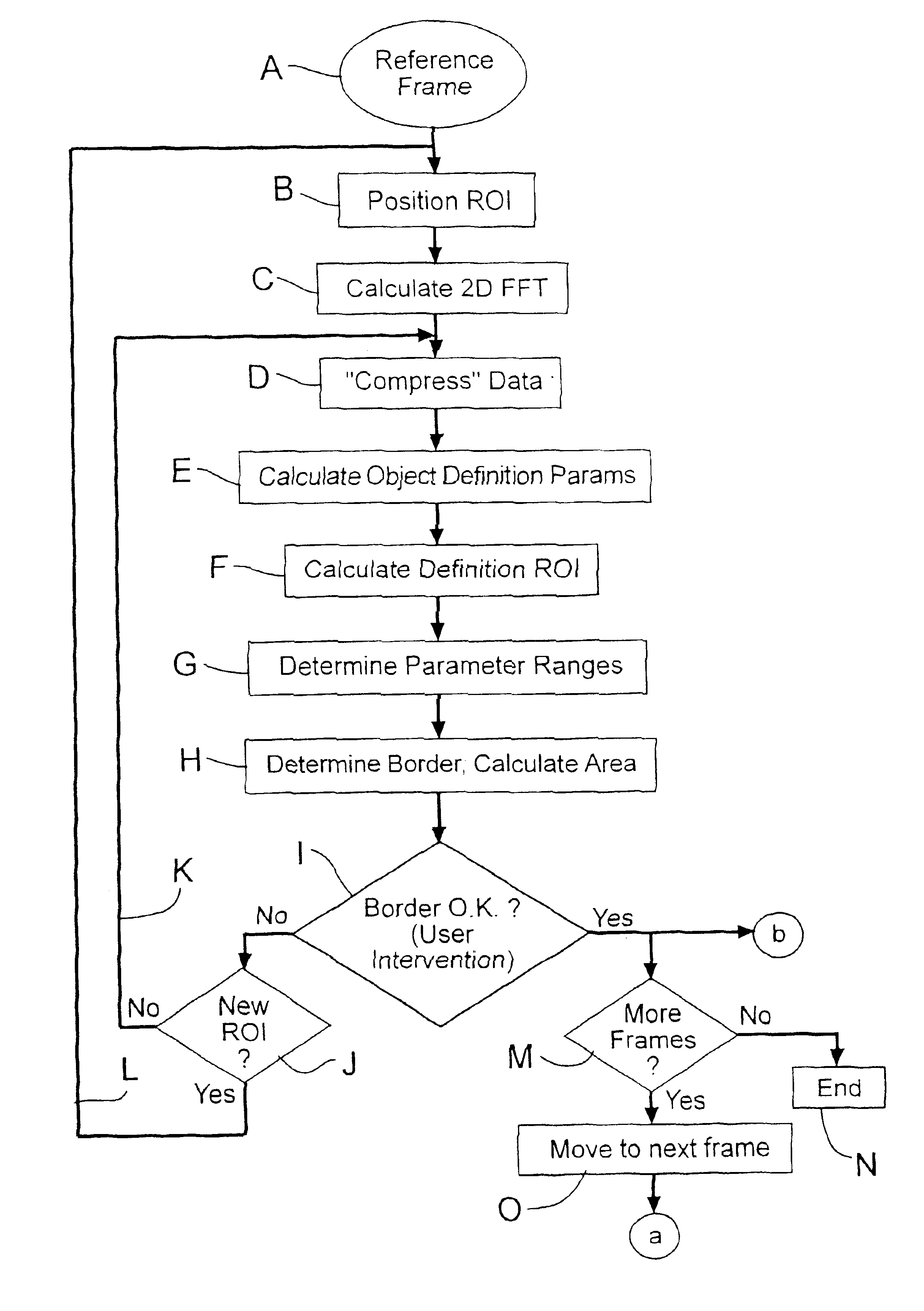
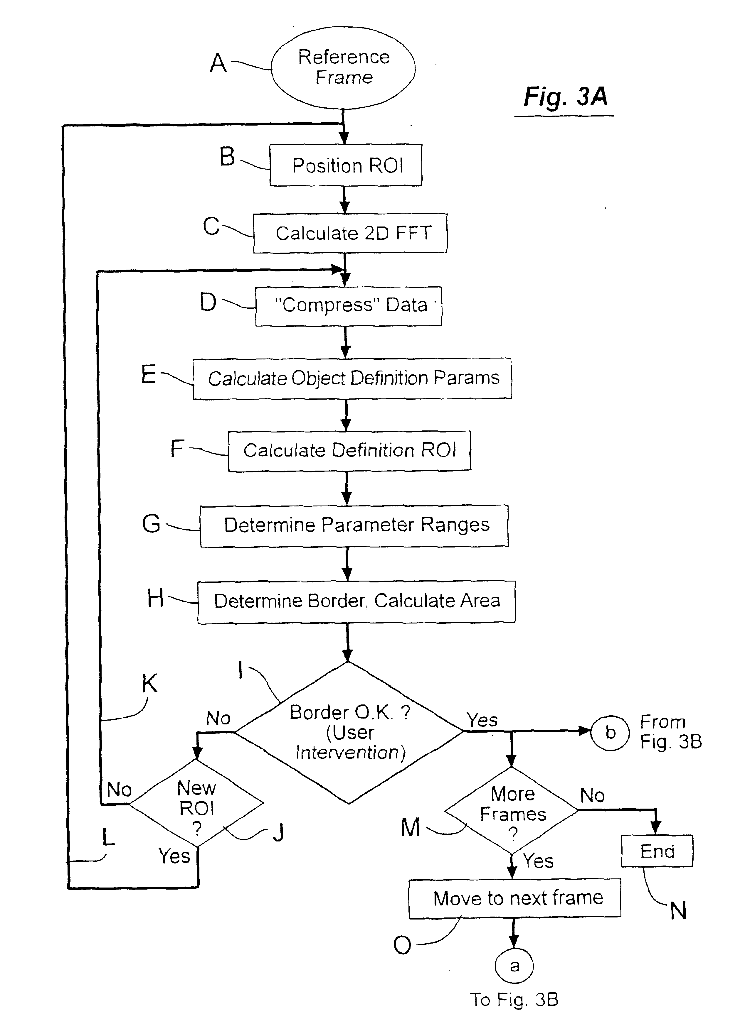
[0016]Typically, the ultrasound image is defined by multiple frames of time-domain data, and the object boundary is constructed in one of the frames (conveniently referred to as a first one of the frames). Another one of the frames is then selected and an object boundary is constructed around the object in the second frame and an area is calculated. This process is repeated for each frame having the object. Hence, one advantage of the invention is that the area of the object in subsequent frames may proceed with essentially no user interaction. Once the areas have been calculated, a volume of the object may be computed based on the areas of the objects in the frames and the distances between the frames.
[0017]In one aspect, the object boundary around the object in the-second and subsequent frames are constructed by placing a definition region of interest at-a center of mass of the object as determined from the first (or a previous) frame and repeating the steps that follow the determination of the range of acceptable object identifying parameters.
[0018]In one particularly preferable aspect, the area of the object in the first frame and the second frame are compared to determine if the areas differ by more than a predetermined amount. If so, the area of the object in the second frame is recomputed using varied criteria. For example, the starting point of the definition region of interest in the object of the second frame may be adjusted. Alternatively, the size of the definition region of interest may be changed. Further, the range of acceptable object identifying parameters may be varied.
[0019]The invention will be better understood by reference to the following detailed description in connection with the accompanying drawings.

Problems solved by technology

Heretofore there have been few automated approaches in the field of in-vivo ultrasonic object definition and identification.
These approaches are limited because algorithms provide little information about the parameters characterizing the object under observation.
In addition, the algorithms are computational and time intensive in cross-sectional area computation, since they must completely calculate the object border in each frame of the volume.
However, such models are inherently limited in that by their nature they cannot accommodate variations in object properties from patient to patient, or even from frame to frame.
This limited approach is subject to significant errors because it yields a model which only partially describes the object behavior and does not take into account variations from case to case.
This procedure is slow and is subject to errors and variations between users.
Moreover, it does not allow for the characterization of the object within the border.
Such an approach is limited by both the differences between in-vitro and in-vivo conditions and between in-vivo cases.

Method used


Image

  • Systems and methods for evaluating objects with an ultrasound image
  • Systems and methods for evaluating objects with an ultrasound image
  • Systems and methods for evaluating objects with an ultrasound image

Examples


Embodiment Construction

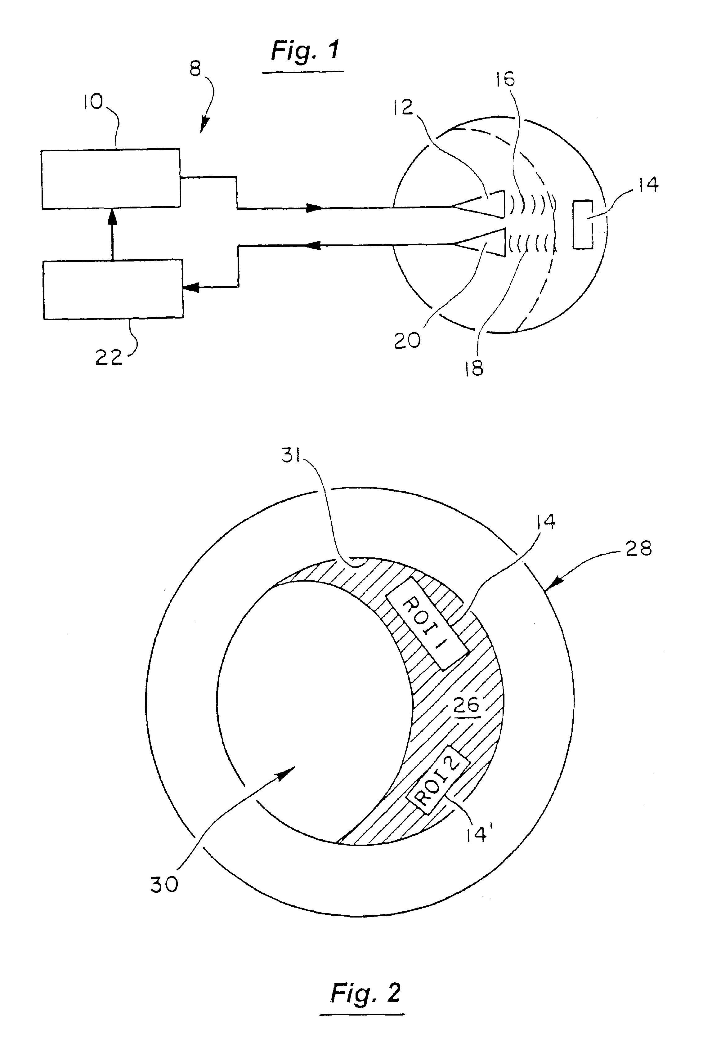
[0027]The invention provides exemplary systems and methods for evaluating objects within ultrasonic images. The objects to be evaluated are preferably representative of various physical features within the anatomy. Merely by way of example, such features may include tissue, plaque, blood, and the like.

[0028]The invention is particularly useful in constructing a border around the object so that the area of the object may easily be calculated. Importantly, the invention is also useful in that it is able to model the object using parameters which are representative of various physical characteristics of the object. These parameters are obtained from in-vivo image data. As one example, if the physical object is constructed at least partially from plaque, the parameters produced by the invention convey information on the nature of the plaque, e.g. its hardness, homogeneity, and the like. In this way, the parameters may be used to more appropriately define a proscribed treatment. Further,...


PUM


Abstract

According to the invention, an object in an ultrasound image is characterized by considering various in-vivo object parameters and their variability within the ultrasonic imaging data. Specifically, it is assumed that the object may be defined in terms of statistical properties (or object identifying parameters), which are consistently different from properties of the environment. Such properties are referred to as the object's signature. The statistical properties are calculated at selected locations within the image to determine if they fall within a predetermined range of values which represents the object. If within the range, the locations are marked to indicate they are positioned within the object. A border may then be drawn around the object and the area calculated.

Description

[0001]This application is a continuation of Ser. No. 09 / 840,591 filed May 23, 2001 and now U.S. Pat. No. 6,514,202, which is a continuation of Ser. No. 09 / 506,513 filed Feb. 17, 2000 and now U.S. Pat. No. 6,287,259, which is a continuation of Ser. No. 09 / 165,670 filed Oct. 02, 1998 and now U.S. Pat. No. 6,120,445.BACKGROUND OF THE INVENTION[0002]This invention relates to the automated characterization and identification of objects, including automated detection of their borders, in intravascular ultrasonic imaging.[0003]The value of ultrasonic imaging can be enhanced if models can be developed which accurately correlate properties of-ultrasound objects in an in-vivo environment. Heretofore there have been few automated approaches in the field of in-vivo ultrasonic object definition and identification. Previously proposed approaches may be classified in two categories. First, the defining of an object as an area surrounded by a detected border. Detection of the border in turn is base...

Claims


Application Information

Patent Timeline
Patent Type & Authority Patents(United States)
IPC IPC(8): A61B8/00G01S7/52A61B8/12
CPCA61B8/00G01S7/52036A61B8/12
Inventor GRUNWALD, SORIN
Owner BOSTON SCI SCIMED INC
Learn More