Apparatus and method for cone beam volume computed tomography breast imaging

a computed tomography and volume technology, applied in the field of apparatus and method for breast imaging, can solve the problems of limited specificity and positive predictive value of mammography, significant health problems of breast cancer, and relatively low sensitivity of mammography to detect small breast cancers, etc., to achieve accurate characterization of breast lesions, high spatial and temporal resolution, and good correlation

Inactive Publication Date: 2006-01-17
UNIVERSITY OF ROCHESTER
View PDF3 Cites 187 Cited by
  • Summary
  • Abstract
  • Description
  • Claims
  • Application Information

AI Technical Summary

Benefits of technology

[0018]It is therefore a primary object of the invention to provide a clinically useful three-dimensional imaging technique for accurate detection, diagnostics and treatment planning of breast cancer.
[0065]With the present invention, a CBVCTBI scan can be completed rapidly, and several sets of scans can be performed continuously for dynamic contrast studies and angiogenesis studies.

Problems solved by technology

Breast cancer represents a significant health problem.
However, mammography has relatively low sensitivity to detect small breast cancers (under several millimeters).
Specificity and the positive predictive value of mammography remain limited owing to an overlap in the appearances of benign and malignant lesions.
Limited sensitivity and specificity in breast cancer detection of mammography are due to its poor contrast detectability, which is common for all types of projection imaging techniques (projection imaging can only have up to 10% contrast detectability).
Thus conventional mammography is often not able to directly detect tumors of a few millimeters due to poor low contrast resolution.
In addition, not all breast cancers detected with mammography will be found early enough to cure.
Relatively low specificity of mammography results in biopsy for indeterminate cases despite the disadvantages of higher cost and the stress it imposes on patients.
Second, carcinoma has a substantially higher volume growth rate compared to a benign tumor which lacks growth.
Since mammography is a two-dimensional static imaging technique, it cannot provide any information regarding characteristics 2, 4, or 5.
The specificity and positive predictive value of mammography, however, remain limited, owing to an overlap in the appearances of benign and malignant lesions and to poor contrast detectability, which is common for all projection imaging techniques.
Biopsy is therefore often necessary in indeterminate cases, despite the disadvantages of higher cost and the stress it imposes on patients.
However, the integration of MR into routine clinical practice has been hampered by a number of limitations, including long scanning times and the high cost of MR examinations.
Additionally, many patients cannot undergo MR because of MR contraindications (e.g., aneurysm clips, pacemaker) or serious claustrophobia.
The constant trade-off between spatial and temporal resolution in MR has made it difficult to achieve the spatial resolution necessary for improved lesion detection and characterization.
That technique, however, suffers from a number of disadvantages including significantly increased radiation exposure due to the fact that standard CT can not be used to target only the breast, so that the majority of x-rays are wasted on whole body scanning.
Ultrasound has poor resolution in characterizing lesion margins and identifying microcalcifications.
Ultrasound is also extremely operator dependent.
However, patients are uncomfortable even though compression may not be harmful to them.

Method used


Image

  • Apparatus and method for cone beam volume computed tomography breast imaging
  • Apparatus and method for cone beam volume computed tomography breast imaging
  • Apparatus and method for cone beam volume computed tomography breast imaging

Examples


Embodiment Construction

[0091]A preferred embodiment of the present invention and variations thereon will now be set forth in detail with reference to the drawings, in which the same reference numerals refer to the same components throughout.

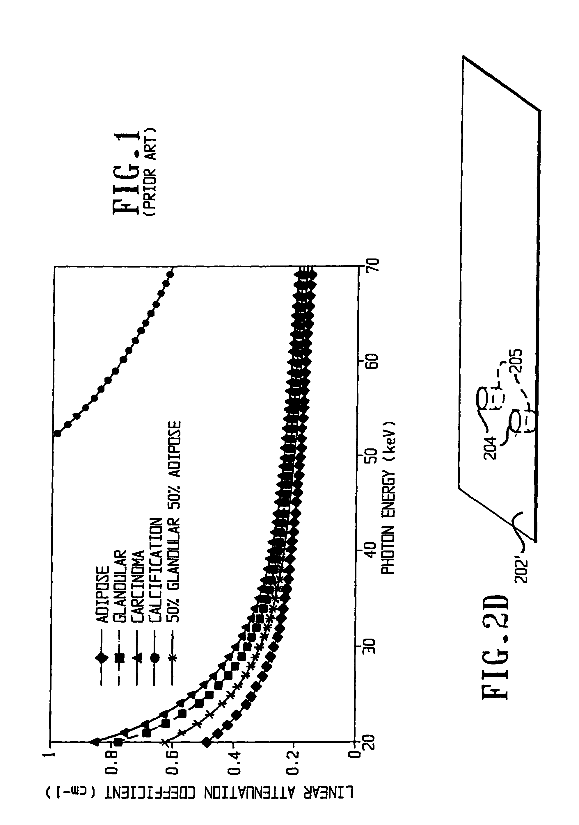
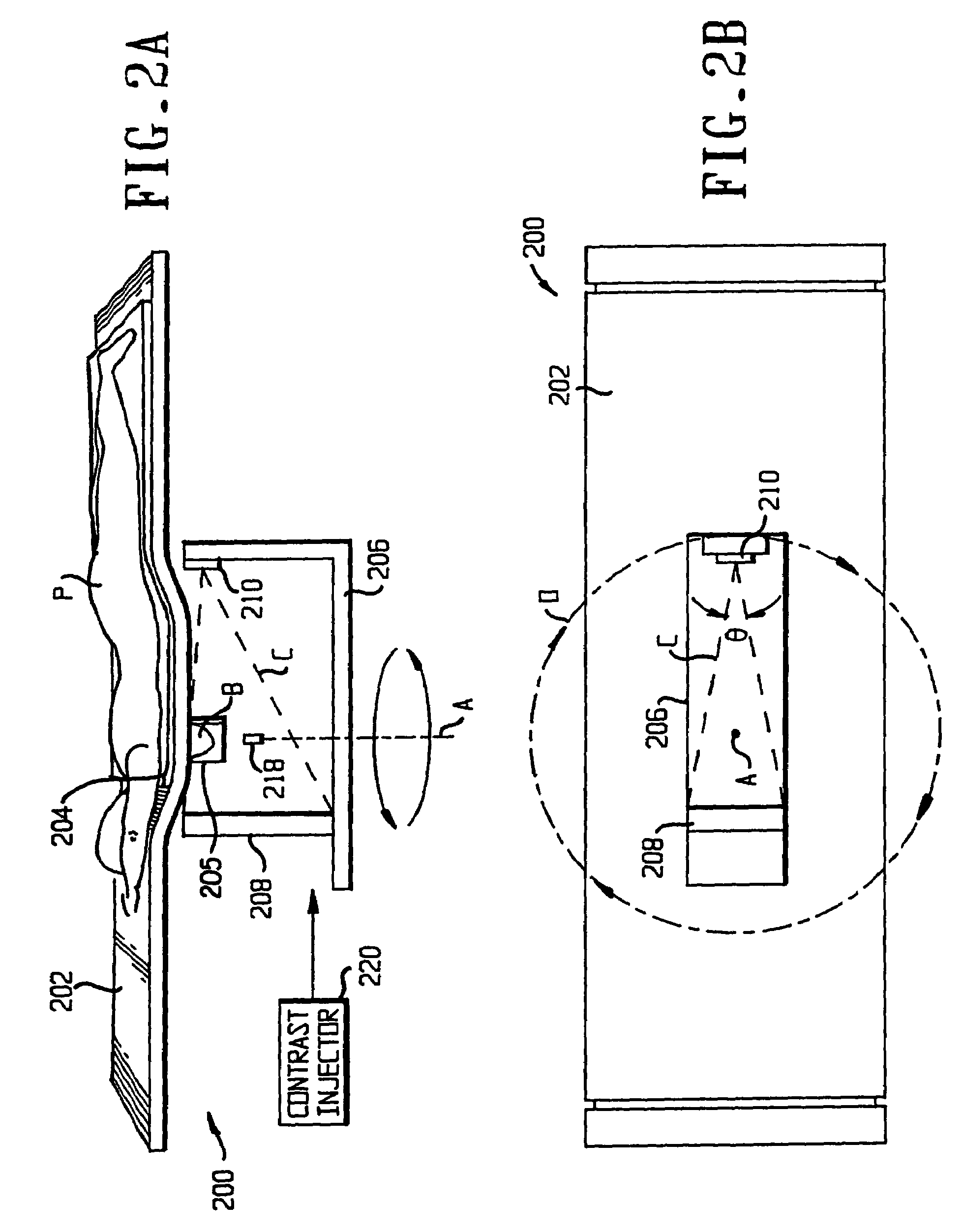
[0092]The limitations accompanying conventional mammography are addressed by incorporating a cone beam volume CT reconstruction technique with a flat panel detector. With cone beam geometry and a flat panel detector, a flat panel-based cone beam volume computed tomography breast imaging (CBVCTBI) system can be constructed as shown in FIGS. 2A–2F, and three-dimensional (3D) reconstructions of a breast from a single fast volume scan can be obtained. In contrast to conventional mammography, the flat panel-based CBVCTBI system provides the ability to tomographically isolate an object of interest (e.g. a lesion) from the other objects in adjacent planes (e.g. other lesion or calcification). The 3D tomographic reconstructions eliminate lesion overlap and provide a complete, ...


PUM


Abstract

Cone beam volume CT breast imaging is performed with a gantry frame on which a cone-beam radiation source and a digital area detector are mounted. The patient rests on an ergonomically designed table with a hole or two holes to allow one breast or two breasts to extend through the table such that the gantry frame surrounds that breast. The breast hole is surrounded by a bowl so that the entire breast can be exposed to the cone beam. Spectral and compensation filters are used to improve the characteristics of the beam. A materials library is used to provide x-ray linear attenuation coefficients for various breast tissues and lesions.

Description

REFERENCE TO RELATED APPLICATION[0001]This application is a continuation-in-part of U.S. patent application Ser. No. 10 / 291,745 filed Nov. 12, 2002 now abandoned, which is a continuation-in-part of U.S. patent application Ser. No. 09 / 640,713, filed Aug. 18, 2000, now U.S. Pat. No. 6,480,565, which claims the benefit of U.S. Provisional Application No. 60 / 166,223, filed Nov. 18, 1999. The disclosures of all of those applications are hereby incorporated by reference in their entireties into the present disclosure.BACKGROUND OF THE INVENTION[0002]1. Field of the Invention[0003]The present invention relates to the use of cone beam volume computed tomography (CBVCT) in breast imaging and in particular to improvements therein that make better use of the radiation from the radiation source, the data output by the detector, or both. Throughout the present disclosure, it will be understood that references to breast imaging (mammography) are illustrative rather than limiting, as the invention...

Claims


Application Information

Patent Timeline
Patent Type & Authority Patents(United States)
IPC IPC(8): A61B6/04A61B6/00A61B6/03A61B6/06A61N
CPCA61B6/032A61B6/0435A61B6/06A61B6/4085A61B6/466A61B6/502A61B6/04Y10S378/901A61B6/0414A61B6/12A61B6/481A61B6/504A61B10/0233A61B2019/5242A61B6/027A61B2090/3764
Inventor NING, RUOLA
Owner UNIVERSITY OF ROCHESTER
Why Eureka
Try Eureka