
Method, apparatus and system for complete examination of tissue with hand-held imaging devices having mounted cameras

a handheld imaging device and camera technology, applied in the field of medical imaging, can solve the problems of large size, more expensive imaging devices, and large volume of breasts, and achieve the effect of reducing the review tim

Inactive Publication Date: 2015-12-24
TRACTUS
View PDF0 Cites 48 Cited by
  • Summary
  • Abstract
  • Description
  • Claims
  • Application Information

AI Technical Summary

Benefits of technology

The patent describes a hand-held imaging probe that can accurately and dynamically compute the position and orientation of its imaging elements during a scan of tissue. This allows the device to determine the actual spatial position and computed orientation of each image along the tissue surface. By measuring the distance between the boundaries of each scan sequence, the device can determine if the scanned tissue is complete and if there are any areas that need further scanning. The device can also modify the scanned images to remove redundancy and improve the overall quality of the scan. Overall, this technology enables the accurate and efficient mapping of tissue structures using a hand-held imaging probe.

Problems solved by technology

The portability and small size of the device means that it can be used in locations, both geographic and anatomic, that are difficult for larger, more expensive imaging devices such as X-ray and MRI.
There may even be cancers in the other seven quadrants, but it is not the purpose of the diagnostic examination, however, to find those possible, but previously not identified, lesions.
The process requires significant patient manipulation and tissue distortion to pull the mammary tissue as far into the field of view of the X-ray radiation emitting and detecting imaging device as is possible.
These “spot compressions” are often accompanied by magnification, with the result that only a portion of the breast appears in the image.
The ability to map the images is critical because the device is not effective in practice if an abnormality is identified, but the physician does not know where it is within the patient's anatomy.
It is not possible to “map” all of the structures a single two-dimensional view, however, because the human anatomy and human tissue structures are three dimensional.
For example, if the X-ray reveals two shadows, or regions of interest, the device cannot determine which of two shadows is closest to the energy emitter and which is closer to the energy detector.
The device does not know where the imaging component is in space if the device does not know where the hand holding the device is in space.
One drawback of this approach, however, is that there is no quality control to assure that the user responded to the prompts appropriately and that the images are actually being recorded at regular intervals.
Another drawback of this approach is that it can be annoying to the operator to be prompted continually to adjust parameters on the scan.
If one wishes to reconstruct a three-dimensional map of a set of images, however, then the relative positioning information is critical.
Under these conditions, the ability of these images to distinguish, or “resolve”, smaller structures, such as a human hair (0.2 mm) is not possible.
Even when the resolution is sufficient to present small objects in some fashion, the operator may not be able to distinguish the exact nature of that small object unless the resolution can also present more details (that is smaller features) on the shape and texture of that object.
The earlier ultrasound devices packaged 64 imaging elements in a linear array and could not resolve features smaller than 2 mm.
Although any single X-Y slice could resolve lesions as small as a millimeter, the inter-slice spacing made resolution of lesions smaller than 8.6 mm unreliable.
Hand-held imaging devices rely on a human operator to translate the imaging probe over the tissue to be examined and present resolution challenges that are very different from the robotic devices.
The primary challenges in the efficacy of a hand-held device are the ability to map individual images, the ability to resolve between the discrete images in the image set, and to determine whether the family of image sets represents complete coverage of the structure.
Conversely, a blurry mammogram of the entire breast “covers” the entire breast, but may not do so with adequate resolution to be a useful examination.
For example, if the operator scans too quickly, the images in a scan sequence may be spaced too far apart to show a potential cancerous region.
The primary challenges in the three-dimensional reconstruction are the spacing between adjacent pixels in the third axis of the XYZ Cartesian coordinate system, viz., the Z-axis and the relative location of the families of sets of discrete images obtained during the scanning process.
As important as the imaging requirements are to achieving a practical screening technology, time constraints can also affect practicality, thus the utility, of the device.
As described earlier, although it is not possible for a skilled and trained operator to objectively determine the completeness of the area covered, and the resolution (in terms of the relative spacing between adjacent images) of a scan when they are personally performing a manual examination, they may believe, subjectively, that the coverage and resolution are adequate.
If the reviewer is observing a set of images that were recorded by another operator, however, it is not possible for the reviewer to have any defendable means of determining whether the area covered represents the entire structure or that the resolution, in terms of spacing between images, meets the minimal standards that the user requires.
Although there is additional information in those 900 additional images, the incremental improvement in patient care may not be warranted for the additional 1.5 minutes of physician time to review the track.

Method used


Image

  • Method, apparatus and system for complete examination of tissue with hand-held imaging devices having mounted cameras
  • Method, apparatus and system for complete examination of tissue with hand-held imaging devices having mounted cameras
  • Method, apparatus and system for complete examination of tissue with hand-held imaging devices having mounted cameras

Examples


Embodiment Construction

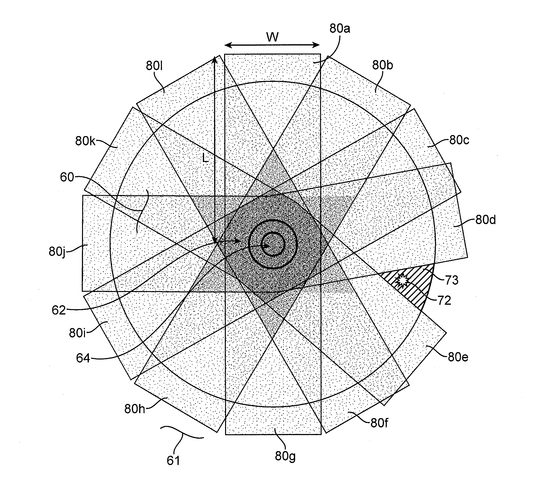
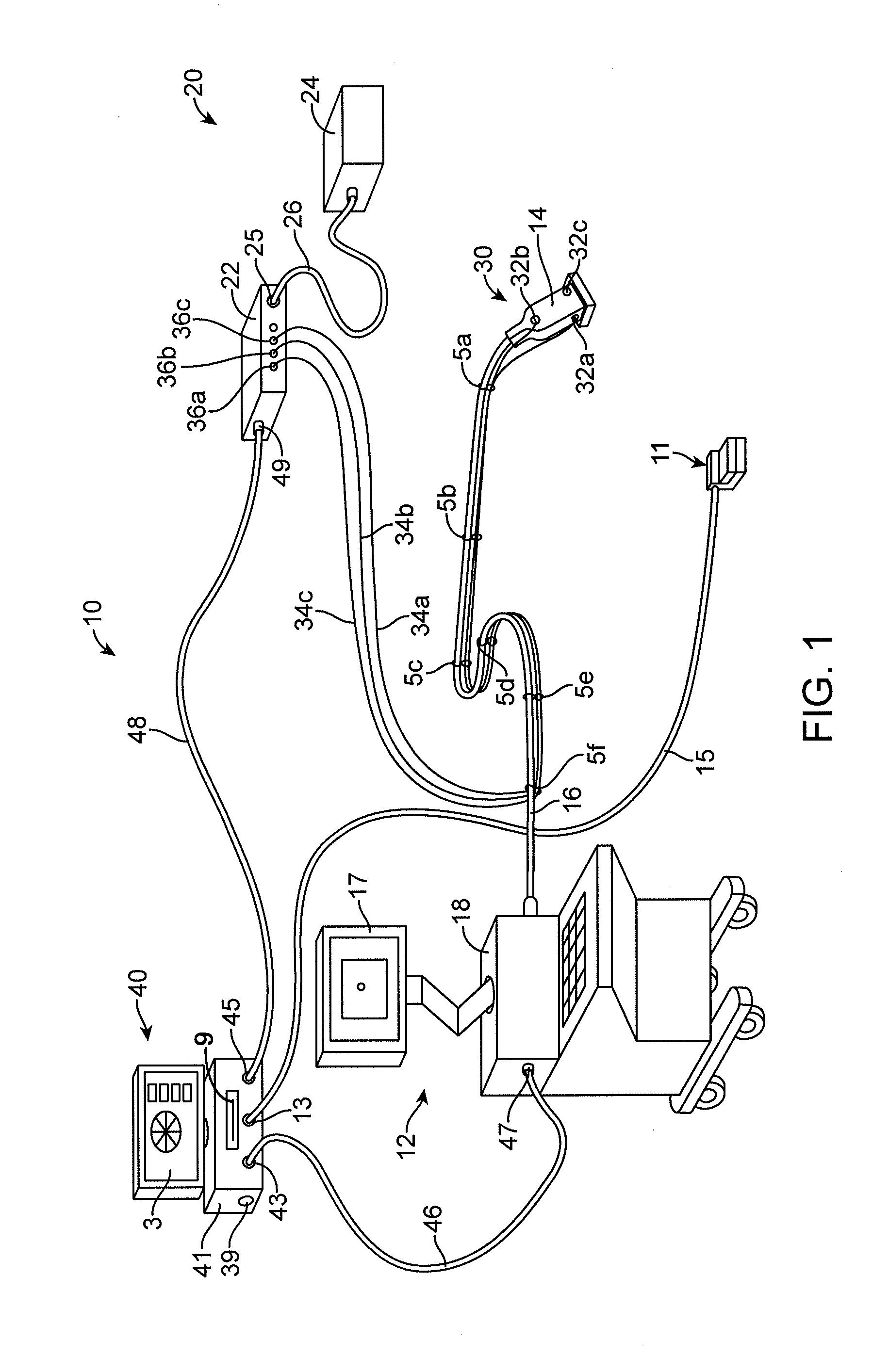
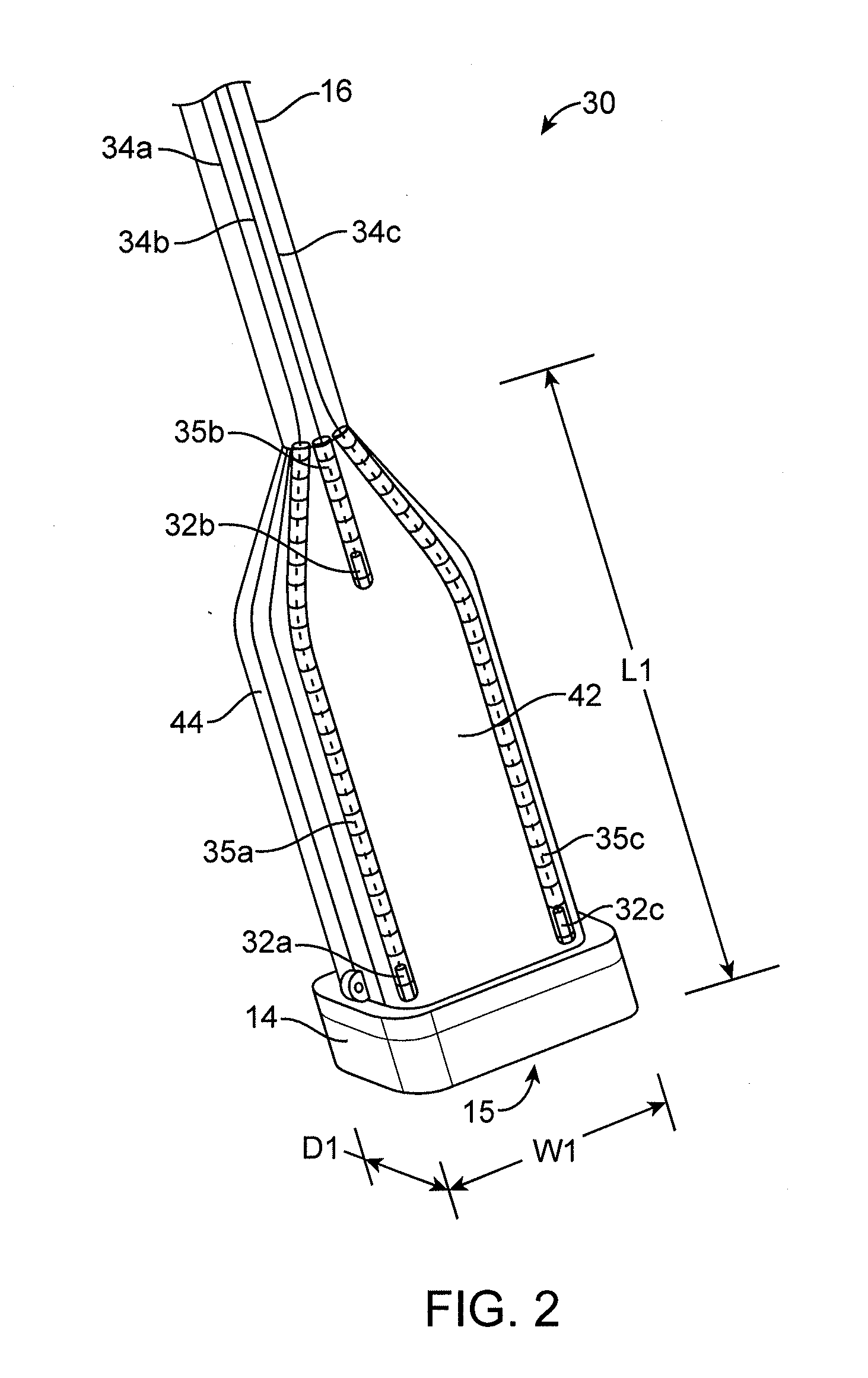
[0166]As described briefly above, embodiments contemplated provide for methods, devices, systems that can be used with manual imaging techniques to ensure satisfactory quality and adequate completeness of a scanning procedure for a patient's target region. Some embodiments employ rapid-response position sensors or rapidly imaged optical registers affixed to an existing hand-held imaging system, for example, a diagnostic ultrasound system, and associated hand-held imaging probes. By way of example, one type of ultrasound system that can be used with some embodiments described is the Phillips iU22 xMatrix Ultrasound System with hand-held L12-50 mm Broadband Linear Array Transducer (Andover, Mass.). Also, a commercially available system which provides accurate x, y, z position coordinates for multiple sensors as a function of time, providing said position information at a rapid tracking rate, is, by way of example, the Ascension Technology 3D Guidance trakSTAR (Burlington, Vt.).

[0167]R...


PUM


Abstract

A scan completeness auditing system for use with an imaging console in screening a volume of tissue comprising a position tracking system configured to track and record a position of a manual imaging probe. The position tracking system com prises a plurality of cameras adapted to couple to the manual imaging probe and configured to provide position data for the manual imaging probe. The scan completeness auditing system includes a receiver comprising a controller-configured to electronically receive position data for the manual ultrasonic imaging probe from the position tracking system and to electronically receive and record a first scan sequence comprising a first set of scanned images representing cross-sections of the tissue from the manual imaging probe. The controller can be configured to compute an image-to-image spacing between successive images within the first scan sequence and to determine whether the computed image-to-image spacing exceeds a maximum limit. An alert when the computed image-to-image spacing exceeds the maximum limit.

Description

CROSS REFERENCE TO RELATED APPLICATIONS[0001]This application claims the benefit of U.S. Patent Appl. No. 61 / 753,832, filed Jan. 17, 2013, the disclosure of which is incorporated herein by reference. This application may also be related to U.S. Patent Appl. No. 61 / 545,278, filed Oct. 10, 2011 and International Application No. PCT / US2012 / 059176, filed on Oct. 8, 2012, the disclosures of each of which are incorporated herein by reference.INCORPORATION BY REFERENCE[0002]All publications and patent applications mentioned in this specification are herein incorporated by reference to the same extent as if each individual publication or patent application was specifically and individually indicated to be incorporated by reference.FIELD[0003]Embodiments described relate generally to medical imaging and methods and devices for ensuring adequate quality and coverage of scanned and recorded images. In another aspect, embodiments described relate to reducing review time of scanned and recorded ...

Claims


Application Information

Patent Timeline
Patent Type & Authority Applications(United States)
IPC IPC(8): A61B8/00A61B6/00A61B5/06G06T7/00G06T15/08
CPCA61B8/4245G06T7/0012G06T15/08A61B5/06A61B6/547G06T2207/10048G06T2207/10132G06T2207/10081G06T2207/10088G06T2207/30068G06T2211/412G06T2200/04A61B5/064A61B6/502A61B8/08A61B8/0825A61B8/4263A61B8/483
Inventor EGGERS, PHILIP E.HUNTLEY, SCOTT P.EGGERS, ERIC A.ROBINSON, BRUCE A.
Owner TRACTUS
Learn More