Method of detecting single gene copies in-situ
a single gene and gene copy technology, applied in the field of nucleic acid detection and localization techniques, can solve the problems of radioactive toxicity and environmental concerns, other viral diseases are also frequently difficult to detect or distinguish clinically, and the signal generated by fluorescent markers is typically faded
- Summary
- Abstract
- Description
- Claims
- Application Information
AI Technical Summary
Problems solved by technology
Method used
Image
Examples
example 1
HER-2 / neu Brightfield Assay for Embedded Breast Tissue Specimens
[0068]Paraffin embedded tissue specimen slides were baked for 1 to 20 hours at 65° C. + / −2° C. The slides were then deparaffinized in three washes of xylene for 5 minutes each and washed in 100% ethanol, 2 times for 2 minutes each and air dried label end down, propped at an angle.
[0069]Next, the specimens were digested in 0.5 mg / ml Proteinase K / 2×SSC (pH 7.0) at 45° C. (Proteinase K solution was prepared by adding 1 ml deionized water to a 25 mg vial of Proteinase K, shaking the vial to suspend the Proteinase K and transferring the 1 ml of solution to 50 ml of 0×SSC prewarmed to 45° C.). Cell line controls were digested for 12 minutes, and tissue specimens for 35 minutes. The slides were rinsed for 10 seconds in 2×SSC at room temperature followed by dehydration in a room temperature ethanol series of 70%, 80%, and 100% ethanol for 1 minute each. The slides were air dried with the label end down.
[0070]10 μl digoxigenin-l...
example 2
Detection of Single Gene Copies
[0077]The slides produced in Example 1 were mounted with Permount (Fisher Scientific, NJ) and coverslipped.
[0078]Using brightfield microscopy, microphotographs were made on Kodak Ektachrome color slide film (EL 400), using a Zeiss Axiophot 20 epi-fluorescence microscope (Zeiss, West Germany) equipped with a 100×Plan-Neofluar oil immersion objective or a 100×Plan-APO oil immersion objection (Zeiss, West Germany), a Zeiss MC-100 camera (Zeiss, West Germany), and a blue or neutral density filter.
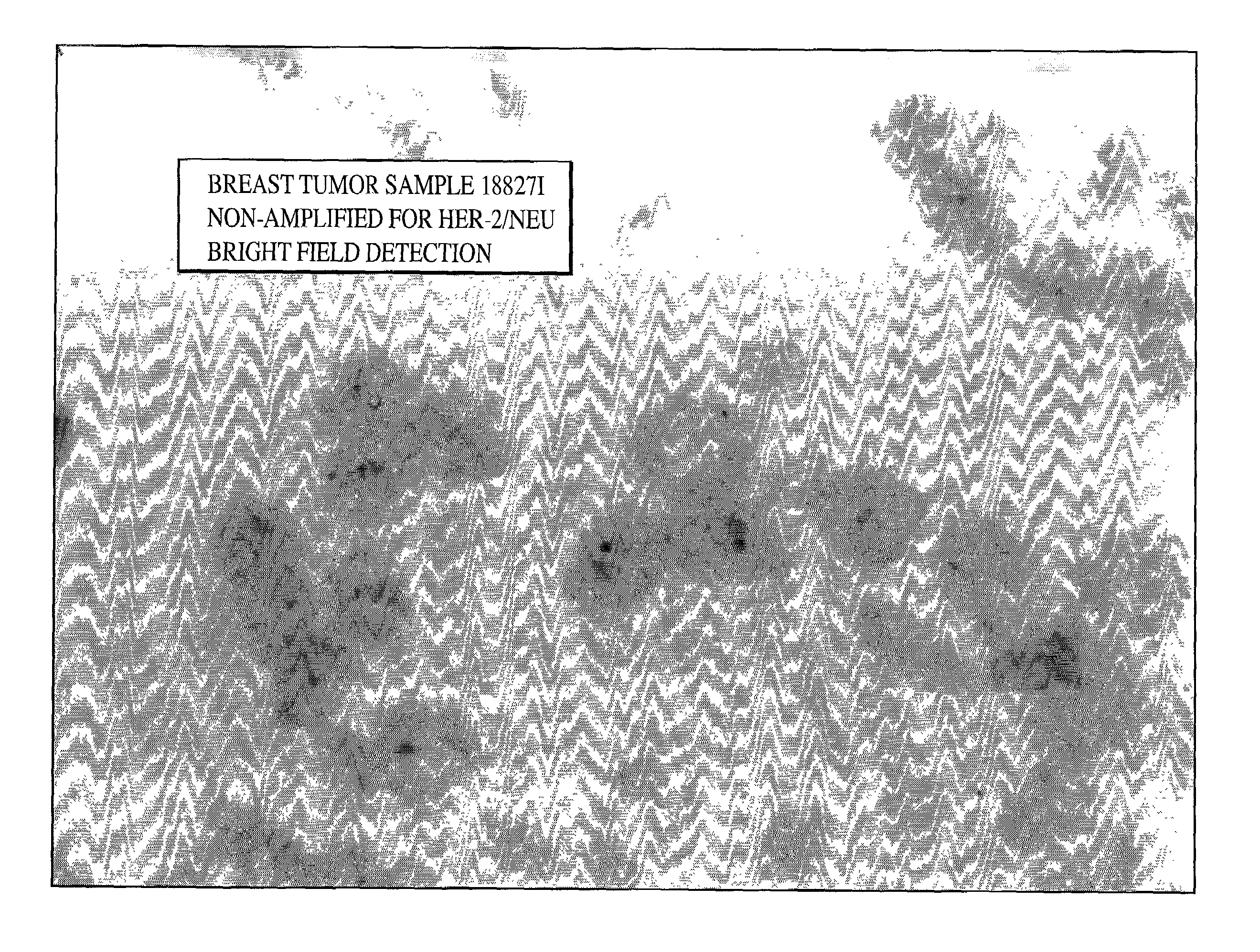
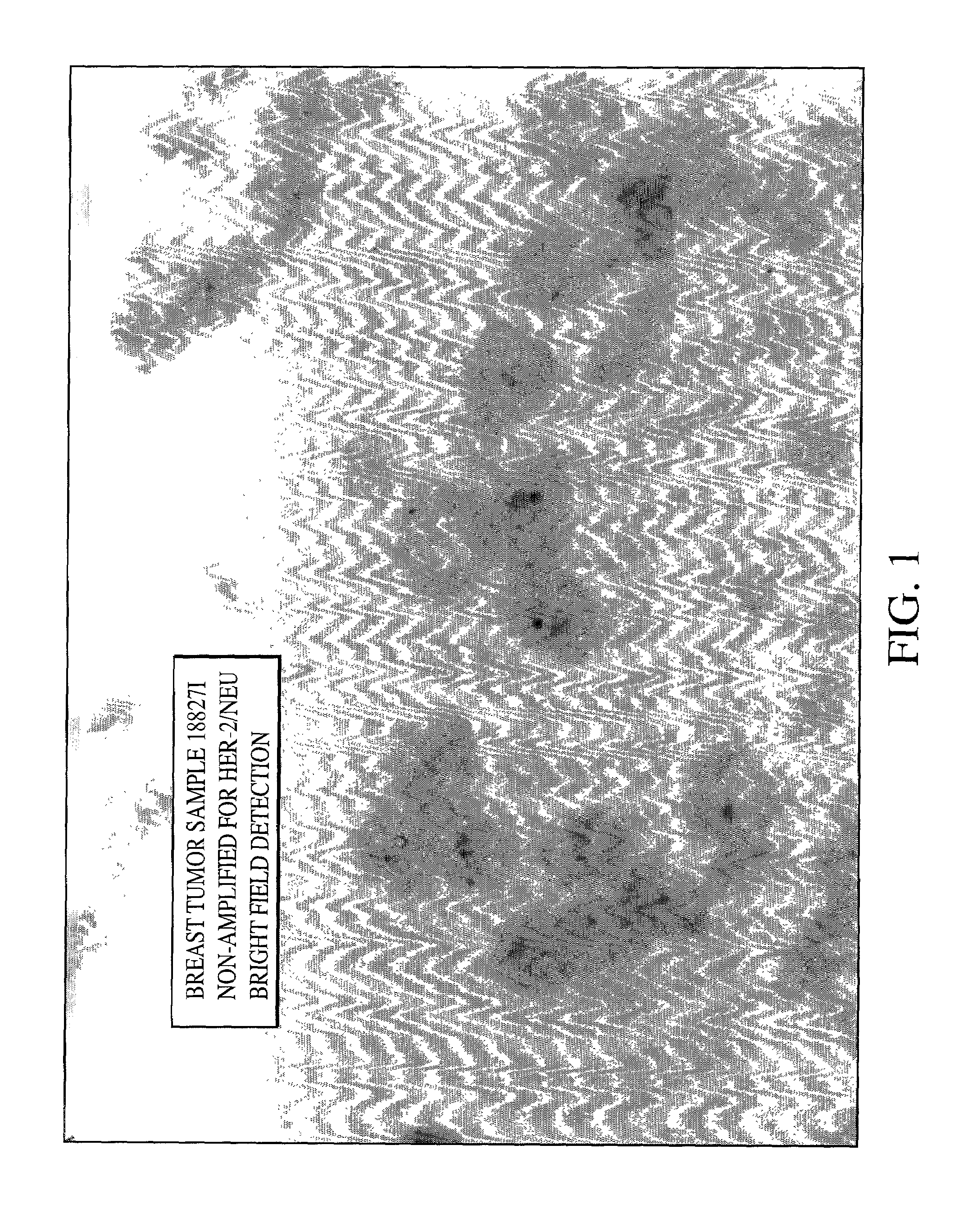
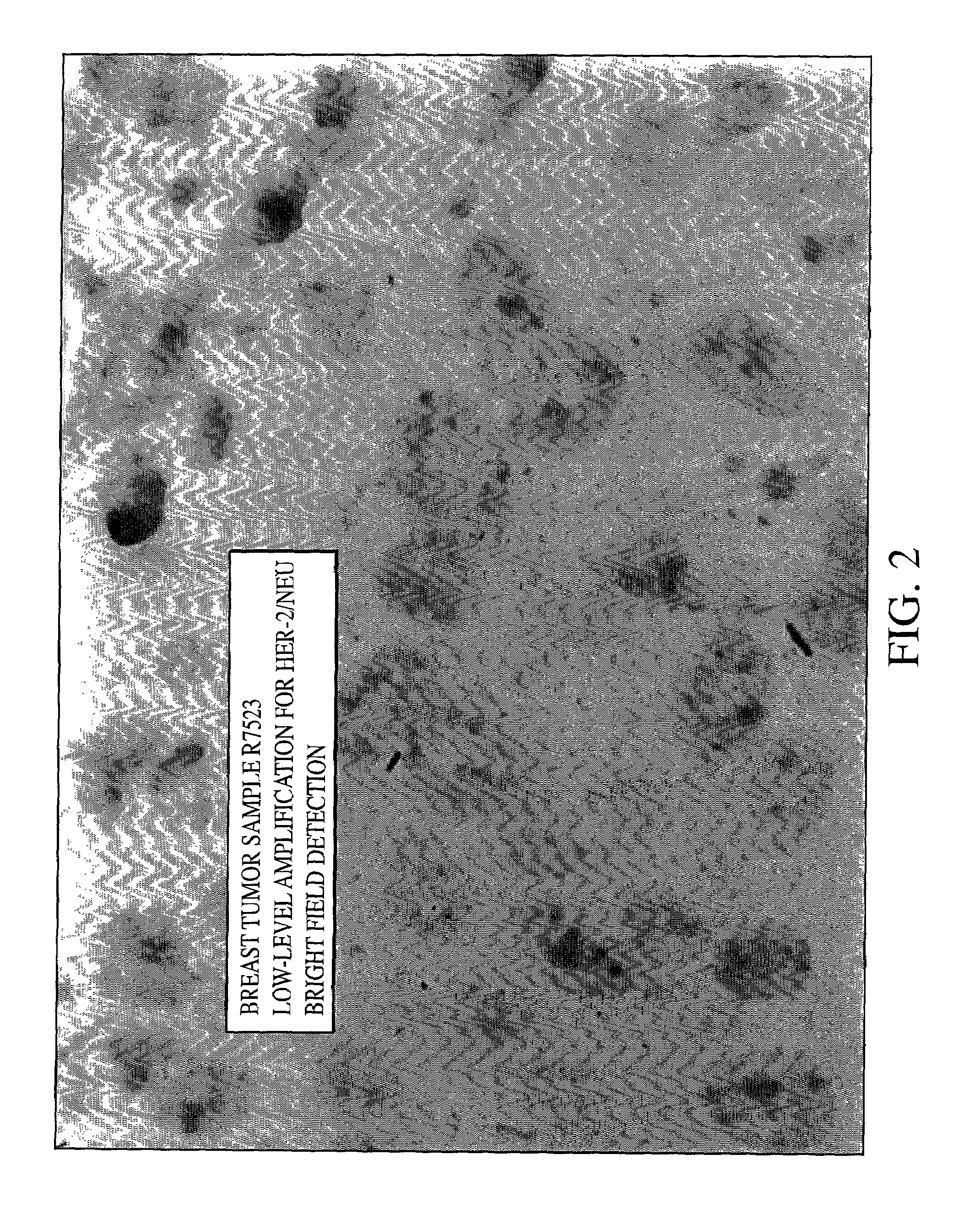
[0079]As can be seen from FIGS. 1, 2 and 3 single blue signal was visible at the location of each copy of the HER-2 / neu gene. The expected results for visualization of the HER-2 / neu gene in normal metaphase chromosomes from the MDA-MB468 breast tumor cell line is two; one on each chromatin, and the number of the blue signals in normal interphase nuclei is also two, or possibly two pairs if the cell is in G2 phase. In the cultured breast tumor cell lines MDA-MB-361...
example 3
Detection of High Risk Human Papilloma Virus in Gynecologic Tissue Specimen
[0080]Six separate commercially available plasmids, i.e., pGem2, pUC13, pGem1, pLINK322, pGEM1 and pUCI3 containing entire genomes of HPV types 16 (DNA sequence available from GenBank, Accession No. K02718), 18 (DNA sequence available from GenBank, Accession No. X05015), 31 (DNA sequence available from GenBank, Accession No. J04353), 33 (DNA sequence available from GenBank, Accession No. M12732), 35 (DNA sequence available from GenBank, Accession No. M74117) and 51 (DNA sequence available from GenBank, Accession No. M62877) respectively, were labeled by nick translation with digoxigenin dCTP. Alternatively, one may clone the HPV into a plasmid by standard molecular biology techniques within the skill of the art. The labeled plasmids were then mixed together to form a single reagent. Incorporation of the digoxigenin nucleotide into the labeled DNA was verified by dot-blot procedure. DNA fragment size was deter...
PUM
| Property | Measurement | Unit |
|---|---|---|
| temperature | aaaaa | aaaaa |
| temperature | aaaaa | aaaaa |
| brightfield microscopy | aaaaa | aaaaa |
Abstract
Description
Claims
Application Information
 - R&D
- Intellectual Property
- Life Sciences
- Materials
- Tech Scout
- Unparalleled Data Quality
- Higher Quality Content
- 60% Fewer Hallucinations
Browse by: Latest US Patents, China's latest patents, Technical Efficacy Thesaurus, Application Domain, Technology Topic, Popular Technical Reports.
© 2025 PatSnap. All rights reserved.Legal|Privacy policy|Modern Slavery Act Transparency Statement|Sitemap|About US| Contact US: help@patsnap.com


