Method of optimizing cardiac resynchronization therapy using sensor signals of septal wall motion

a septal wall and sensor signal technology, applied in the field of cardiac therapy, can solve the problems of not being used to directly measure deflection, detrimental effects, and cardiac output (co) drop, and achieve the effect of increasing cardiac output and reducing uncoordinated motion

Inactive Publication Date: 2006-08-15
MEDTRONIC INC
View PDF16 Cites 227 Cited by
  • Summary
  • Abstract
  • Description
  • Claims
  • Application Information

AI Technical Summary

Benefits of technology

[0008]The present invention addresses the above described needs by providing means for detecting uncoordinated ventricular motion, monitoring or measuring septal wall deflection due to ventricular uncoordinated motion, and / or reducing said uncoordinated motion with appropriately timed electrical stimulation coupled to both ventricular chambers to decrease said uncoordinated motion and increase cardiac output.
[0009]That is, the present invention provides novel methods and apparatus for measuring septal wall deflection in a closed-loop system including an IPG to thereby provide automatic optimization of inter-atrial (A—A) pacing intervals and / or interventricular (V—V) pacing intervals to minimize uncoordinated cardiac motion and improve CO, among other advantages. The present invention thus provides means for reducing: intra-atrial uncoordinated motion and intra-ventricular uncoordinated motion (A—A and V—V uncoordinated motion, respectively), as well as atrio-ventricular uncoordinated motion (i.e., A-V and V-A uncoordinated motion).
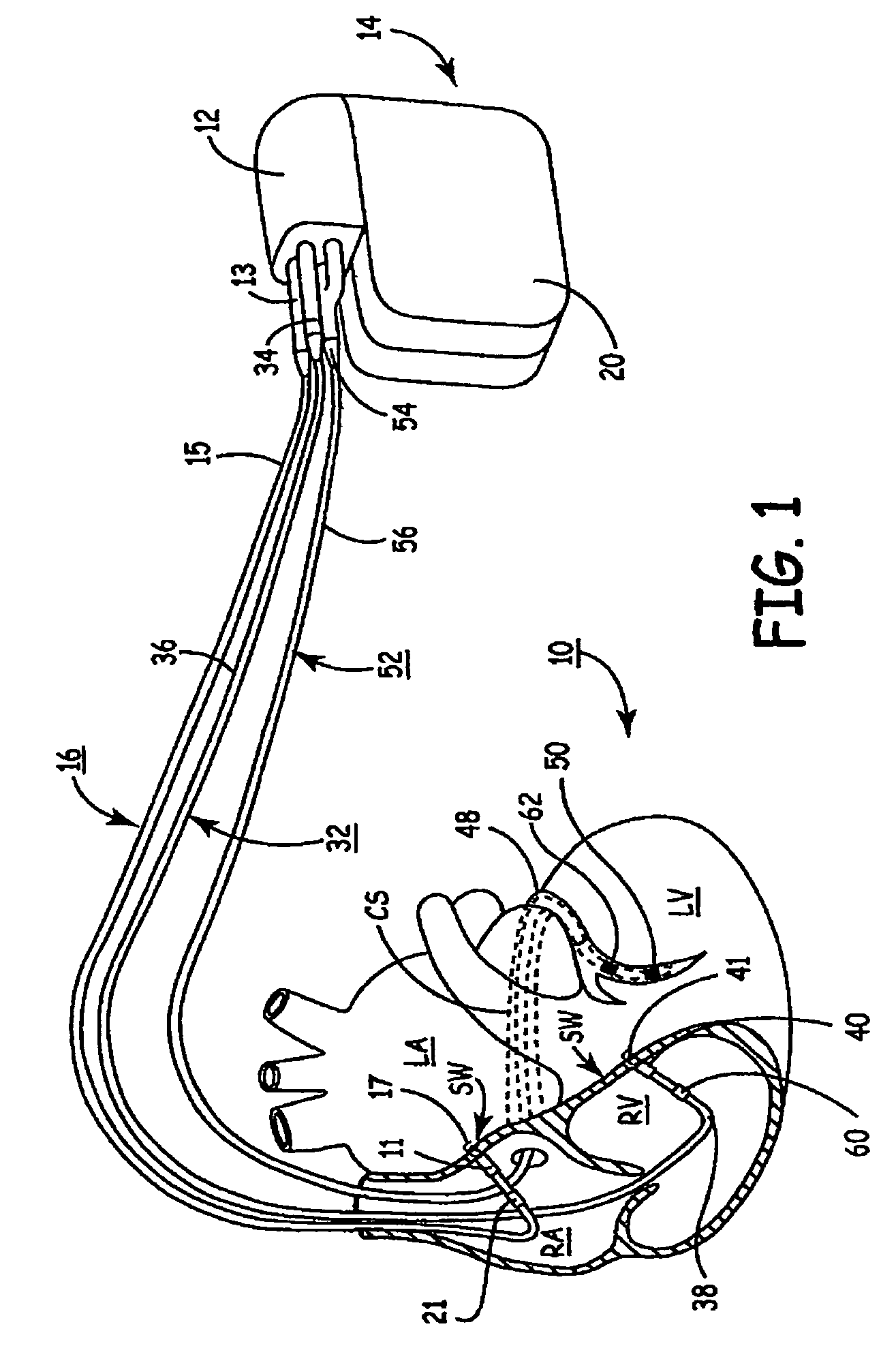
[0010]In embodiments of the present invention directed to reducing A—A uncoordinated motion an additional lead should be electrically coupled to the left atrium (LA). In one implementation of the present invention, if desired, a variety of respective intraventricular timing can be implemented so that an LV depolarization is timed to slightly precede an RV depolarization (or vice versa). That is, if due to physiologic cardiac characteristics—or other confounding factors—for a particular patient the timing of a discrete cardiac pacing pulse to the LV may need to be slightly offset from a corresponding cardiac pacing pulse delivered to the RV to minimize deflection of the ventricular septal wall. This optimization modality is believed to be highly effective in promoting beneficial, synchronized cardiac depolarization events, especially for HF patients.
[0012]A temporal window during which a useful signal may be obtained should be defined. If the window is accurately defined the duty cycle of the transducer and attendant computational overhead (and related energy drain) can be minimized. Alternatively, constant measuring, signal filtering, mathematical integrating (if applicable) and comparing the resulting signals to choose optimum pacing intervals may be employed. However, one reliable technique for defining the temporal window is to simply trigger the motion measurements from detected cardiac cycle activity. An atrial or ventricular electrogram, or other sensing vector(s) may be used. For example, a trigger event may comprise a sensed, intrinsic or evoked atrial or ventricular event with a programmable or predetermined motion-sensing-delay interval added so that the temporal motion-sensing window can begin and end when the septal wall motion is most likely to be of interest. Another technique to define the temporal window is to trigger from a maximum motion signal and, optionally, add a motion-sensing-delay before beginning to track signals related to motion of septal wall tissue. Since the present invention is directed at minimizing septal wall motion, a fiducial or reference point may effectively comprise a maximum magnitude acceleration signal.
[0015]According to the present invention, multiple-axis accelerometers may be used in conjunction with or in lieu of single-axis accelerometers. If a three-axis accelerometer is coupled to tissue opposing the septal wall tissue then an essentially three-dimensional (3D) representation or motion model may be determined. For example, if a single-axis accelerometer coupled to septal wall tissue indicates relatively limited displacement while a three-axis accelerometer coupled to the LV free wall indicates a non-linear “loop motion” displacement of the septal wall tissue, such mechanical uncoordinated motion may be more accurately determined. With additional empirical study, such motion may provide a strong correlation to one or more New York Heart Association (NYHA) HF classifications (e.g., Class I, II, II, IV and / or so-called “end-stage heart failure”).
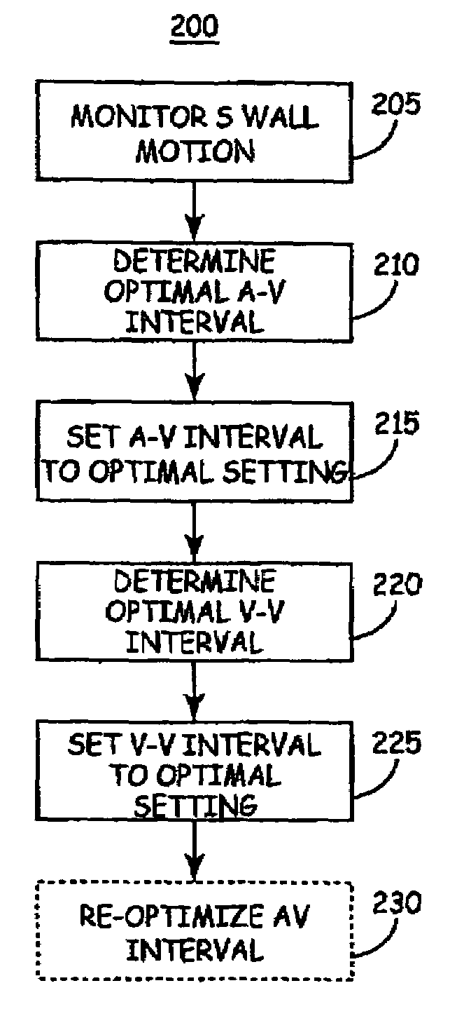
[0016]With respect to the closed-loop CRT optimization methods and apparatus, in addition to detecting (diagnosing) cardiac mechanical dysfunction and / or providing a correlation between sensed linear or non-linear acceleration (i.e., motion due to displacement) of the septal wall of a patient and NYHA Class of said patient, the present invention also provides automatically optimized, dynamically-adjustable CRT pacing modalities. In essence, one basic embodiment of the present invention provides A—A, A-V interval timing and / or V—V interval timing to minimize atrial and / or ventricular septal wall displacement.

Problems solved by technology

Such uncoordinated motion causes a decrease in cardiac output (CO), among other detrimental effects.
However, to the best knowledge of the inventors such technique has not been used to directly measure deflection of the ventricular or atrial septal wall in patients afflicted with uncoordinated ventricular motion and the like.

Method used


Image

  • Method of optimizing cardiac resynchronization therapy using sensor signals of septal wall motion
  • Method of optimizing cardiac resynchronization therapy using sensor signals of septal wall motion
  • Method of optimizing cardiac resynchronization therapy using sensor signals of septal wall motion

Examples


Embodiment Construction

[0026]As indicated above, the present invention is directed toward providing a method and apparatus for optimizing ventricular function and selecting cardiac pacing intervals for the purposes of restoring normal ventricular synchrony based on monitoring septal wall motion. The present invention is useful in optimizing atrial-ventricular, inter-atrial and inter-ventricular pacing intervals during cardiac resynchronization therapy (CRT) used for treating heart failure. The present invention is also useful in selecting pacing parameters used during temporary pacing applied for treating post-operative uncoordinated cardiac chamber (e.g., atrial and / or ventricular) motion. As such, the present invention may be embodied in an implantable cardiac pacing system including a dual chamber or multichamber pacemaker and associated set of medical electrical leads. Alternatively, the present invention may be embodied in a temporary pacing system including an external pacing device with associated ...


PUM


Abstract

The present invention relates to monitoring septal wall motion of the atrial and / or ventricular chambers of a heart for optimizing cardiac pacing intervals based on signals derived from the monitored wall motion. At least one lead of medical device is equipped with a motion sensor adapted to couple to septal tissue. The device receives and may post-process (e.g., suitably filter, rectify and / or integrate) motion signals to determine acceleration, velocity and / or displacement. During pacing interval optimization the wall motion is measured for those pacing intervals and the pacing interval setting(s) that produce minimal wall motion for chronic therapy delivery. In addition, methods for periodically determining whether to cease or resume delivery of a bi-ventricular pacing therapy to a patient that may have experienced beneficial reverse remodeling of the heart.

Description

CROSS REFERENCE TO RELATED APPLICATIONS[0001]The present patent disclosure relates to and hereby incorporates by reference two non-provisional U.S. patent applications previously invented by E. Chinchoy; namely, application Ser. No. 10 / 376,981 filed 28 Feb. 2003 entitled, “Method and Apparatus for Assessing Left Ventricular Function and Optimizing Cardiac Pacing Intervals Based on Left Ventricular Wall Motion” and application Ser. No. 10 / 377,207 filed 28 Feb. 2003 now U.S. Pat. No. 6,885,889, entitled, “Method and Apparatus for Optimizing Cardiac Resynchronization Therapy Based on Left Ventricular Acceleration.”FIELD OF THE INVENTION[0002]The present invention relates to the field of cardiac therapy. In particular, the present invention provides methods and apparatus for measuring displacement of cardiac tissue which measurement may be used to optimize certain forms of bi-ventricular cardiac therapy, such as cardiac resynchronization therapy (CRT).BACKGROUND OF THE INVENTION[0003]Th...

Claims


Application Information

Patent Timeline
Patent Type & Authority Patents(United States)
IPC IPC(8): A61N1/365A61N1/368A61N1/362
CPCA61N1/3627A61N1/3684A61N1/3682A61N1/36542A61N1/36843
Inventor NEHLS, ROBERT J.SHELDON, TODD J.
Owner MEDTRONIC INC
Why Eureka
Try Eureka