
Capture management in multi-site pacing

Inactive Publication Date: 2006-11-21
MEDTRONIC INC
View PDF24 Cites 90 Cited by
  • Summary
  • Abstract
  • Description
  • Claims
  • Application Information

AI Technical Summary

Benefits of technology

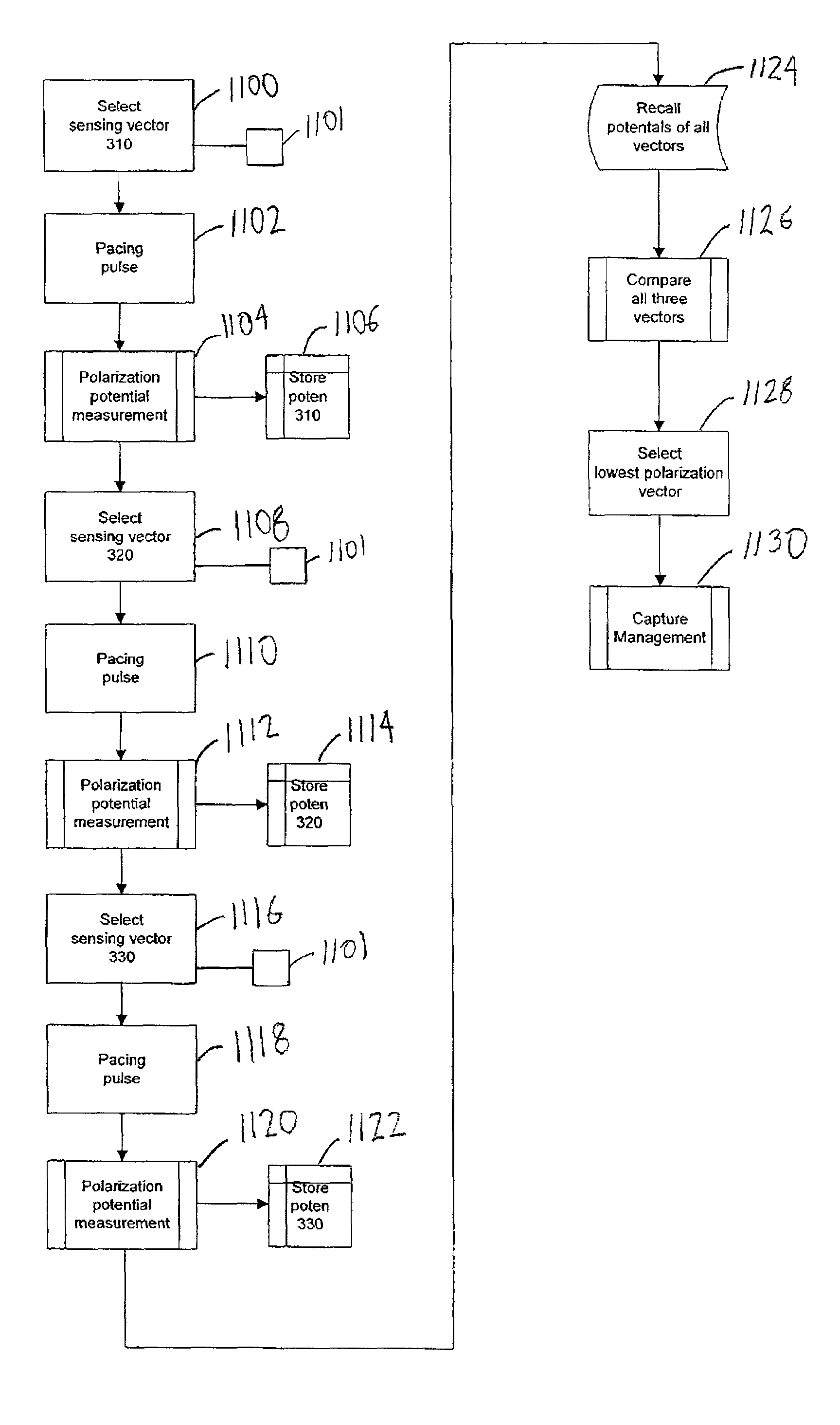
[0025]In comparison to known multi-site cardiac pacing techniques, various embodiments of the present invention may provide one or more of the following advantages. For instance, pacing methodologies in accordance with embodiments of the present invention may select a sensing vector between two or more electrodes of a plurality of electrodes that records the lowest polarization potential. As a result, the evoked response signal indicative of capture detection is more pronounced. Further, devices and methods of the present invention may permit atrial capture detection across both atria by utilizing one or more electrodes normally used for delivering atrial or ventricular pacing pulses. Moreover, methods of the present invention permit discrimination of loss of capture of a single ventricle in bi-ventricular pacing systems.

Problems solved by technology

One such problem involves the need to more accurately discern polarization potential from evoked response polarization signals after delivery of a pacing pulse.
Another problem involves the need to determine loss of capture of a single ventricle in bi-ventricular pacing systems.

Method used


Image

  • Capture management in multi-site pacing
  • Capture management in multi-site pacing
  • Capture management in multi-site pacing

Examples


Embodiment Construction

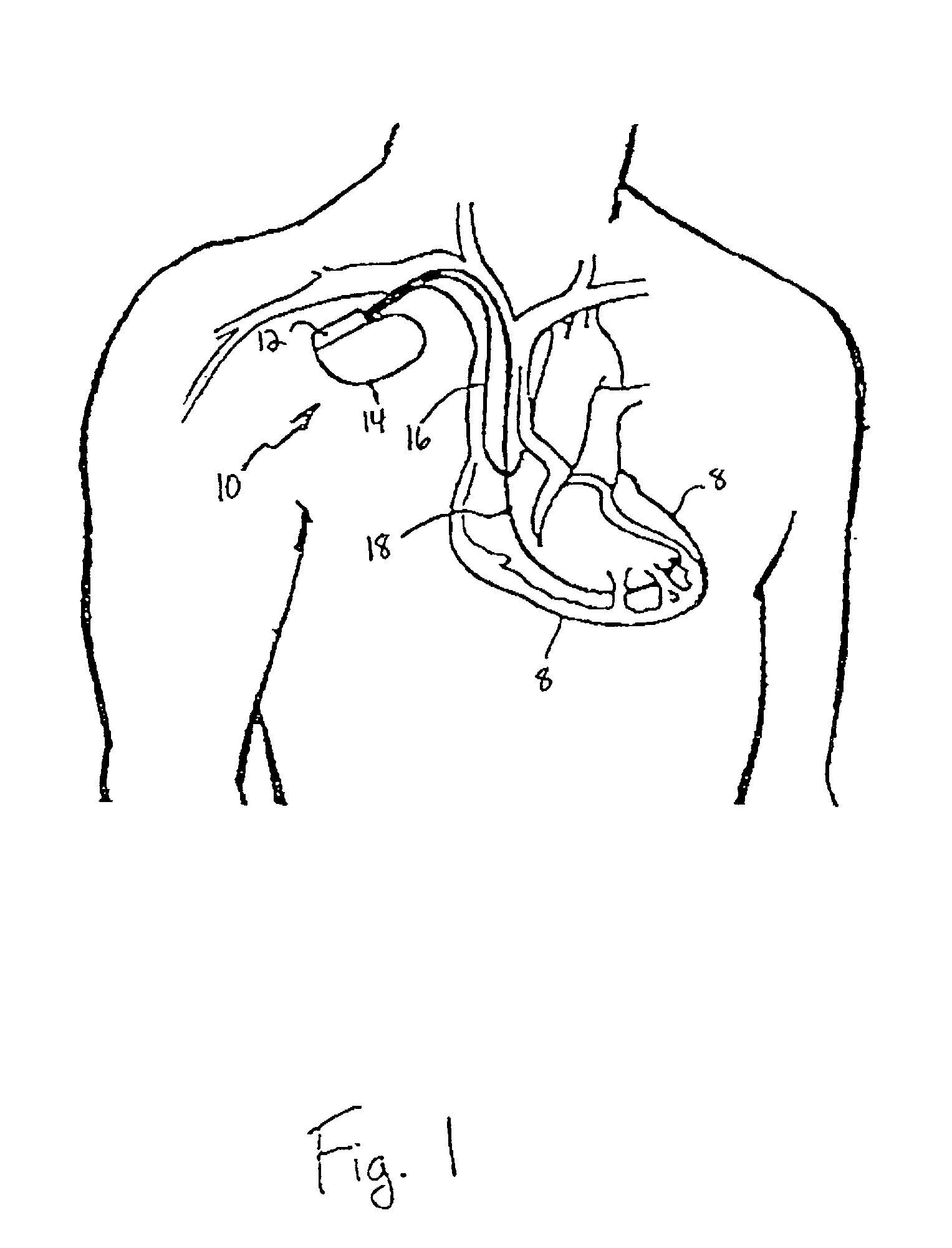
[0049]FIG. 1 is a simplified schematic view of one embodiment of implantable medical device (“IMD”) 10 of the present invention. IMD 10 shown in FIG. 1 is a pacemaker comprising at least one of pacing and sensing leads 16 and 18 attached to hermetically sealed enclosure 14 and implanted near human or mammalian heart 8. Pacing and sensing leads 16 and 18, sense electrical signals attendant to the depolarization and re-polarization of the heart 8, and further provide pacing pulses for causing depolarization of cardiac tissue in the vicinity of the distal ends thereof. Leads 16 and 18 may have, for example, unipolar or bipolar electrodes disposed thereon, as is well known in the art. Examples of IMD 10 include implantable cardiac pacemakers disclosed in U.S. Pat. No. 5,158,078 to Bennett et al., U.S. Pat. No. 5,312,453 to Shelton et al. or U.S. Pat. No. 5,144,949 to Olson.

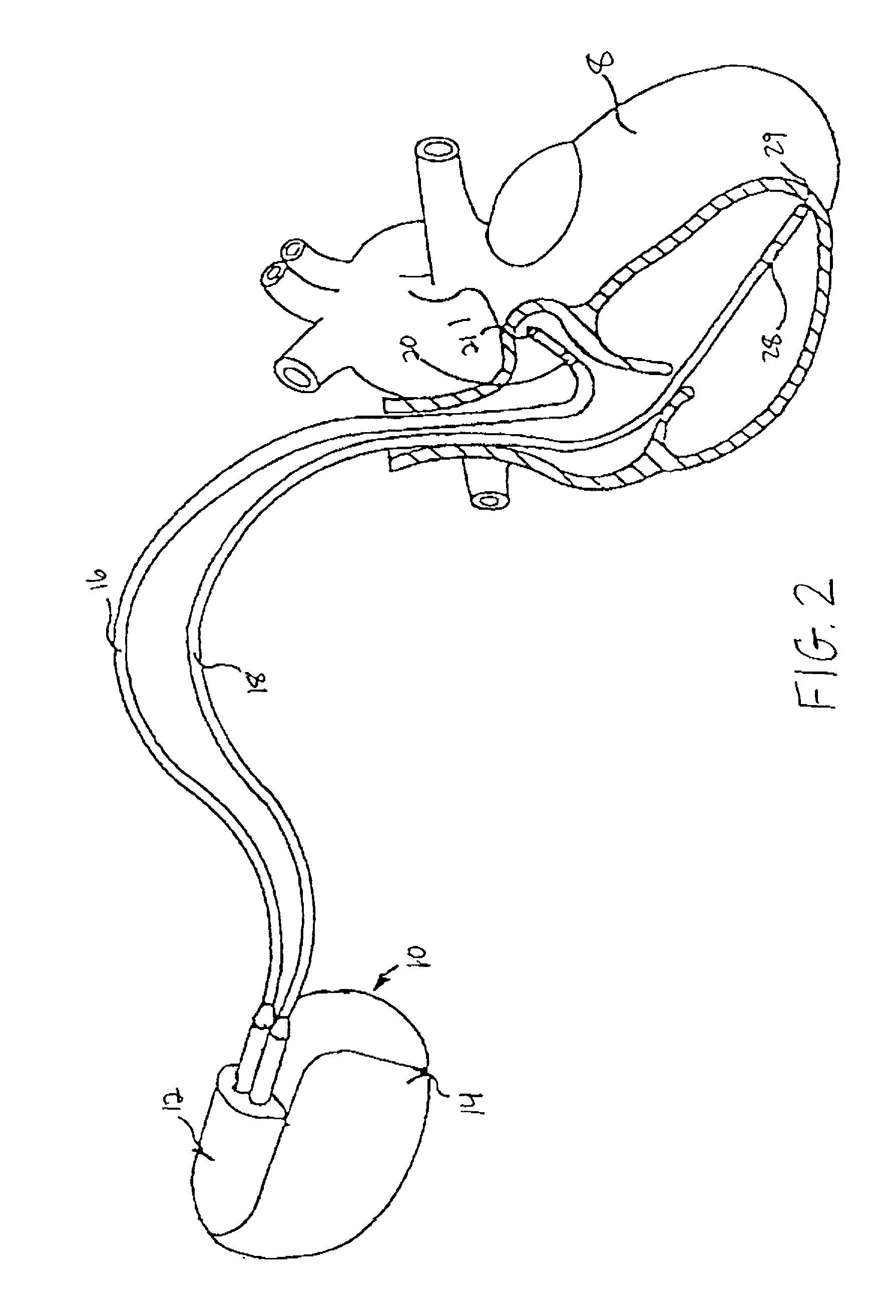
[0050]FIG. 2 shows connector module 12 and hermetically sealed enclosure 14 of IMD 10 located near human or mammali...


PUM


Abstract

Methods and apparatus for capture management in multi-chamber pacing are disclosed. In one embodiment, the invention includes determining a combination of electrodes from a plurality of electrodes that yields the lowest polarization potential immediately following delivery of an electrical stimulus to a heart; and performing capture detection using that combination of electrodes. In order to distinguish loss of capture in one ventricle in bi-ventricular pacing, certain embodiments may also include measuring a width of a QRS complex and determining when the width is greater than a predetermined value. A method for detecting single ventricular loss of capture in bi-ventricular pacing is also described utilizing comparison of evoked QRS complex morphology to a predefined waveform.

Description

FIELD OF THE INVENTION[0001]The present invention relates to implantable medical devices such as implantable cardiac pacemakers and cardioverter / defibrillators. More particularly, the present invention pertains to implantable medical devices for use in multi-site pacing.BACKGROUND OF THE INVENTION[0002]Implantable medical devices (IMDs) for use in cardiac pacing and defibrillation are well known. Most of these devices include sense amplifier circuitry for detecting intrinsic cardiac electrical activity so that the devices may be inhibited from generating unnecessary stimulating pulses when a heart is functioning properly. While the present invention is not limited to any one IMD, it will, for the sake of brevity, be described with respect mainly to pacemaker-cardioverter-defibrillators (PCDs).[0003]Dual-chamber cardiac pacemakers typically have separate sense amplifiers and associated circuitry for atrial and ventricular sensing. The sense amplifiers detect the presence of intrinsic...

Claims


Application Information

Patent Timeline
IPC IPC(8): A61N1/37A61N1/362
CPCA61N1/36185A61N1/3686A61N1/371A61N1/3627A61N1/3714A61N1/3716
Inventor FEREK-PETRIC, BOZLDER
Owner MEDTRONIC INC
Learn More