CT image production method and X-ray CT system

a production method and image technology, applied in tomography, applications, instruments, etc., can solve problems such as too long processing tim

Active Publication Date: 2007-05-08
GE MEDICAL SYST GLOBAL TECH CO LLC
View PDF11 Cites 2 Cited by
  • Summary
  • Abstract
  • Description
  • Claims
  • Application Information

AI Technical Summary

Benefits of technology

[0010]Therefore, an object of the present invention is to provide a CT image production method and an X-ray CT system in which production of a large amount of data that is not actually used to construct a CT image can be avoided and a common procedure can be adapted to reconstruction planes whose positions relative to a multi-channel detector are different from one another.

Problems solved by technology

However, since a large amount of data that is not actually used to construct a CT image is produced on a projection plane, a processing time is too long.

Method used


Image

  • CT image production method and X-ray CT system
  • CT image production method and X-ray CT system
  • CT image production method and X-ray CT system

Examples


first embodiment

[0073][First Embodiment]

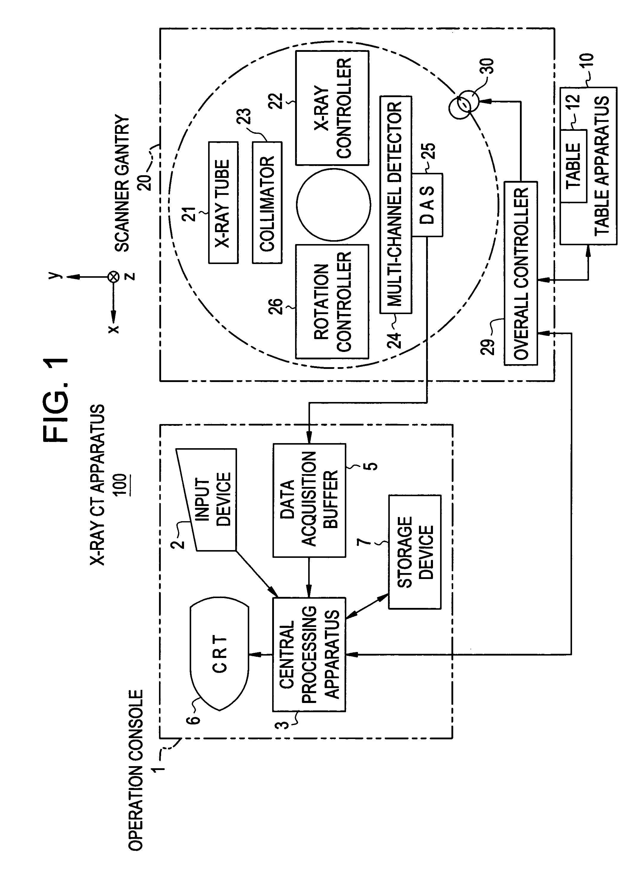
[0074]FIG. 1 is a block diagram showing the configuration of an X-ray CT system 100 in accordance with the first embodiment.

[0075]The X-ray CT system 100 comprises an operator console 1, a radiographic table assembly 10, a scanner gantry 20.

[0076]The operator console 1 comprises an input device 2 that receives an operator's entry, a central processing unit 3 that executes a scan control process or image reconstruction, a data collection buffer 5 in which data acquired by the scanner gantry 20 is recorded, a CRT 6 on which a reconstructed CT image is displayed, and a storage device 7 in which programs, data, and CT images are stored.

[0077]The radiographic table assembly 10 includes a radiographic table 12 on which an object of radiography lies down and which carries the object of radiography into or out of a bore of the scanner gantry 20. The radiographic table 12 is raised or lowered and rectilinearly moved by a motor incorporated in the radiographic table as...

second embodiment

[0112][Second Embodiment]

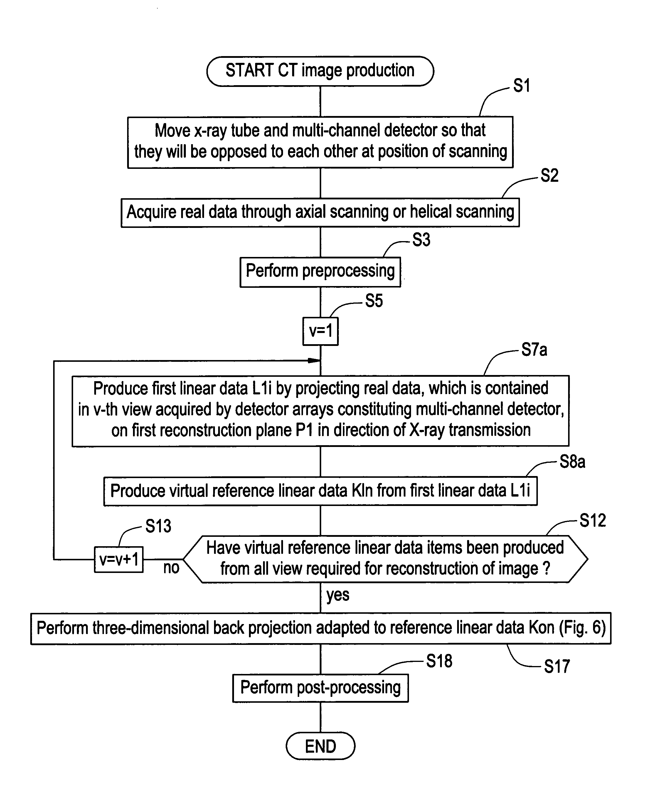
[0113]FIG. 16 is a flowchart describing CT image production in accordance with the second embodiment.

[0114]At step S1, the X-ray tube 21 and multi-channel detector 24 are moved to be opposed to each other at a desired position of scanning (in practice, the radiographic table 12 is rectilinearly moved).

[0115]At step S2, axial scanning or helical scanning is performed in order to acquire real data.

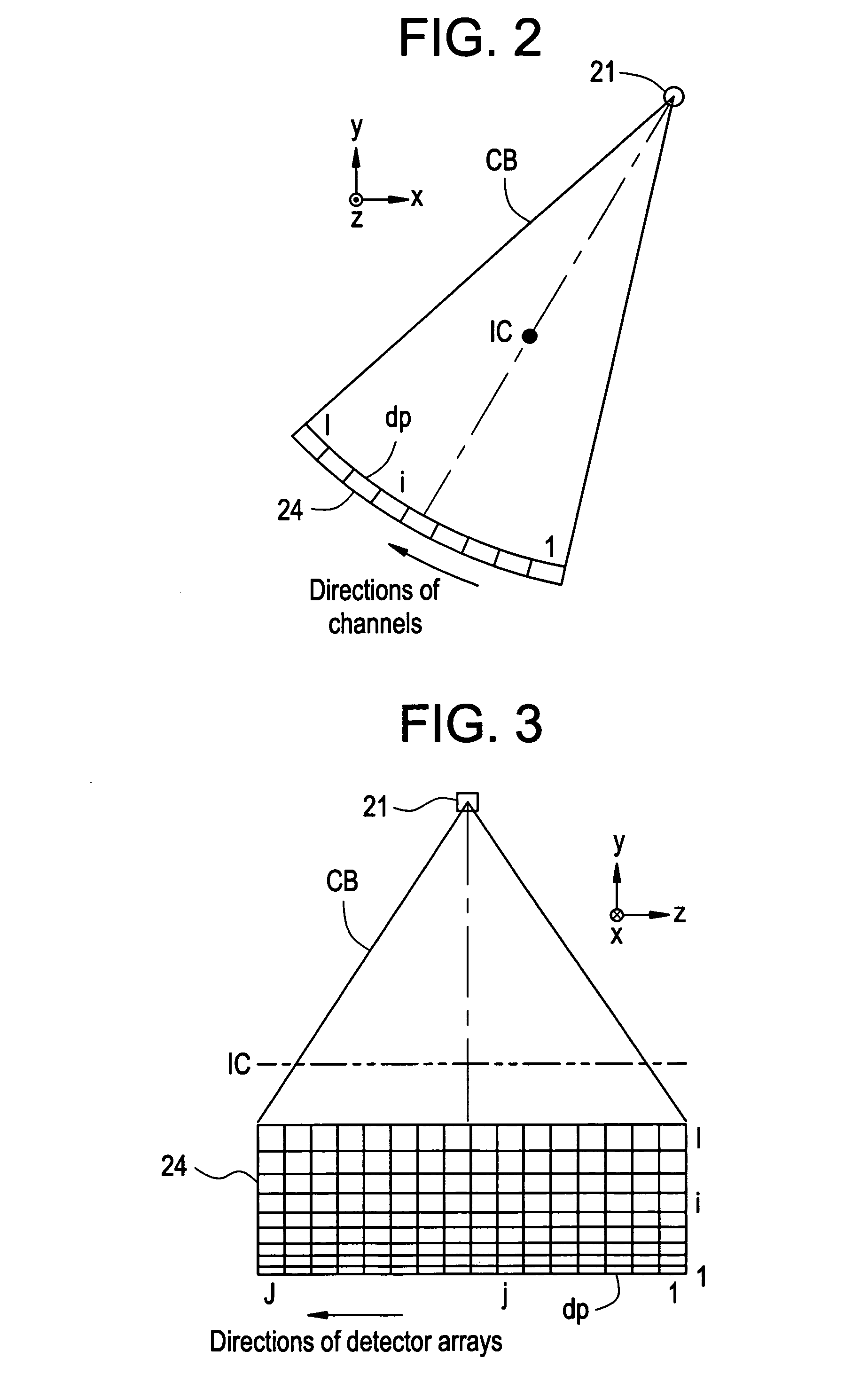
[0116]Herein, as shown in FIG. 17, the X-ray tube 21 and multi-channel detector 24 are moved so that when the view angle view is 0°, they will be opposed to each other at the reference position Z=Z0. Real data shall be acquired in this state. Moreover, a CT image shall be constructed by weighting and summating a first CT image that expresses a first reconstruction plane P1 located at a first position Z=Z1, and a second CT image that expresses a second reconstruction plane P2 located at a second position Z=Z2.

[0117]Referring back to FIG. 16, at step S3, the acquired r...

third embodiment

[0130][Third Embodiment]

[0131]FIG. 18 is a flowchart illustrating CT image procedure in accordance with the third embodiment.

[0132]At step S1, the X-ray tube 21 and multi-channel detector 24 are moved to be opposed to each other at a desired position of scanning (in practice, the radiographic table 12 is rectilinearly moved).

[0133]At step S2, axial scanning or helical scanning is performed in order to acquire real data.

[0134]Herein, as shown in FIG. 19(a) and FIG. 19(b), the X-ray tube 21 and multi-channel detector 24 are moved so that when the view angle view is 0°, they will be opposed to each other at the reference position Z=Z0 or Z=Z3. Real data shall be acquired in this state. Consequently, if axial scanning is designated, it must be performed twice or more. Moreover, a CT image expressing a first reconstruction plane P1 located at a first position Z=Z1 shall be produced.

[0135]Referring back to FIG. 18, at step S3, the acquired real data is subjected to preprocessing (includin...


PUM


Abstract

A method to make a common procedure adaptable to reconstruction planes whose positions relative to a multi-channel detector are different from one another. A reference procedure required for construction of a CT image based on reference linear data representing one line or a plurality of lines formed on a reference reconstruction plane is preserved. Real data associated with a first reconstruction plane different from the reference reconstruction plane is used to produce virtual reference linear data equivalent to the reference linear data. A CT image is reconstructed based on the virtual reference linear data using the reference procedure.

Description

CROSS REFERENCE TO RELATED APPLICATIONS[0001]This application claims the benefit of Japanese Application No. 2004-140668 filed May 11, 2004.BACKGROUND OF THE INVENTION[0002]The present invention relates to a computed tomography (CT) image production method and an X-ray CT system. More particularly, the present invention relates to a CT image production method and an X-ray CT system in which production of a large amount of data that is not actually used to construct a CT image can be avoided and a common procedure can be adapted to reconstruction planes whose positions relative to a multi-channel detector are different from one another.[0003]One of the CT image production methods that have been known in the past is such that: real data that is acquired by detector arrays included in a multi-channel detector and that is contained in one of the views required for reconstruction of an image is projected onto a projection plane, which is a virtual plane, in a direction of X-ray transmiss...

Claims


Application Information

Patent Timeline
Patent Type & Authority Patents(United States)
IPC IPC(8): A61B6/00A61B6/03G06T11/00
CPCG06T11/005Y10S378/901A61B6/03
Inventor HAGIWARA, AKIRA
Owner GE MEDICAL SYST GLOBAL TECH CO LLC
Why Eureka
Try Eureka