Ultrasonic diagnostic apparatus

a diagnostic apparatus and ultrasonic technology, applied in the field of ultrasonic diagnostic apparatus, can solve the problems of inability to observe the cavity portion inside the organ, inability to observe the cavity region which has been eliminated, and inability to obtain accurate volum

Inactive Publication Date: 2007-05-29
HITACHI LTD
View PDF22 Cites 14 Cited by
  • Summary
  • Abstract
  • Description
  • Claims
  • Application Information

AI Technical Summary

Benefits of technology

[0011]Also, it is possible to calculate and provide the volume of the cavity portion of the organ based on a three-dimensional image thereof. Further, it is possible to provide data for supporting diagnosis, such as a change of the volume with time and a ratio between the minimum and maximum values of the volume.

Problems solved by technology

In this case, however, a cavity portion inside an organ cannot be observed.
Accordingly, when a cavity is observed in such a manner that a cutting plane is specified and information concerning regions in front of the cutting plane is eliminated, as conventionally performed, it is not possible to observe the regions of the cavity which have been eliminated.
Therefore, an accurate volume cannot be obtained.

Method used


Image

  • Ultrasonic diagnostic apparatus
  • Ultrasonic diagnostic apparatus
  • Ultrasonic diagnostic apparatus

Examples


Embodiment Construction

[0019]A preferred embodiment of the present invention will be described in further detail with reference to the accompanying drawings. Although a heart, particularly a left ventricle, is described as the object of observation in the following example, the present invention is clearly also applicable to other cardiac cavities and to cavity portions of other organs.

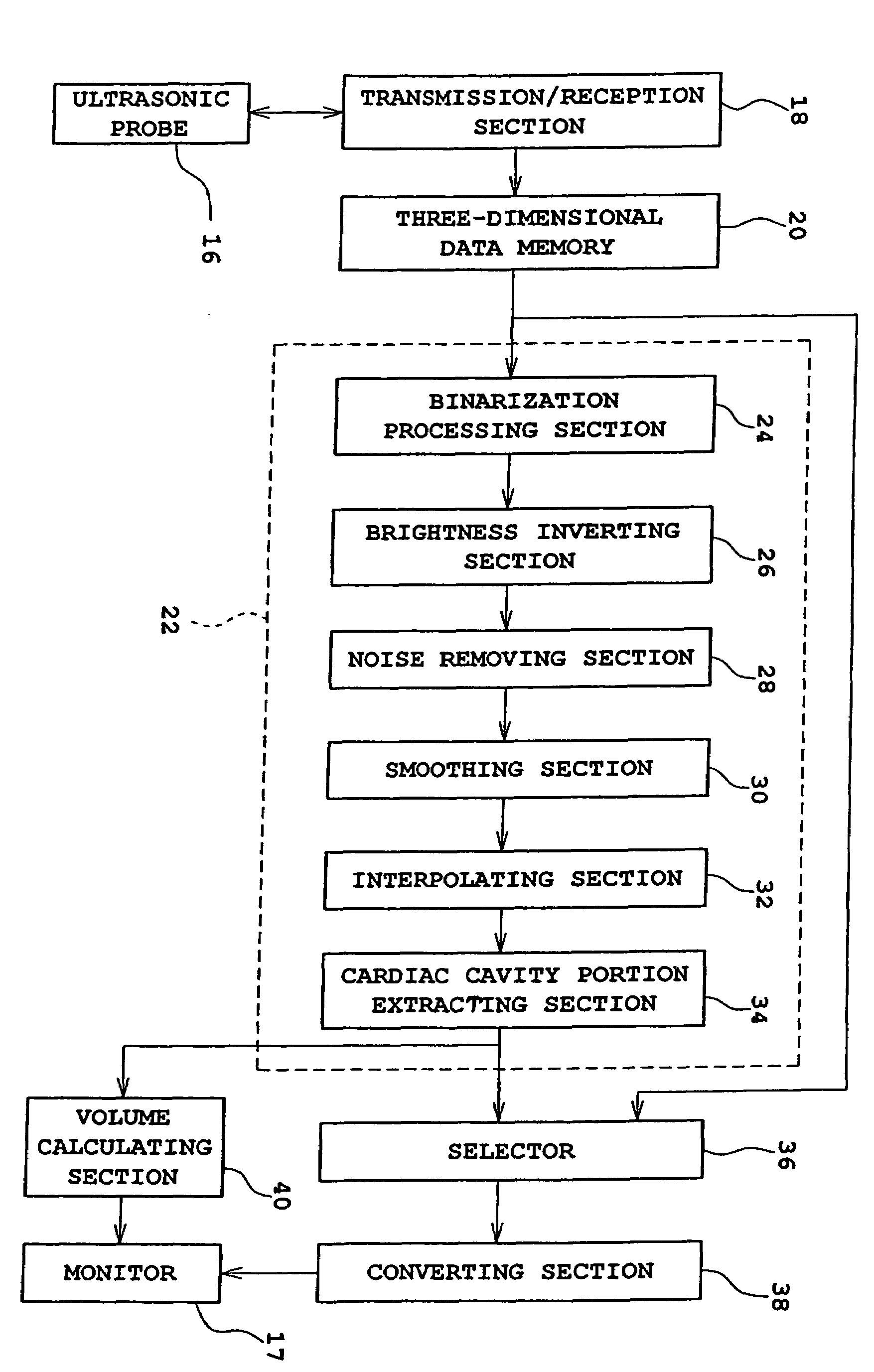
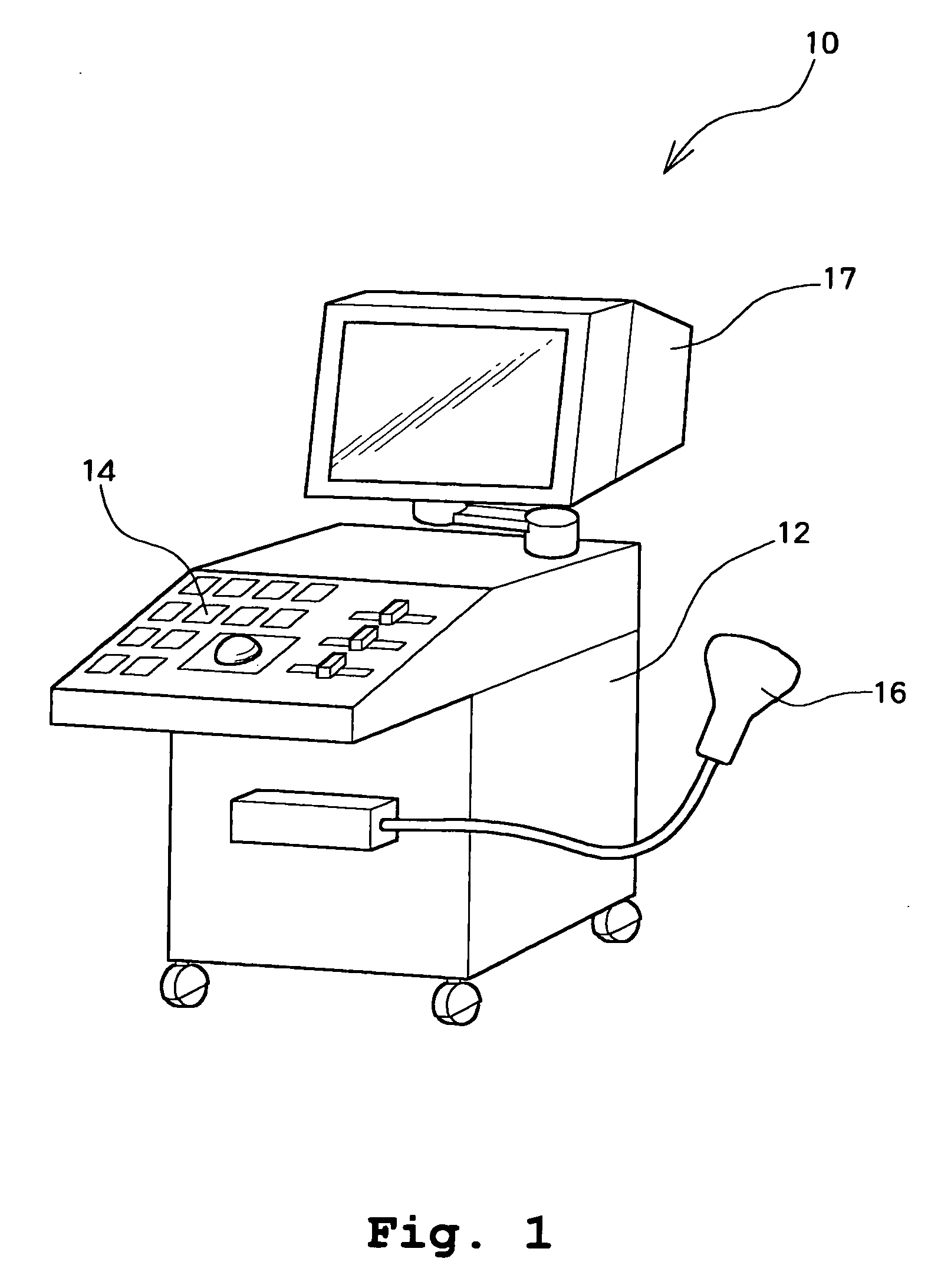
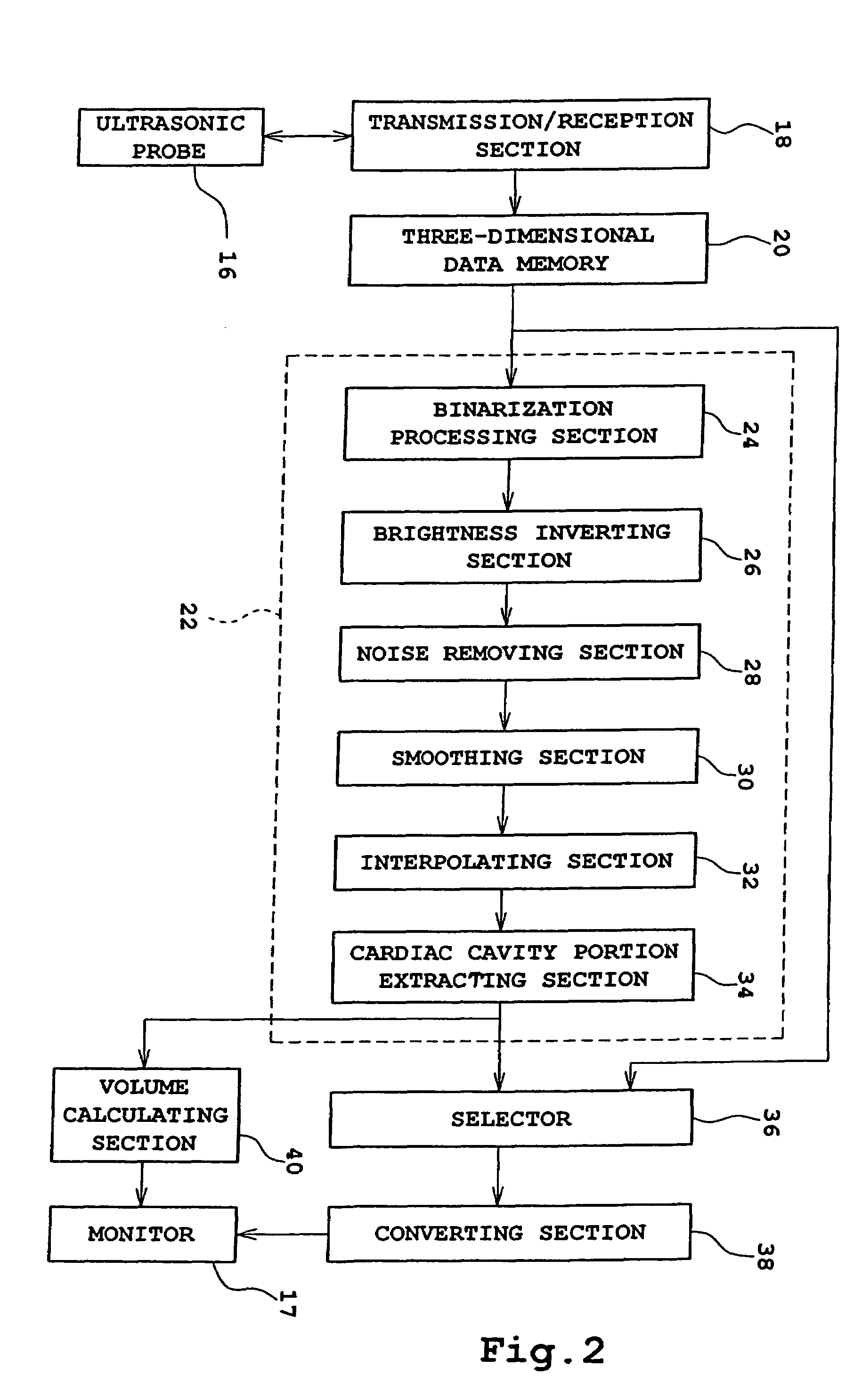
[0020]FIG. 1 schematically shows an appearance of an ultrasonic diagnostic apparatus 10 according to an embodiment of the present invention. The ultrasonic diagnostic apparatus 10 has a function of forming a three-dimensional ultrasonic image. The ultrasonic diagnostic apparatus 10 includes an operation panel 14 for performing various operations at a front surface of a main body 12 of the apparatus 10. Further, an ultrasonic probe 16 for transmitting and receiving ultrasound with regard to an object is connected via a cable to the apparatus main body 12. A monitor 17 is provided above the apparatus main body 12 for displayi...


PUM


Abstract

In an ultrasonic diagnostic apparatus capable of forming a three-dimensional ultrasonic image, an entire cavity portion of an organ can be displayed without any part being invisible. Three-dimensional ultrasonic image data obtained based on an ultrasonic wave transmitted and received with respect to an object organ is stored in a memory 20. With regard to the stored data, a brightness value of each voxel is binarized in a binarization processing section 24 using a predetermined threshold value. Then, the binarized brightness value is further inverted in a brightness value inverting section 26. Consequently, the cavity portion of the organ which has low brightness before inversion has high brightness, and the wall portion of the organ which has high brightness before inversion and which obstruct observation of the inner cavity portion has now low brightness. Thus, solid representation of the cavity portion to be observed can be obtained, thereby facilitating observation.

Description

BACKGROUND OF THE INVENTION[0001]1. Field of the Invention[0002]The present invention relates to an ultrasonic diagnostic apparatus for providing an image of a cavity portion of an organ as a three-dimensional image.[0003]2. Description of Related Art[0004]Ultrasonic diagnostic apparatuses which transmit and receive ultrasound with respect to a living body and which then provide a three-dimensional image of inside the living body based on a received ultrasonic signal have been known. In general, the three-dimensional image thus obtained is a solid representation of information concerning a brightness value of each voxel. In this case, however, a cavity portion inside an organ cannot be observed. When observing a heart, for example, a cardiac muscle portion is brightly displayed because ultrasound is reflected by the cardiac muscle of the heart, and no information can be obtained regarding a cavity portion inside the organ, such as the characteristics or behavior of a ventricle. Ther...

Claims


Application Information

Patent Timeline
Patent Type & Authority Patents(United States)
IPC IPC(8): A61B8/00A61B8/06A61B8/08A61B19/02G06T1/00G06T5/00G06T15/08G06T19/00H04N7/18
CPCA61B8/065A61B8/483A61B19/0248A61B2019/025A61B50/10A61B50/13
Inventor MURASHITA, MASARU
Owner HITACHI LTD
Why Eureka
Try Eureka