Methods and devices for automated biopsy and collection of soft tissue

a tissue and automatic technology, applied in the field of tissue sampling methods and devices, can solve the problems of no single procedure is ideal for all cases, and inability to accurately measure, etc., to achieve convenient and inexpensive fabrication, improve and more versatile operation

Inactive Publication Date: 2007-06-05
DEVICOR MEDICAL PROD
View PDF121 Cites 289 Cited by
  • Summary
  • Abstract
  • Description
  • Claims
  • Application Information

AI Technical Summary

Benefits of technology

[0009]This invention addresses the aforementioned needs by providing a number of important new features and innovations for the active capture type of biopsy instrument which each collectively or singly contribute to improved and more versatile operation For example, such innovations include a molded tissue cassette housing, permitting easy and inexpensive fabrication while also permitting the handling and viewing of multiple tissue samples without physical contact by the instrument operator. The housing is interconnected with the piercing needle using a thumbwheel which permits the needle to rotate relative to the housing, thereby preventing the vacuum tube from wrapping about the housing. Several variant vacuum port embodiments are disclosed, each of which have advantages in certain tissue environments. Also disclosed is a method for backflushing biological debris from the instrument which builds up after repeated sampling procedures, without removing the instrument from the selected tissue location.
[0010]With respect to the single-use type of biopsy instrument, several tissue capture embodiments are disclosed for improving the capture process, so that complete and well preserved samples are obtained. Many of these embodiments are also applicable for use with the active capture instrument type.
[0012]Surprisingly, the inventors have found that preferably, in order to minimize tissue clogging of the cutter, the knock-out pin should have an effective diameter or cross-sectional area of at least 0.030 inches, and the ratio of the effective diameter of the knock-out pin to the internal diameter of the cannular cutter should be at least approximately one-half.

Problems solved by technology

The type of biopsy utilized depends in large part on circumstances present with respect to the patient, and no single procedure is ideal for all cases.
However, they also have disadvantages.
An important disadvantage is that the small core size makes it necessary to accurately place the needle when sampling small lesions.
This action is tedious and time consuming.
A further disadvantage of such single-use guns is the needle typically used in such a device, e.g. the True Cut® needle manufactured by Travenol Laboratories.
Unfortunately, the True Cut® needle is rough on organs and lesions, often only obtaining small fragments of tissue, and is quite operator dependent—some individuals are good at operating the device and some are not.
However, it does not operate equally well in all procedures and in all bodily environments.

Method used


Image

  • Methods and devices for automated biopsy and collection of soft tissue
  • Methods and devices for automated biopsy and collection of soft tissue
  • Methods and devices for automated biopsy and collection of soft tissue

Examples


second modified embodiment

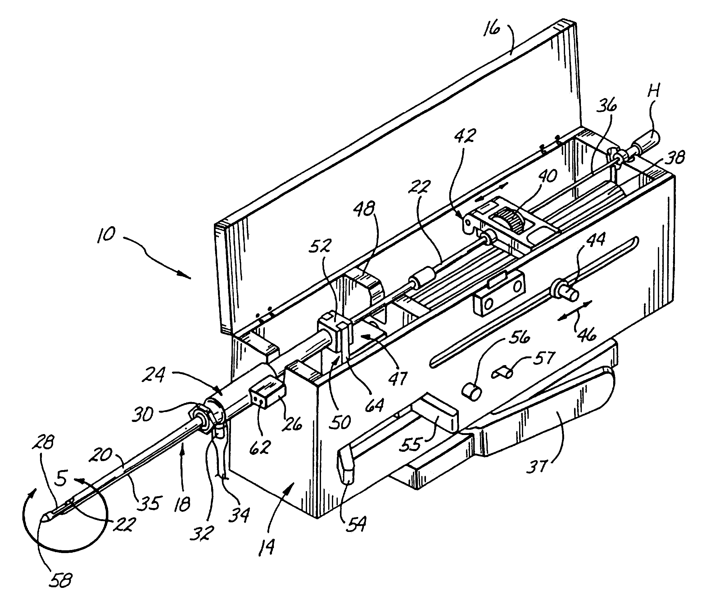
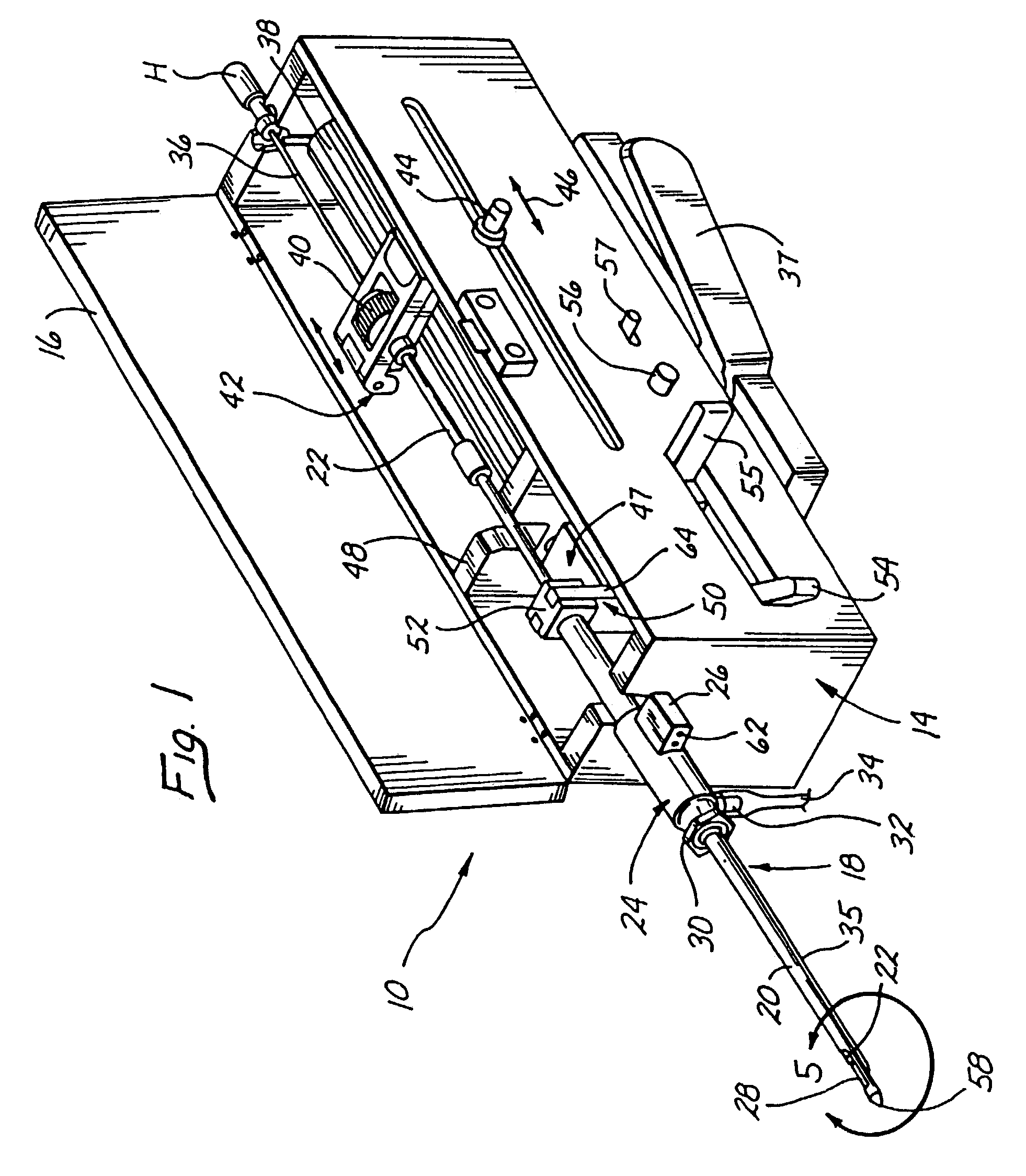
[0090]FIGS. 29 and 30 illustrate the needle assembly in the FIG. 25 device. Again, like elements are designated by like reference numerals, followed by a b. In this embodiment, the inner needle 100b has been modified to include an “alligator” tip 140, which includes jaws 142, 144 and teeth 146. When the spring 116 is released, the inner needle 100b shoots distally and captures tissue in the opening 148 within the jaws 142, 144. Then, when the spring 114 is released, the outer cannula 98b shoots distally, severing tissue along the sides of the tissue sample opening 148 as it moves distally, and also forcing the jaws 142, 144 shut, so that they “bite off” the end of the tissue sample 138b, as illustrated in FIG. 30. This embodiment also may be adapted for use with the device of FIG. 1, if desired.

third modified embodiment

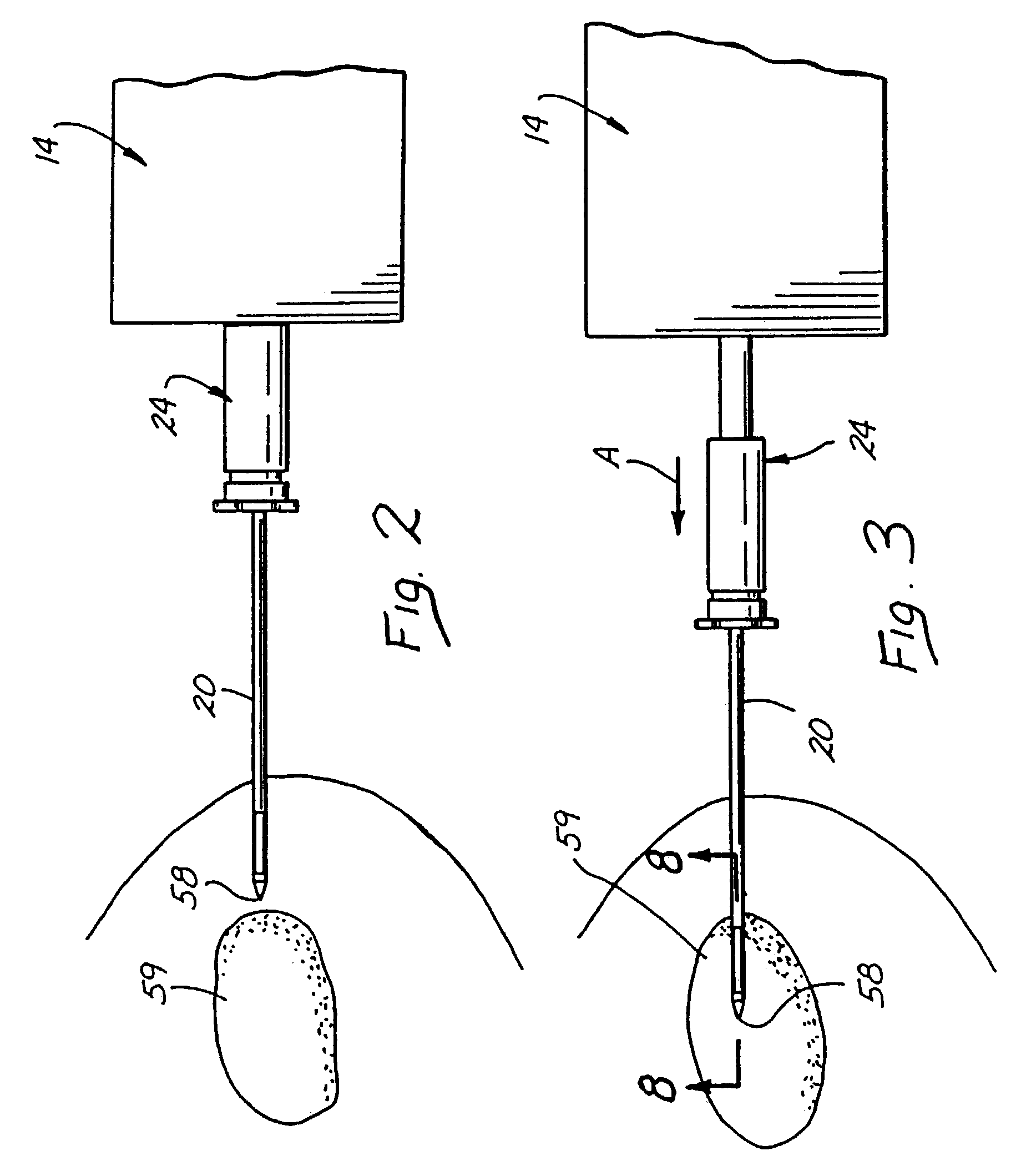
[0091]Finally, FIGS. 31–34 illustrate the needle assembly in the FIG. 25 device. In this embodiment, like elements are designated by like reference numerals, followed by a c. Like the FIG. 29 embodiment, the inner needle or “grabber”100c has been modified, this time to include a plurality of hooked extractors 150 extending from its distal end The outer cannula 98c includes a sharpened cutter point 152. In operation, initially the grabber 100c is retracted into the cutter 98c while the device is in its energized state, the point 152 being used to pierce the body wall 154 as the device is guided to the desired tissue sample 138c (FIG. 32). Then, as illustrated in FIG. 33, the grabber 100c is shot distally by means of the release of spring 116. As it travels distally, the hooked extractors 150 become extended and latch onto the tissue sample 138c. Then, once the second spring 114 is released, the cutter 98c shoots distally, collapsing the hooked extractors 150 and severing the tissue s...


PUM


Abstract

Instruments for performing percutaneous biopsy procedures are disclosed, which have advantageous features for improving functionality and performance over prior art devices. These instruments comprise two types, single-use devices, and multiple-use devices having active tissue capture capability. Improved features include the ability to retrieve and evaluate multiple tissue samples during a single insertion procedure, without physical handling of the samples, as well as constructional features, such as a molded tissue cassette housing, variant vacuum port embodiments suited for different tissue environments, and a method for backflushing the instrument to remove biological debris, among others.

Description

CROSS-REFERENCE TO RELATED APPLICATIONS[0001]This is a divisional of pending application U.S. Ser. No. 09 / 734,787 filed Dec. 13, 2000, which is a continuation of U.S. Ser. No. 08 / 825,899 filed Apr. 2, 1997 now abandoned; which is a divisional of U.S. Ser. No. 08 / 386,941 filed Feb. 10, 1995 now U.S. Pat. No. 5,649,547; which is a continuation in part of U.S. Ser. No. 08 / 217,246, filed Mar. 24, 1994 (issued as U.S. Pat. No. 5,526,822).FIELD OF THE INVENTION[0002]The present invention relates to methods and devices for tissue sampling, and more specifically to improved biopsy intents and methods for acquiring subcutaneous biopsies and for removing lesions.BACKGROUND OF THE INVENTION[0003]It is often desirable and frequently necessary to sample or test a portion tissue from humans and other animals, particularly in the diagnosis and treatment of patients with cancerous tumors, pre-malignant conditions, and other diseases or disorders. Typically, in the case of cancer, when the physician...

Claims


Application Information

Patent Timeline
Patent Type & Authority Patents(United States)
IPC IPC(8): A61B10/00A61B10/02A61B17/22A61B17/30A61B17/34
CPCA61B10/0266A61B10/0275A61B10/0283A61B8/0841A61B6/12A61B17/221A61B2010/0225A61B2017/303A61B2010/0208
Inventor RITCHART, MARK A.STUART, J. MICHAELBURBANK, FRED H.GALT, KENNETH M.
Owner DEVICOR MEDICAL PROD
Why Eureka
Try Eureka