System and Method for Detecting Pathogens

a pathogen detection and pathogen technology, applied in the field of pathogen detection systems and methods, can solve the problems of reducing the efficacy of second-line art regimens, shortening the clinical durability of available art in developing countries, and false positive diagnosis, etc., to achieve the effects of convenient use, low cost, and convenient manufacturing

Active Publication Date: 2015-10-29
THE BRIGHAM & WOMEN S HOSPITAL INC
View PDF16 Cites 5 Cited by
  • Summary
  • Abstract
  • Description
  • Claims
  • Application Information

AI Technical Summary

Benefits of technology

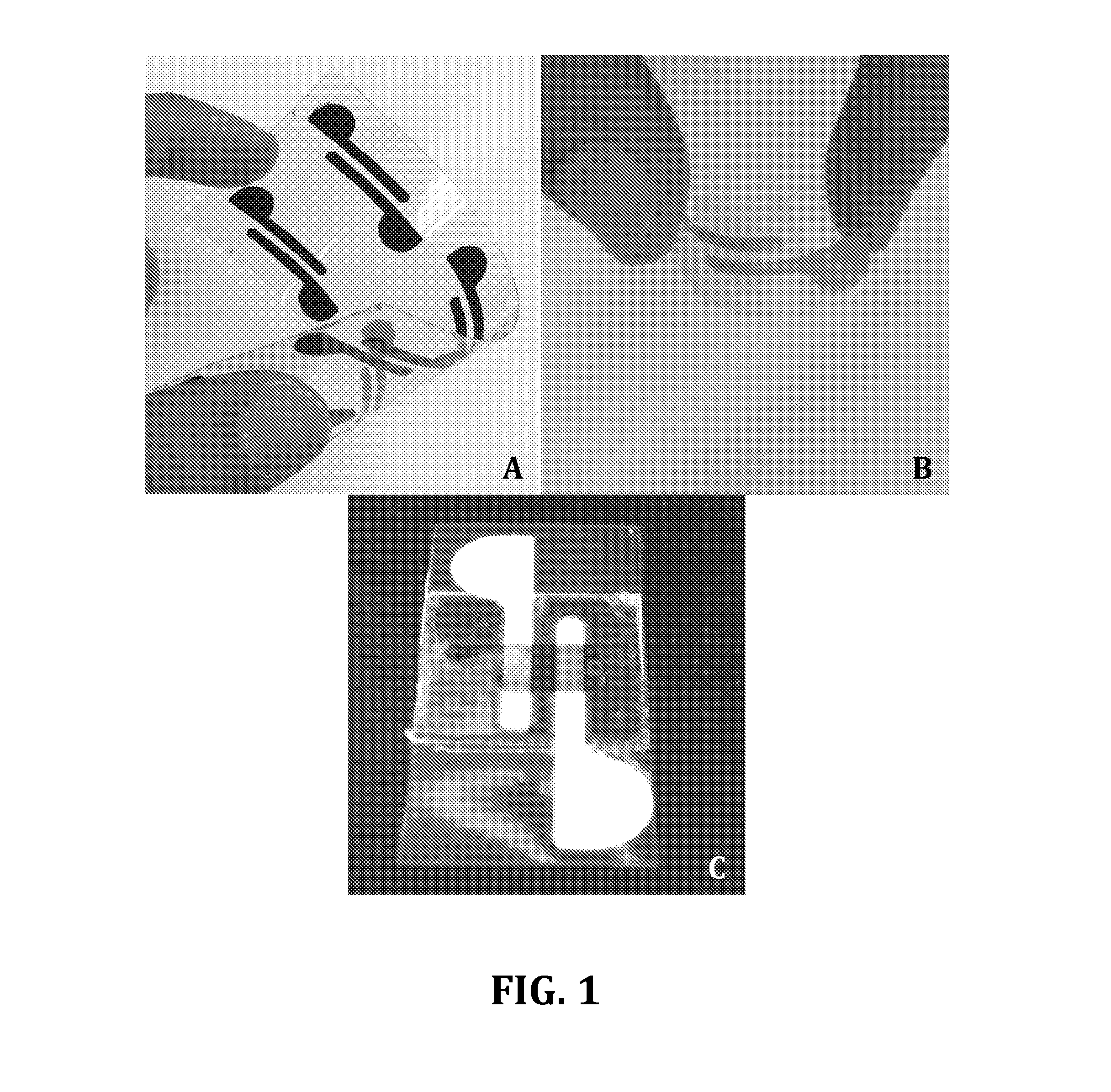
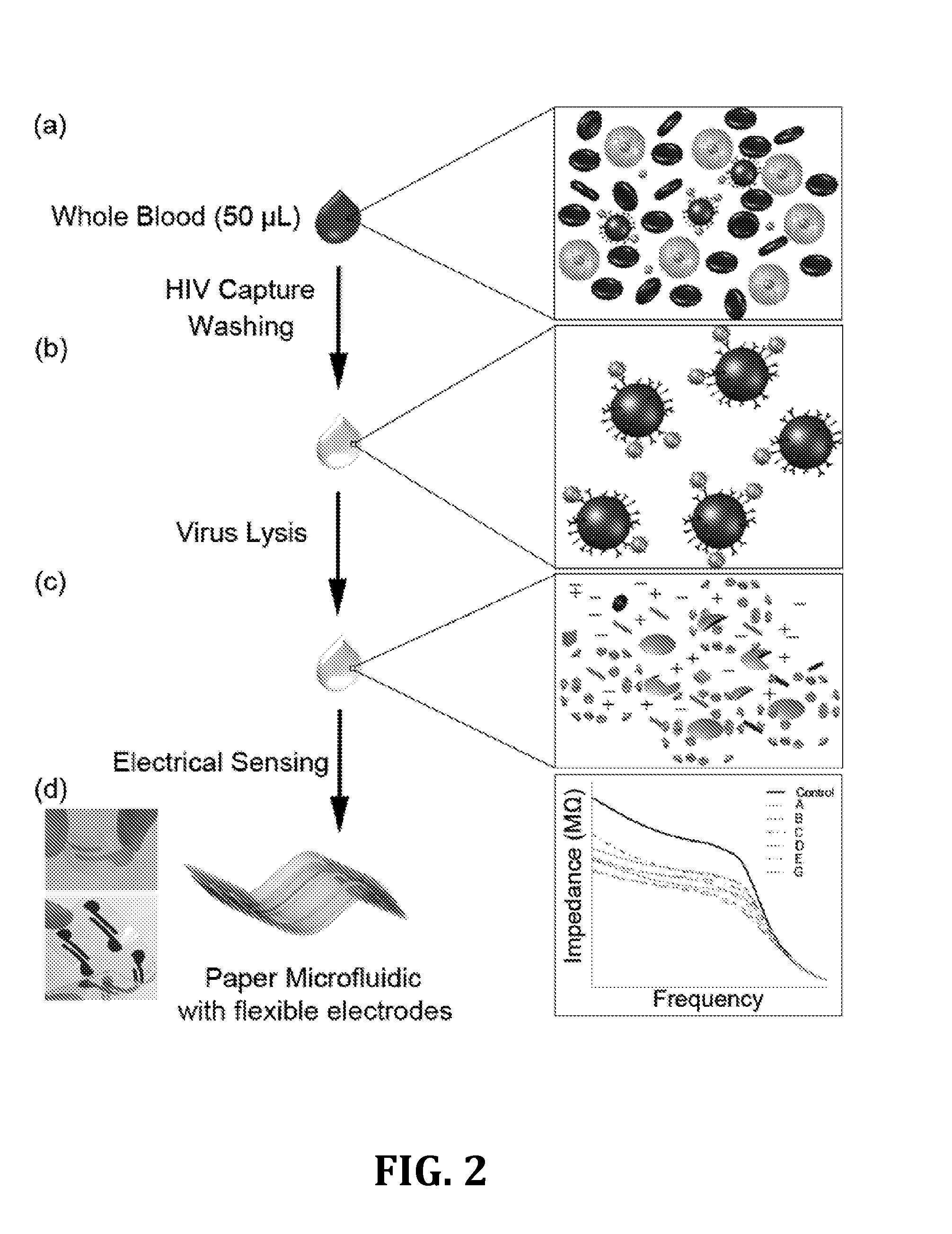
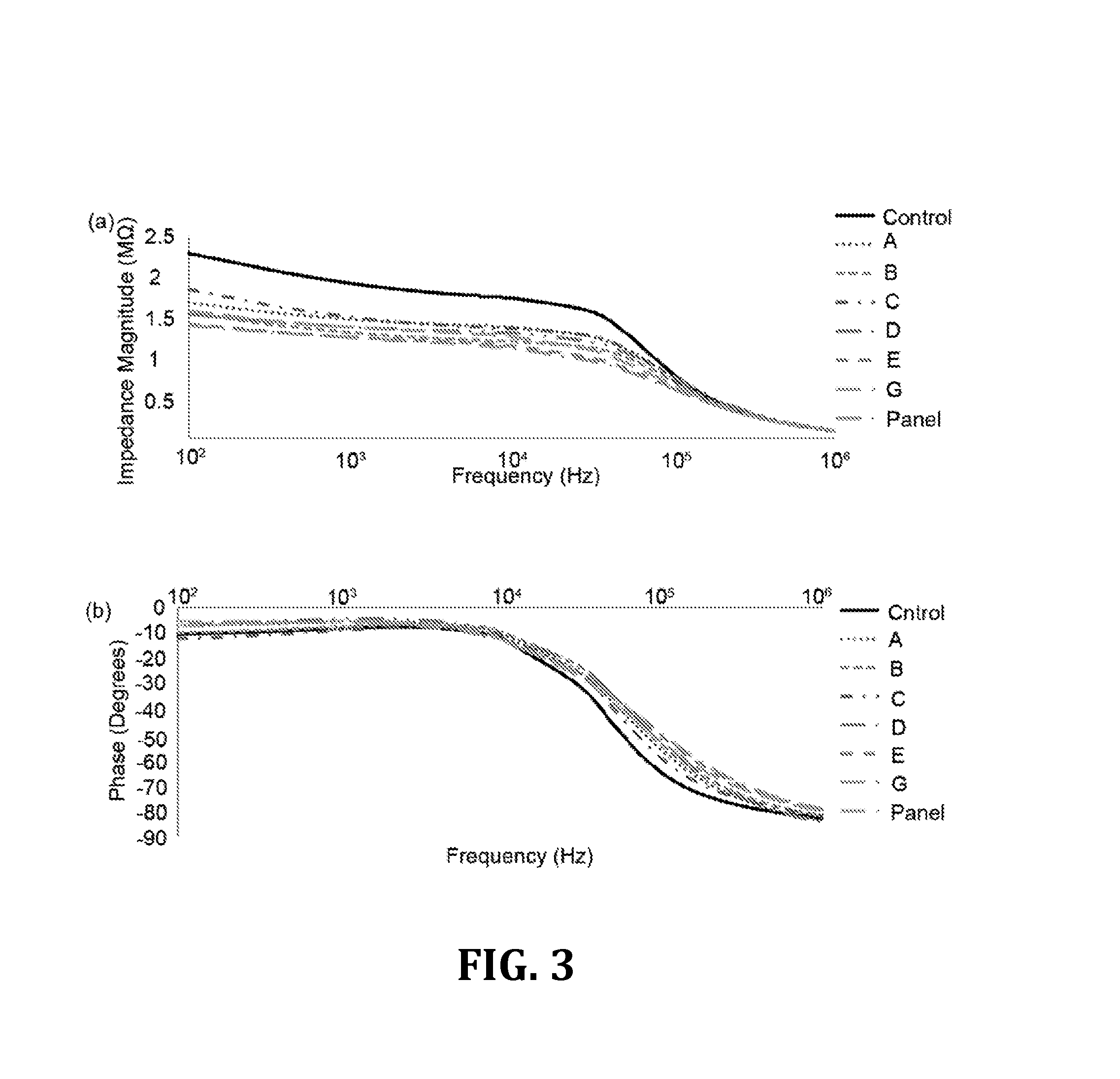
[0010]Diagnostic devices for infectious diseases detection and treatment monitoring in resource-constrained settings should be affordable, sensitive, specific, user-friendly, rapid and robust, equipment free, and deliverable to end-users (ASSURED) according to World Health Organization (WHO) guidelines. The combination of electrical sensing and paper-based microfluidics described below offers a promising opportunity to create such diagnostic tools for developing world. A portable paper-based microchip platform has been developed with simple and mass-producible fabrication technology that can selectively capture and detect HIV-1 utilizing impedance spectroscopy of viral lysate. Multiple HIV subtypes at clinically relevant viral loads that occur during the seroconversion stage (acute HIV) were detected in biological samples including whole blood and plasma using this microchip technology. The specificity of the microchip was also evaluated in the presence of a non-HIV virus (EBV) and the results were evaluated using reverse transcriptase quantitative polymerase chain reaction (RT-qPCR) method as the gold standard method for viral load measurement in developed countries.
[0011]Electrical sensing of small biomolecules, proteins, DNA, and pathogens in biological samples including whole blood, plasma, and sputum has offered a sensitive, specific, and robust diagnostic method for a variety of diseases including cancer and infectious diseases. Paper-based microfluidics has also opened an exciting and promising avenue in the area of diagnostics at the POC. Paper-based microchips are inexpensive, easy to fabricate, mass-producible, easy-to-use, and disposable. Paper is readily available around the world and a paper-based microchip can be made for less than one cent. Paper is thin, light (approximately 10 mg / cm2) and flexible and can absorb fluid samples through capillary effect. Therefore, paper-based microfluidics is a new attractive method for developing affordable tools for broad applications such as drug development, monitoring water and environment quality, and infectious diseases diagnostic at the POC in resource-constrained settings.
[0012]In this application, paper-based microfluidic technology has been integrated with electrical sensing to develop an inexpensive, rapid, and disposable microchip for virus detection at the POC. Multiple HIV-1 subtypes (A, B, C, D, E, G, and panel) have been successfully captured and detected utilizing impedance spectroscopy of viral lysate on a paper-based microfluidic device. The microchip has demonstrated the ability to detect HIV at viral loads (106-108 copies / mL) that occur at the early stage of the infection (acute HIV) where current rapid POC tests and ELISA methods such as OraQuick® cannot detect. This microchip technology is shown to detect HIV-1 spiked in whole blood and plasma samples with viral loads between 106 and 108 copies / mL. This method offers a simple, rapid, and broadly applicable technology to detect pathogens and viruses such as pox, hepatitis, influenza, and tuberculosis in biological samples including whole blood and plasma.
[0017]In some forms of the method, there may be other steps to help improve impedance measurement and improve detection accuracy. For example, after the step of capturing the pathogen from the sample with the recognition element(s), the sample may be washed to remove electrically conductive solution. Additionally, the pathogen may be lysed to release one or more electrically conductive chemical entities inside the pathogen after the step of capturing the pathogen from the sample with the recognition element(s). The one or more electrically conductive chemical entities may include one or more of ions, proteins, antigens, enzymes, and biomolecules inside the captured pathogen. In one specific form, Triton x-100 may be used to lyse the pathogen to release one or more electrically conductive chemical entities inside the pathogen.
[0019]It is contemplated that in some forms of the method, that the detection of the pathogen may occur in the presence of another type of pathogen. In this way, the system may possess the ability to selectively detect the pathogen even in the presence of another type of pathogen.

Problems solved by technology

However, a majority of HIV-infected individuals do not receive ART (46% of HIV-infected patients by the end of 2011) due to lack of affordable, rapid, and sensitive HIV diagnostic tools at the point-of-care (POC).
It has been reported that recommended clinical symptoms criteria might lead to false positive diagnosis when compared with virological criteria.
CD4 cell counting strategy alone is not sufficient to monitor patients efficiently or detect early virological failure, which allow for accumulation of drug-resistant strains and reduce the efficacy of second-line ART regimens and consequently shortening the clinical durability of available ART in developing countries.
Without virological failure confirmation, HIV health care providers may prescribe switching to premature second-line ART and more complex antiretroviral regimen.
Although, these assays offer promising methods for viral load measurements, they are either in the development process, are relatively expensive, require air conditioning, and / or require skilled operators.
Therefore, current viral load technologies are not suitable for POC testing in resource-constrained settings.

Method used


Image

  • System and Method for Detecting Pathogens
  • System and Method for Detecting Pathogens
  • System and Method for Detecting Pathogens

Examples


example i

[0042]For the following example, the following reagents were obtained and used. Triton X-100 (100%), glycerol (100%), and Bovin Serum Albumin (BSA, 10%) were purchased from Sigma-Aldrich® (St Louis, Mo.). Dulbecco's Phosphate Buffered Saline (DPBS, 1×) was bought from Life Technologies' (Grand Island, N.Y.). HyPure™ Molecular Biology Grade Water was obtained from Fisher Scientific (Agawam, Mass.). Ethanol (200 proof) was bought from Sigma Aldrich® (Sheboygan, Wis.). Biotinylated polyclonal goat anti-gp120 antibody (4 g / mL) was obtained from Abcam® (Cambridge, Mass.).

[0043]Magnetic beads conjugated with anti-gp120 antibodies were prepared. Streptavidin-coated magnetic beads (1 μm diameter) were purchased from Thermo Scientific (Rockford, Ill.). After diluting in DPBS by 1:10 (v / v), the beads were washed three times with DPBS (1.5 mL) using a BioMag® multistep magnetic separator from Polyscience Inc. (Warrington, Pa.). Antibody conjugation was done by adding biotinylated anti-gp120 an...

example ii

[0046]Microchips were fabricated in the following manner. Two rail electrodes were patterned on hydrophobic transparency papers using silver ink from Engineered Materials Systems, Inc. (Delaware, Ohio). The electrodes geometry and design were cut on a double-sided-adhesive film (DSA) using a VLS2.3 laser cutter from Universal Laser Systems Inc. (Scottsdale, Ariz.). This DSA used as a mask and taped on top of a paper and conductive ink (graphite or silver) was poured on top of the mask to fill the openings on the DSA. A glass cover slip was used to distribute the ink evenly everywhere in the openings. The paper with the inks were then baked in an oven at 80° C. for one hour. After the ink dried, the protective DSA was removed and the electrodes patterned in the openings of the mask were left on the paper. The thickness of the electrodes was approximately 50 μm. The width and spacing of these two rail electrodes were 2 mm and 1 mm, respectively.

[0047]FIG. 10 shows Scanning Electron Mi...

example iii

[0048]For the results that follow, Tukey's post-hoc test [Analysis of Variance (ANOVA)] was used to analyze the experimental results for multiple comparisons and the statistical significance threshold was set at 0.05 (n=3, p<0.05), unless otherwise indicated. Statistical analyses were performed with Minitab software (V14, Minitab Inc., State College, Pa., USA).


PUM

PropertyMeasurementUnit
frequenciesaaaaaaaaaa

Abstract

A method of detecting a pathogen in a sample. The pathogen from the sample is captured with at least one recognition element. The sample is introduced to a paper-based microfluidic device having spaced electrodes disposed thereon. An impedance magnitude of the sample is measured across the spaced electrodes to detect a presence of the pathogen in the sample. A related paper-based microfluidic device and system are also disclosed.

Description

CROSS-REFERENCE TO RELATED APPLICATIONS[0001]This application claims the priority benefit of U.S. provisional patent application Ser. No. 61 / 736,174 filed on Dec. 12, 2012 and U.S. provisional patent application Ser. No. 61 / 885,244 filed on Oct. 1, 2013. The contents of those applications are incorporated by reference for all purposes as if set forth in their entirety herein.STATEMENT REGARDING FEDERALLY SPONSORED RESEARCH[0002]Not applicable.BACKGROUND OF THE INVENTION[0003]This invention relates to systems and methods for the detection of pathogens. More particularly, the invention relates to paper-based microfluidic systems for pathogen detection.[0004]Antiretroviral therapy (ART) as a successful treatment to suppress human immunodeficiency virus (HIV), prolong life in HIV-infected patients, and reduce HIV infection transmission rates has become even more affordable and accessible in many developing countries. However, a majority of HIV-infected individuals do not receive ART (46...

Claims


Application Information

Patent Timeline
Patent Type & Authority Applications(United States)
IPC IPC(8): G01N33/569B01L3/00G01N27/327
CPCG01N33/56988G01N27/3275B01L3/5027B01L2200/10B01L2300/0816B01L2300/126B01L2300/0645B01L3/5023B01L3/502707C07K16/1063C12M41/36C12N1/02C12Q2565/629G01N27/745G01N33/54326G01N33/5438G01N35/0098
Inventor DEMIRCI, UTKANSHAFIEE, HADI
Owner THE BRIGHAM & WOMEN S HOSPITAL INC
Why Eureka
Try Eureka