Microscopy imaging system and method

Inactive Publication Date: 2007-07-24
SOURCE BIOSCI
View PDF15 Cites 51 Cited by
  • Summary
  • Abstract
  • Description
  • Claims
  • Application Information

AI Technical Summary

Benefits of technology

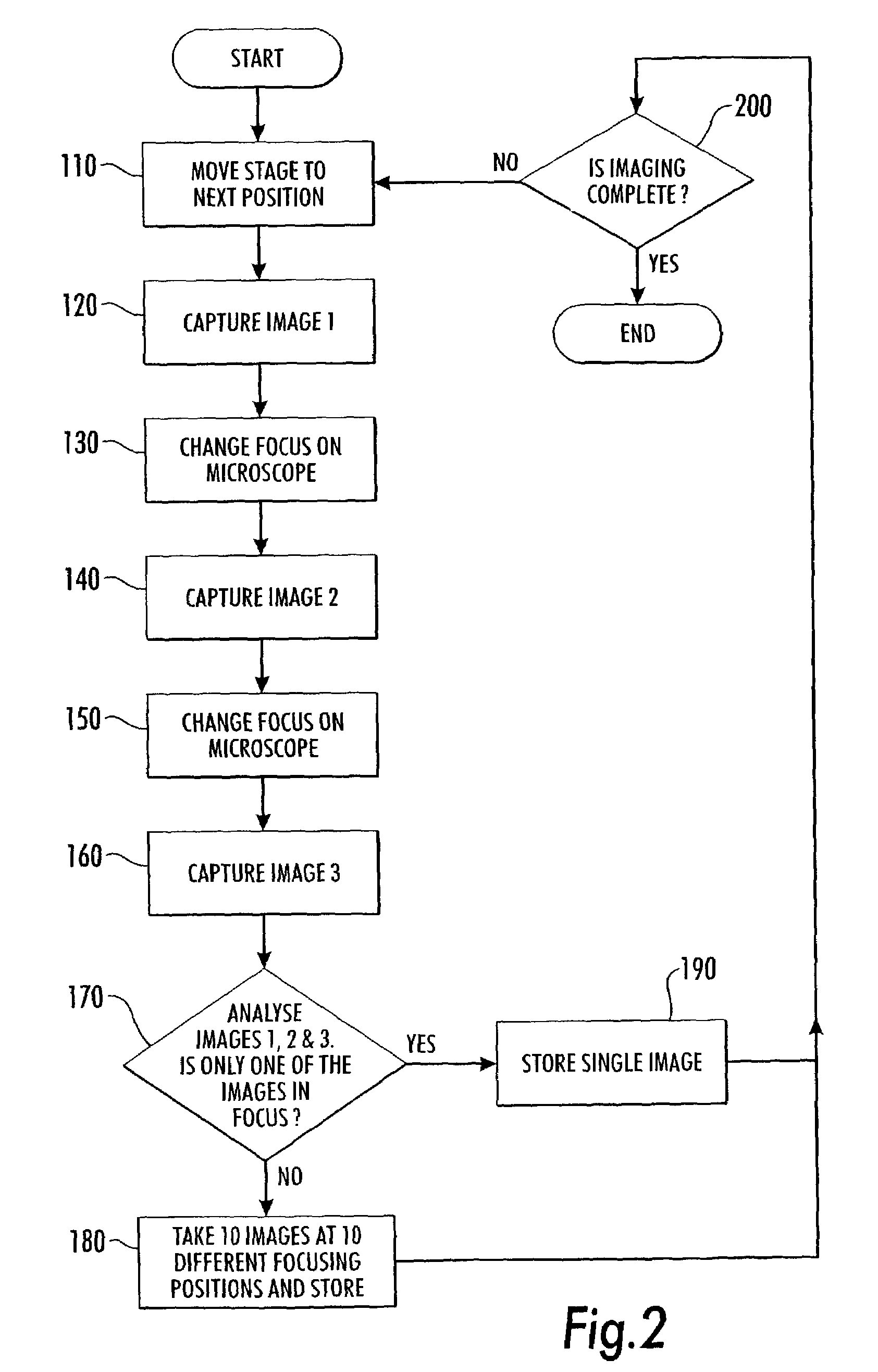
[0014]In this way, digital images at different focusing positions are only acquired in areas of the sp

Problems solved by technology

However, for certain types of specimen sample, there may

Method used


Image

  • Microscopy imaging system and method
  • Microscopy imaging system and method
  • Microscopy imaging system and method

Examples


Embodiment Construction

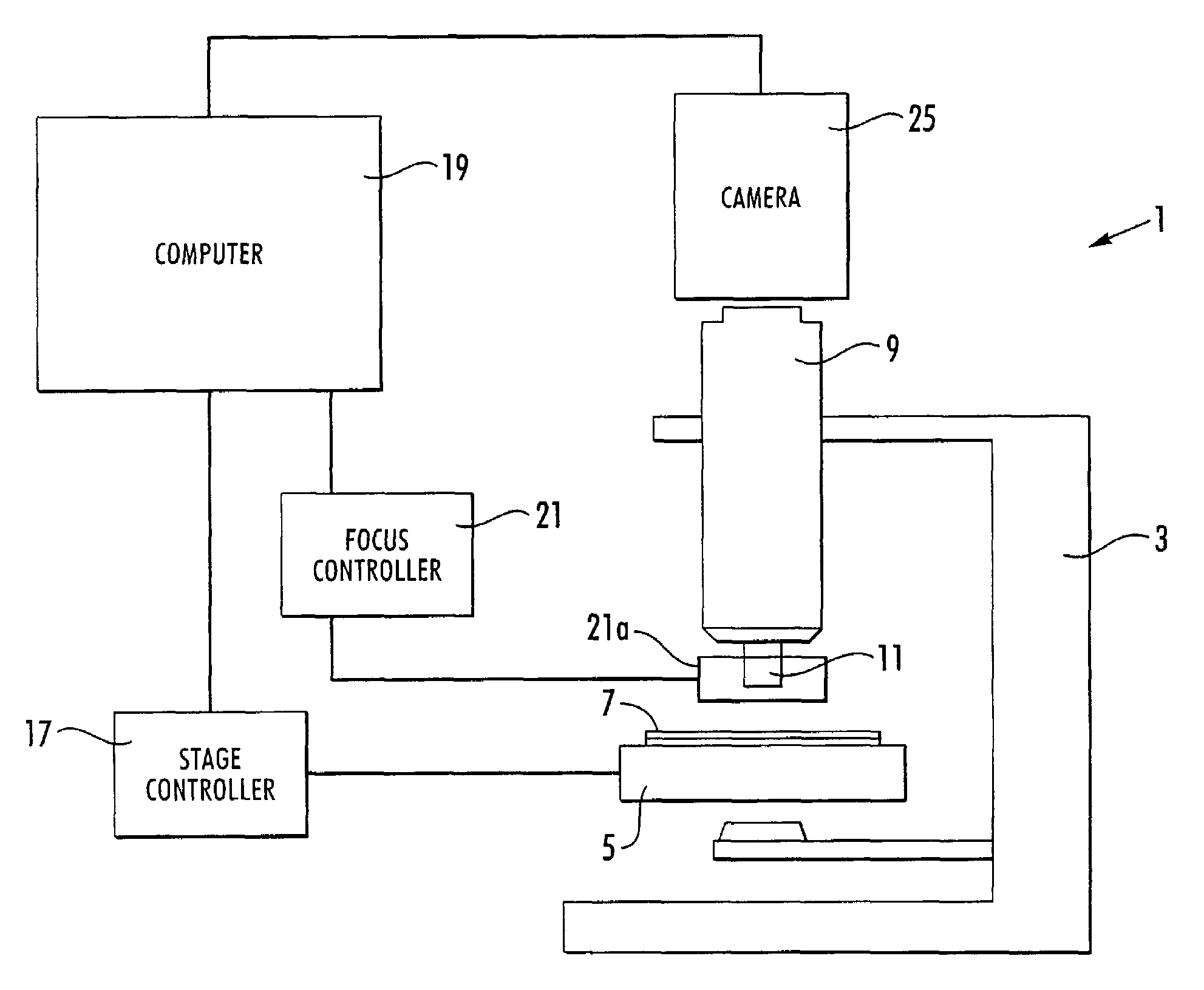
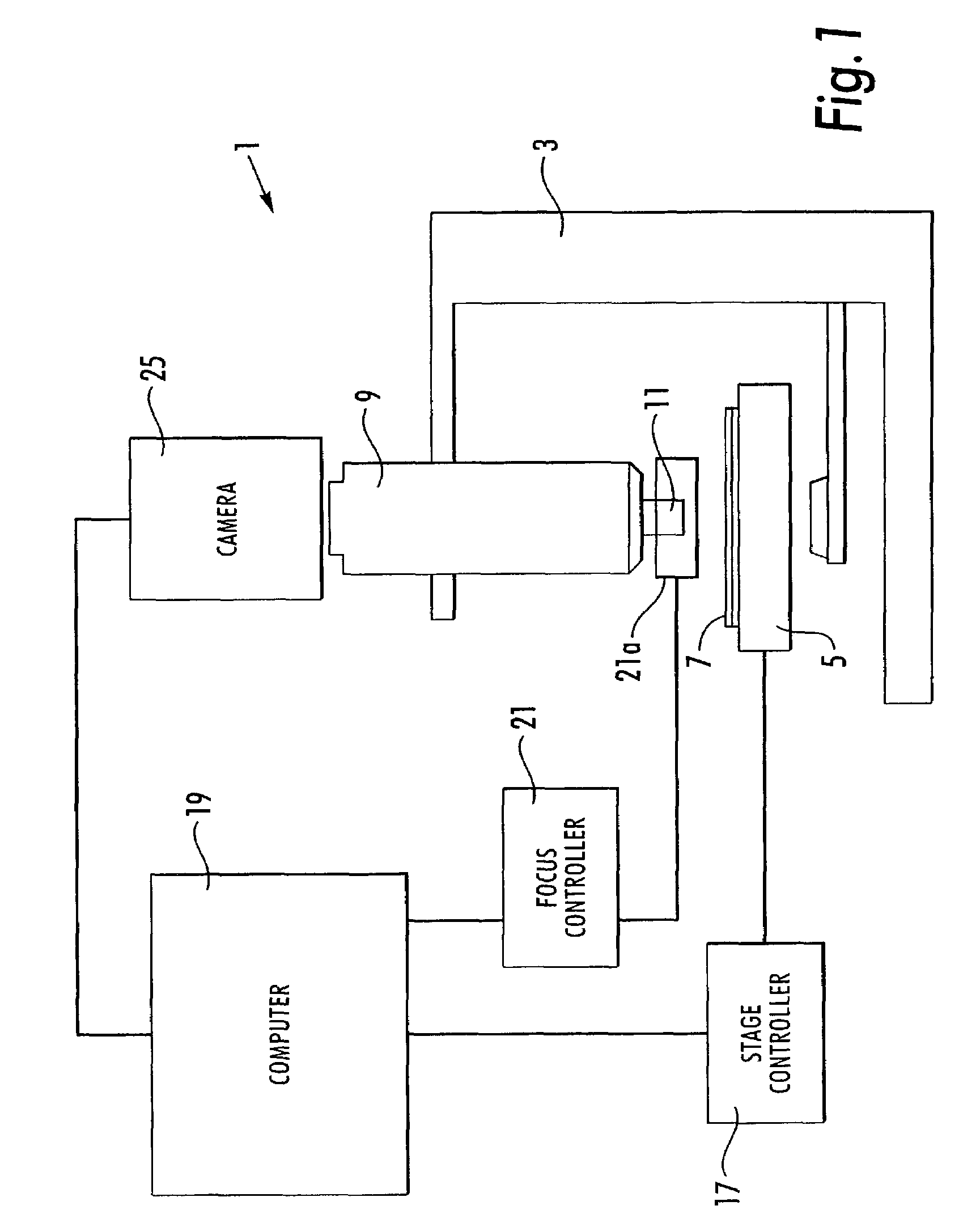
[0023]FIG. 1 illustrates a microscopy imaging apparatus 1 for use in accordance with a preferred embodiment of the present invention. The imaging apparatus 1 comprises a microscope 3 having a motorised microscope stage 5 for holding a microscope slide 7, and an objective lens 11. An example of a suitable microscope is the Axioplan 2 imaging microscope available from Carl Zeiss of Germany, using an Apochromat objective lens. The stage may be any suitable motorised stage having the necessary positioning accuracy, for example the Proscan stage available from Prior Scientific Instruments Limited of Cambridge, UK.

[0024]The motorised stage 5 is driven under the control of a stage controller 17, which controls the stage in response to instructions from computer 19. The motorised stage 5 is typically driven only in the x- and y-directions. In addition, focusing of the microscope is controlled by a piezo-electric controller 21 which controls a focusing device 21a. The focusing device 21a mov...


PUM


Abstract

A microscopy imaging system and method acquires digital images of a microscope specimen using a computer-controlled microscope and digital camera. A specimen on a microscope slide is placed within the microscope. The method comprises the steps of determining whether an area of the specimen contained within a microscope field of view has a thickness, and, if so, capturing digital images of the field of view at a plurality of depths of focus. In a preferred embodiment, the step of determining whether an area of the specimen contained within a microscope field of view has a thickness comprises the steps of: capturing a digital image of the field of view at two or more depths of focus; comparing the captured digital images, and determining that the specimen has a thickness, if the comparing step finds that the images are different. The method may be implemented in the form of a computer program.

Description

PRIORITY CLAIM[0001]The present application claims priority to UK Patent Application No. 02 034 06.4, entitled “Microscopy Imaging System And Method”, which application was filed on 13 Feb. 2002.BACKGROUND OF THE INVENTION[0002]1. Field of the Invention[0003]The present invention relates to a system and method for providing digital images of a microscope specimen, which images can be displayed on a display screen to resemble the images viewed through a microscope.[0004]2. Description of the Related Art[0005]The use of digital images of a microscope specimen, often referred to as “virtual microscope images”, is increasingly common in the field of diagnostic pathology, particular for remote diagnosis and training. The digital images of the specimen are typically acquired by “scanning” the specimen using a microscopy imaging system comprising a computer controlled microscope and digital camera, at one or more magnifications / resolutions.[0006]For example, a method of acquiring digital m...

Claims


Application Information

Patent Timeline
IPC IPC(8): H04N7/18G06K9/00G02B21/24G02B21/36
CPCG02B21/244G02B21/367
Inventor MADDISON, JOHN R
Owner SOURCE BIOSCI
Why Eureka
Try Eureka