Microscopic image capture apparatus and microscopic image capturing method
a microscopic image and capture apparatus technology, applied in the field of microscopic image capture apparatus and microscopic image capture method, can solve the problems of wasting long time in capturing the areas, taking a long time only to capture a high-precision, etc., and achieve the effect of efficiently capturing a high-precision and wide-angle field imag
- Summary
- Abstract
- Description
- Claims
- Application Information
AI Technical Summary
Benefits of technology
Problems solved by technology
Method used
Image
Examples
first embodiment
[0070]FIG. 3 is a flowchart of the actual capturing procedure used according to the basic principle according to the present invention. The capturing procedure is used by the control of the CPU 21 shown in FIG. 1. In FIG. 3, a low-powered objective lens is first used (S301).
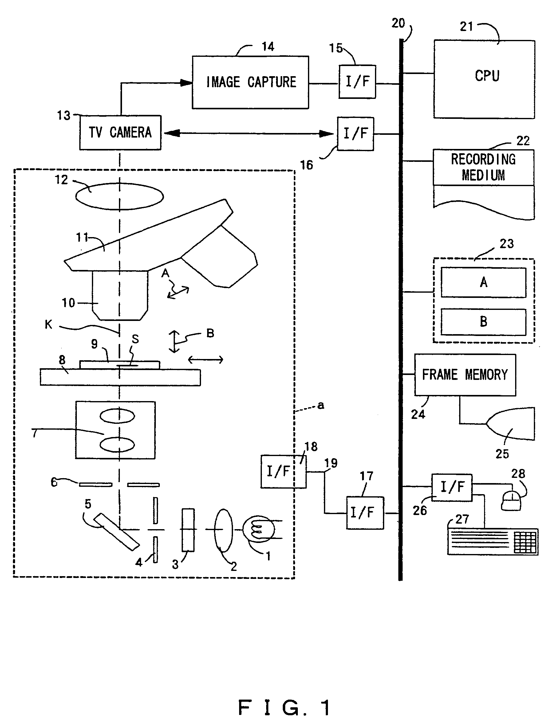
[0071]In this process, when the operator inputs the “start of capture” button (not shown in the attached drawings) on the control screen displayed on the monitor 25, the CPU 21 controls the rotation of the revolver 11 of the microscope unit indicated by the broken lines a shown in FIG. 1 to set the lowest-powered objective lens of the objective lens 10.
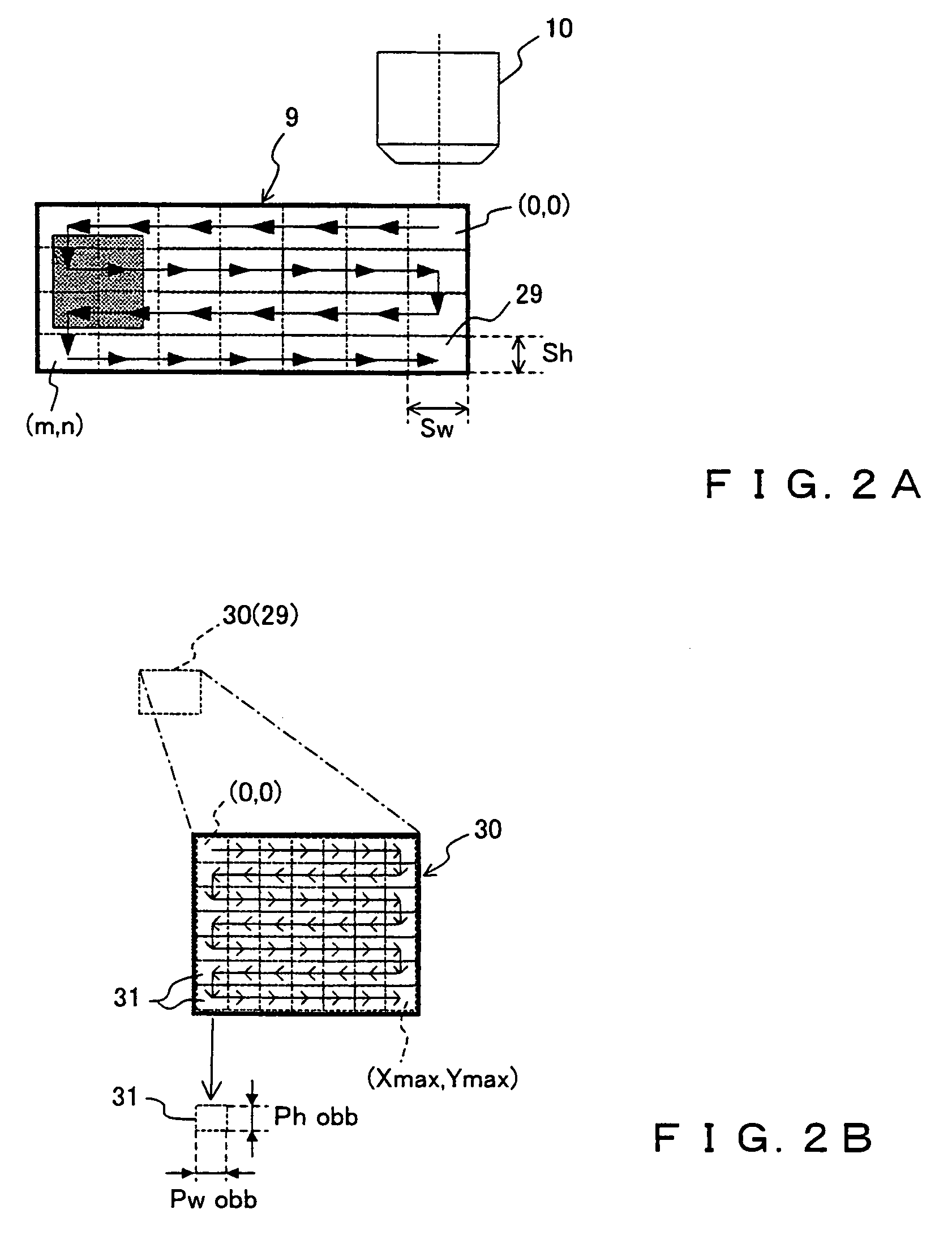
[0072]Then, the entire area of the slide glass 9 is scanned for each actual field size 29 (refer to FIG. 2A) of the low-powered objective lens using the above-mentioned low-powered objective lens to sequentially obtain the image information about a low-magnification actual field size of the stage coordinate sections (0, 0)˜(m, n) depending on the actual field size ...
second embodiment
[0114]In another method, the low-powered objective lens can be replaced with a macro capture device to simultaneously capture the entire area of the slide glass 9 and divide the captured image information into high-powered objective lens conversion pixel size sections. This method is described below as the present invention.
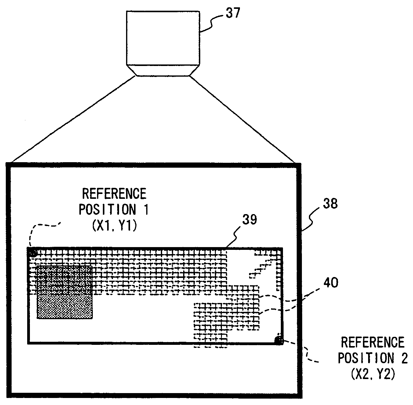
[0115]FIG. 6 is a simple schematic diagram showing the capturing method according to the second embodiment of the present invention. In this embodiment, the entire area of the slide glass 9 is captured using a macro capture device 37 provided separate from the microscope unit indicated by the broken lines a shown in FIG. 1, and a position in which a sample image is contained is detected from image information 39 about the entire area of the slide glass 9 obtained in a macro capture area 38, and the sample image presence / absence coordinates obtained when the position is converted in a field size 40 of a high-powered objective lens are detected, thereby generating ...
third embodiment
[0128]FIGS. 8A and 8B show the method of efficiently generating a, high-precision image in the present invention. FIG. 8A shows the sample S of the slide glass 9 being observed by any objective lens, the central observation position (Xc, Yc) of the sample S, and the field size Wobs×Hobs of the low-powered objective lens around the central observation position (Xc, Yc).
[0129]FIG. 8B(b) is an enlarged view of the field size Wobs×Hobs of a low-powered objective lens around the above-mentioned central observation position (Xc, Yc), and shows the state of dividing the field size Wobs×Hobs of the low-magnification by the field size 40 of a high-powered objective lens.
[0130]Assume that the observer operates an instruction button or a slide button on the control screen area (not shown in the attached drawings) of the monitor 25 using the keyboard 27 or the mouse 28 shown in FIG. 1 while observing the observation moving picture (partial image shown in FIG. 8A moved vertically and horizontall...
PUM
 Abstract
Description
Claims
Application Information
 - R&D
- Intellectual Property
- Life Sciences
- Materials
- Tech Scout
- Unparalleled Data Quality
- Higher Quality Content
- 60% Fewer Hallucinations
Browse by: Latest US Patents, China's latest patents, Technical Efficacy Thesaurus, Application Domain, Technology Topic, Popular Technical Reports.
© 2025 PatSnap. All rights reserved.Legal|Privacy policy|Modern Slavery Act Transparency Statement|Sitemap|About US| Contact US: help@patsnap.com


