Method and apparatus for extracting multi-dimensional structures using dynamic constraints

a dynamic constraint and multi-dimensional structure technology, applied in the field of medical imaging, can solve the problems of large volume datasets, inability to properly integrate background or proximate objects into the segmented structure, and existing techniques that may improperly exclude image data from the segmented structur

Active Publication Date: 2007-12-18
GENERAL ELECTRIC CO
View PDF10 Cites 17 Cited by
  • Summary
  • Abstract
  • Description
  • Claims
  • Application Information

AI Technical Summary

Benefits of technology

[0007]The present technique provides a novel approach to automatically segmenting a structure of interest in a volume data set. Automatic initialization may be performed to identify one or more initiation regions in the image data based upon templates that characterize properties of the structure of interest. The identified regions serve as starting points for the subsequent constrained growth process. Edge boundaries may be computed based on gradient data obtained by comparing each voxel to all adjacent voxels and assigning the maximum absolute gradient value as the gradient for the pixel. Constrained path finding may then be employed using the edge data and the initiation regions, as well as cross-sectional areas for regions in the image data. The constrained path finding process may employ dynamic constraints which are modified based upon local statistics, allowing candidate pixels to be selectively merged into the foreground region. The process may be iterated until the supply of candidate pixels is exhausted. The resulting volume comprises the segmented structure of interest.
[0008]In accordance with a first aspect of the technique, a method for determining edge voxels is provided. According to this aspect, a gradient for each of a plurality of voxels is calculated by determining a maximum absolute gradient component relative to each adjacent voxel. One or more edge voxels from the plurality of voxels is identified based upon a comparison of the gradients of each of the plurality of voxels to a th

Problems solved by technology

Though the manner in which a volume dataset is created may vary, with the above techniques representing only a sample, the analysis of volume datasets raise many common issues.
Existing segmentation techniques, however, may incorrectly incorporate background or proximate objects into the segmented structure due to poor recognition of edges or non-homogeneities in the image data.
Similarly, existing techniques may improperly exclude image data from the segmented structure due to poor edge recognition and/or non-homogeneities.
Such exclusions may potentially result in early or erroneous termination of the segmentation technique.
Furthermore, splits or mergers in the structure of interest may not be properly segmented by existing techniques due to these shortcomings.
In addition, anatomic and pathological variability

Method used


Image

  • Method and apparatus for extracting multi-dimensional structures using dynamic constraints
  • Method and apparatus for extracting multi-dimensional structures using dynamic constraints
  • Method and apparatus for extracting multi-dimensional structures using dynamic constraints

Examples


Embodiment Construction

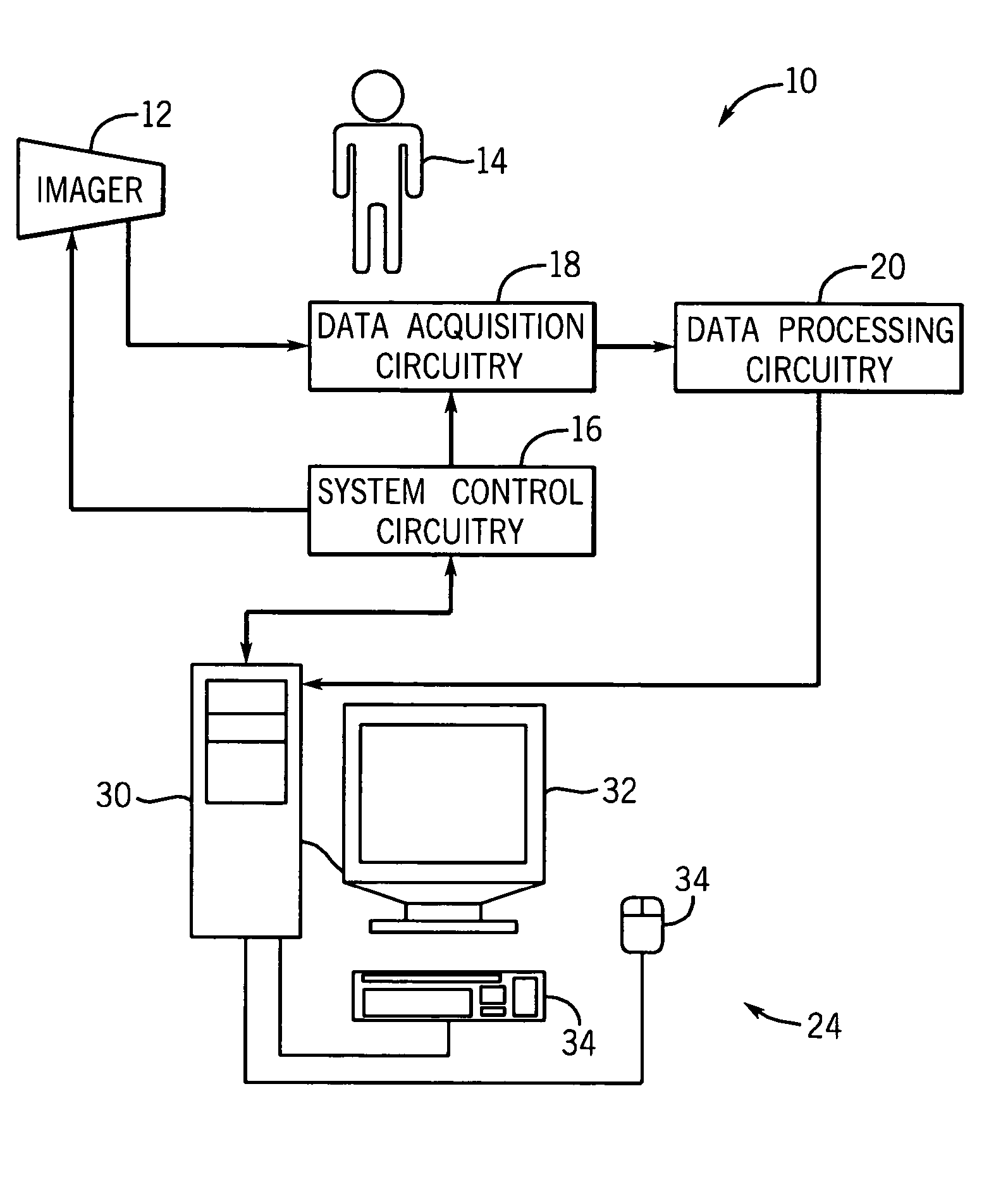
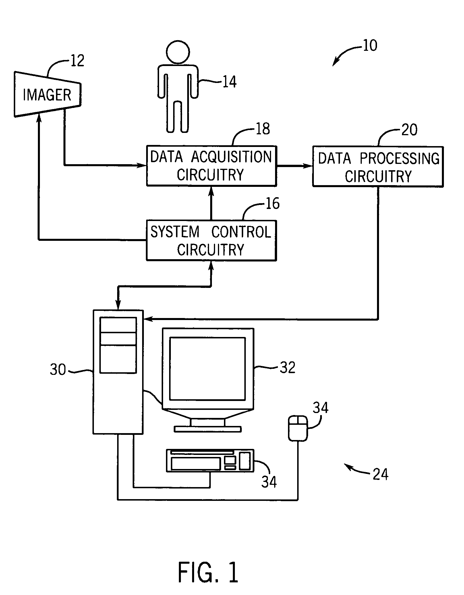
[0017]Turning now to the drawings, and referring first to FIG. 1, an exemplary imaging system 10 suitable for use in a medical context is depicted. Generally, the imaging system 10 includes some type of imager 12 that may operate in accordance with various physical principles for creating image data. In general, the imager 12 creates image data representative of regions of interest in a patient 14 in a digital medium. As will be appreciated by one of ordinary skill in the art, the imaging system 10 may comprise a computer tomography (CT), a magnetic resonance (MR), a positron emission tomography (PET), an electron beam tomography (EBT) or other modality of imaging system capable of generating a volume data set.

[0018]The imager 12 operates under the control of system control circuitry 16. The system control circuitry 16 may include a wide range of circuits, such as radiation source control circuits, timing circuits, circuits for coordinating data acquisition in conjunction with patie...


PUM


Abstract

A technique is provided for segmenting a structure of interest from a volume dataset. The technique identifies regions of the structure using templates having characteristics of the structure of interest. The identified regions may then undergo a constrained growth process using dynamic constraints that may vary based on local statistics associated with the identified structure regions. Edges within the volume may be determined using gradient data determined by evaluating the strongest gradient between each pixel and all adjacent pixels. The edge data may be used to prevent the constrained growing process from exceeding the boundaries of the structure of interest.

Description

BACKGROUND OF THE INVENTION[0001]The present invention relates generally to the field of medical imaging. Specifically, the invention relates to a technique for automatically extracting structures, particularly three-dimensional structures, from a volume dataset.[0002]A volume dataset may be generated by a variety of medical imaging technologies or modalities. For example, magnetic resonance imaging (MRI) modalities generate volume datasets by exposing a patient to magnetic field and by measuring the response and realignment of various magnetically susceptible molecule types within the body. By imposing weak gradient fields along the three-dimensional axes during the process, positional information may be obtained which allows a volume dataset to be constructed. Other technologies, such computed tomography (CT), measure the attenuation of streams of radiation through the body from numerous angles. The attenuation information may be combined and reconstructed to generate a volume dat...

Claims


Application Information

Patent Timeline
IPC IPC(8): G06K9/00G01T1/161A61B5/055A61B6/00A61B6/03G06T1/00G06T5/00G06T7/60
CPCA61B6/00G06T7/0012G06T7/0083A61B6/548G06T2207/20156G06T2207/30101G06T2207/10081G06T7/12
Inventor MALLYA, YOGISHAMULLICK, RAKESHSURYANARAYANAN, SRIKANTH
Owner GENERAL ELECTRIC CO
Why Eureka
Try Eureka