Systems and methods for providing automatic 3D lesion segmentation and measurements

Active Publication Date: 2008-02-19
SIEMENS HEALTHCARE GMBH
View PDF7 Cites 55 Cited by
  • Summary
  • Abstract
  • Description
  • Claims
  • Application Information

AI Technical Summary

Benefits of technology

[0009]Exemplary embodiments of the invention generally include 3D medical imaging systems and methods that provide automatic 3D segmentation of abnormal anatomical structures (lesions) such as colonic polyps, aneurisms, lung nodules, etc, and obtaining accurate size measurements and other descriptive characteristics of 3D segmented lesions that can be used for identification and classification. More specifically, exemplary embodiments of the invention generally incl

Problems solved by technology

In the field of medical imaging, the segmentation of abnormal anatomical structures (lesions) such as colonic polyps, aneurisms or lung nodules is a challenging problem because of the highly variable shape, texture, density and size of such lesions and their attachment to surrounding normal structures.
For example, the problem of colonic polyp segmentation is particularly difficult considering the complex shape of colon wall where prominent or thickened Haustral folds and retained stool often resemble the shape and density of polyps.
The drawback 2D polyp segmentation applied to the sub-images extracted from the axial slices or to triples of perpendicular planes slicing the sub-volume around the polyp candidate is that the three-dimensional

Method used


Image

  • Systems and methods for providing automatic 3D lesion segmentation and measurements
  • Systems and methods for providing automatic 3D lesion segmentation and measurements
  • Systems and methods for providing automatic 3D lesion segmentation and measurements

Examples


Embodiment Construction

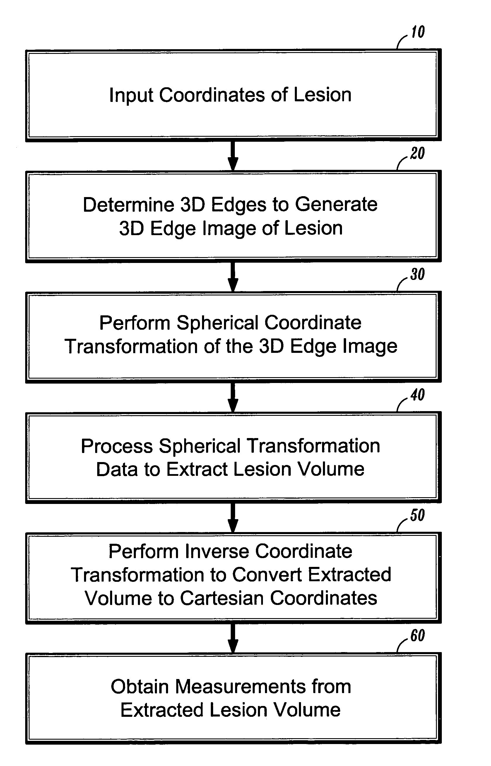
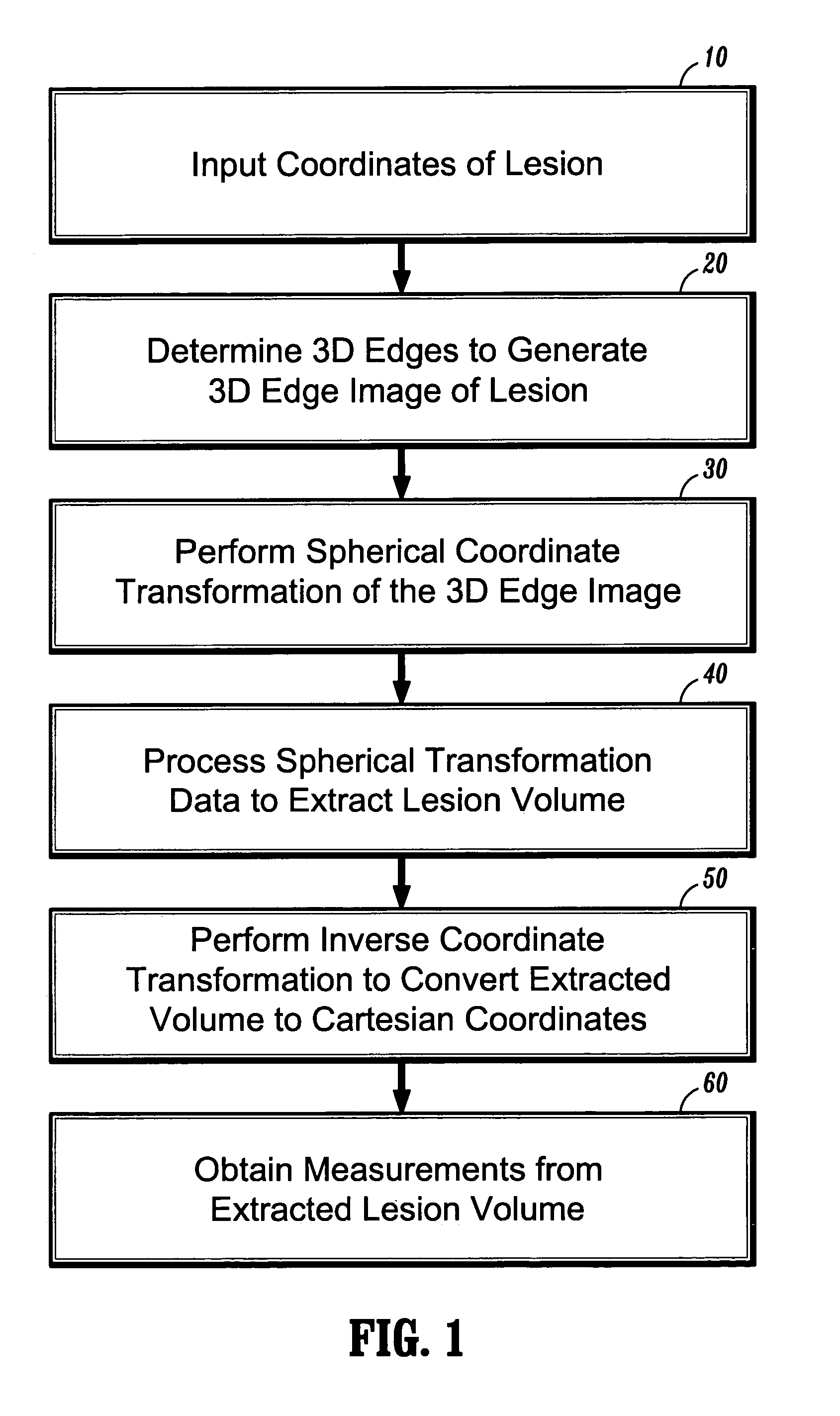
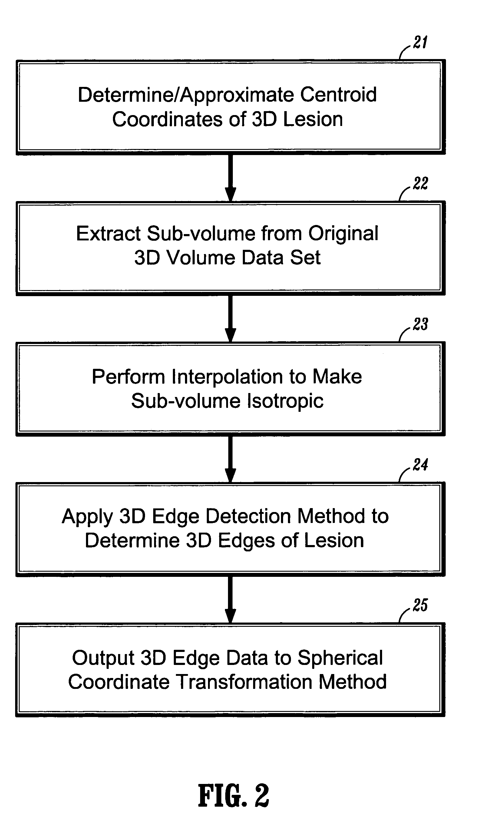
[0024]In general, exemplary embodiments of the invention as described herein include systems and methods for automatic 3D segmentation of abnormal anatomical structures such as colonic polyps, aneurisms or lung nodules, etc., in 3D medical imaging applications. In one exemplary embodiment of the invention described herein, a system and method for 3D lesion segmentation implements automated methods for spherical coordinate transformation of a 3D edge image and subsequent interpolation of a lesion surface, which enables an accurate determination of a boundary between a lesion of interest and surrounding normal anatomical tissue and structures.

[0025]Further, exemplary systems and methods according to the invention provide methods for automatically measuring various dimensions and characteristics of 3D segmented lesions, which can be implemented for purposes of identification or automatic classification based on the extracted lesion volume. In particular, systems and methods according t...


PUM


Abstract

Systems and methods are provided for automatic 3D segmentation of abnormal anatomical structures such as colonic polyps, aneurisms or lung nodules, etc., in 3D medical imaging applications. For example, systems and methods for 3D lesion segmentation implement a centroid-based coordinate transformation (e.g., spherical transformation, ellipsoidal transformation, etc.) to transform a 3D surface of the lesion from an original volume space into, e.g., a spherical or ellipsoidal coordinate space, followed by interpolation of the transformed lesion surface to enable accurate determination of a boundary between a lesion and surrounding normal structures.

Description

CROSS-REFERENCE TO RELATED APPLICATION[0001]This application claims priority to U.S. Provisional application Ser. No. 60 / 453,573, filed on Mar. 11, 2003, which is fully incorporated herein by reference.TECHNICAL FIELD OF THE INVENTION[0002]The present invention relates generally to a systems and methods for three-dimensional (3D) medical image rendering and visualization. More specifically, the present invention relates to medical imaging systems and methods for providing automatic 3D segmentation of abnormal anatomical structures (lesions) such as colonic polyps, aneurisms or lung nodules, as well as methods for obtaining accurate size measurements and other descriptive characteristics of segmented lesions, which are useful for identification and classification.BACKGROUND[0003]In the field of medical imaging, the segmentation of abnormal anatomical structures (lesions) such as colonic polyps, aneurisms or lung nodules is a challenging problem because of the highly variable shape, t...

Claims


Application Information

Patent Timeline
IPC IPC(8): G06K9/00G06K9/34G06T5/00G06T7/00G06T7/60
CPCG06T7/0083G06T7/602G06T2200/04G06T2207/10072G06T2207/20104G06T2207/30032G06T2207/30064G06T7/12G06T7/62
Inventor JEREBKO, ANNAKRISHNAN, ARUNBOGONI, LUCA
Owner SIEMENS HEALTHCARE GMBH
Why Eureka
Try Eureka