Multi modality imaging methods and apparatus

a multi-modal imaging and apparatus technology, applied in the field of image processing and image reconstruction, can solve the problems of complex and computationally intensive associated reconstruction algorithms, inability to perform medical imaging with relatively thick ct slices, and inability to achieve the image quality or resolution required to allow an operator to perform a medical diagnosis of the patient using the generated ct images

Active Publication Date: 2008-03-25
GENERAL ELECTRIC CO
View PDF22 Cites 28 Cited by
  • Summary
  • Abstract
  • Description
  • Claims
  • Application Information

AI Technical Summary

Problems solved by technology

The associated reconstruction algorithms are complex and computationally intensive.
While appropriate for generating the CT attenuation correction factors utilized to generate a PET image, this relatively thick CT slice is generally not useful for medical imaging purposes.
Therefore, while generating a relatively thick CT slice is beneficial to generate the attenuation correction factors used to generate the PET image, the relatively thick CT slice may not have the image quality or resolution to allow an operator to perform a medical diagnosis of the patient using the generated CT image.

Method used


Image

  • Multi modality imaging methods and apparatus
  • Multi modality imaging methods and apparatus
  • Multi modality imaging methods and apparatus

Examples


Embodiment Construction

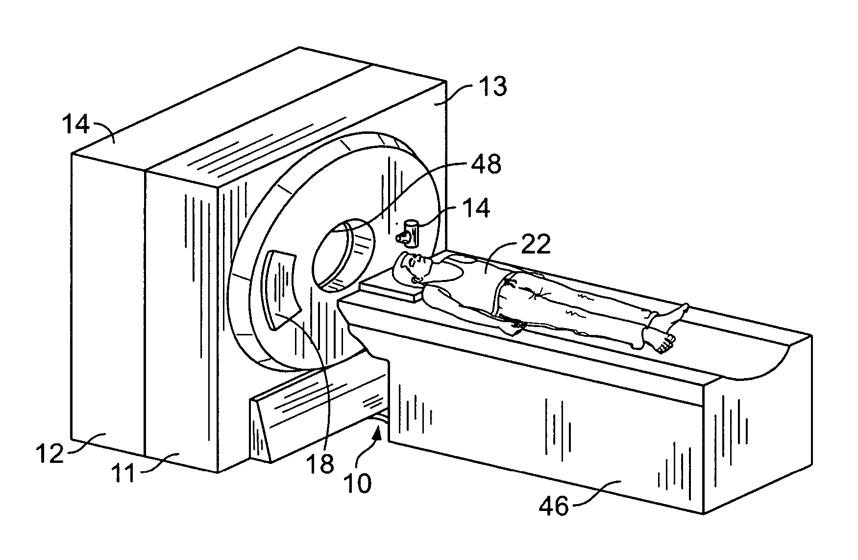
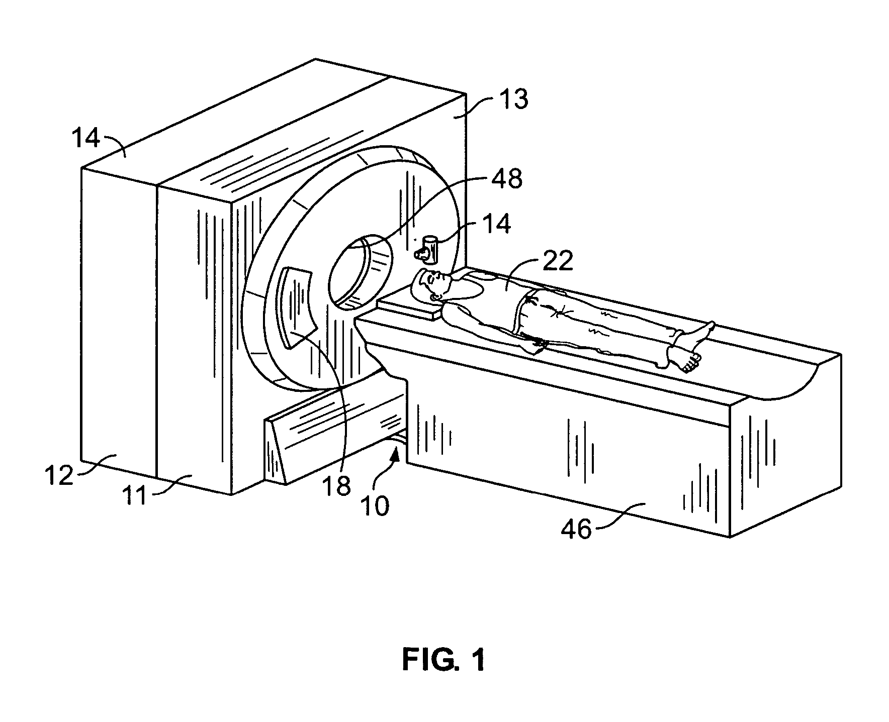
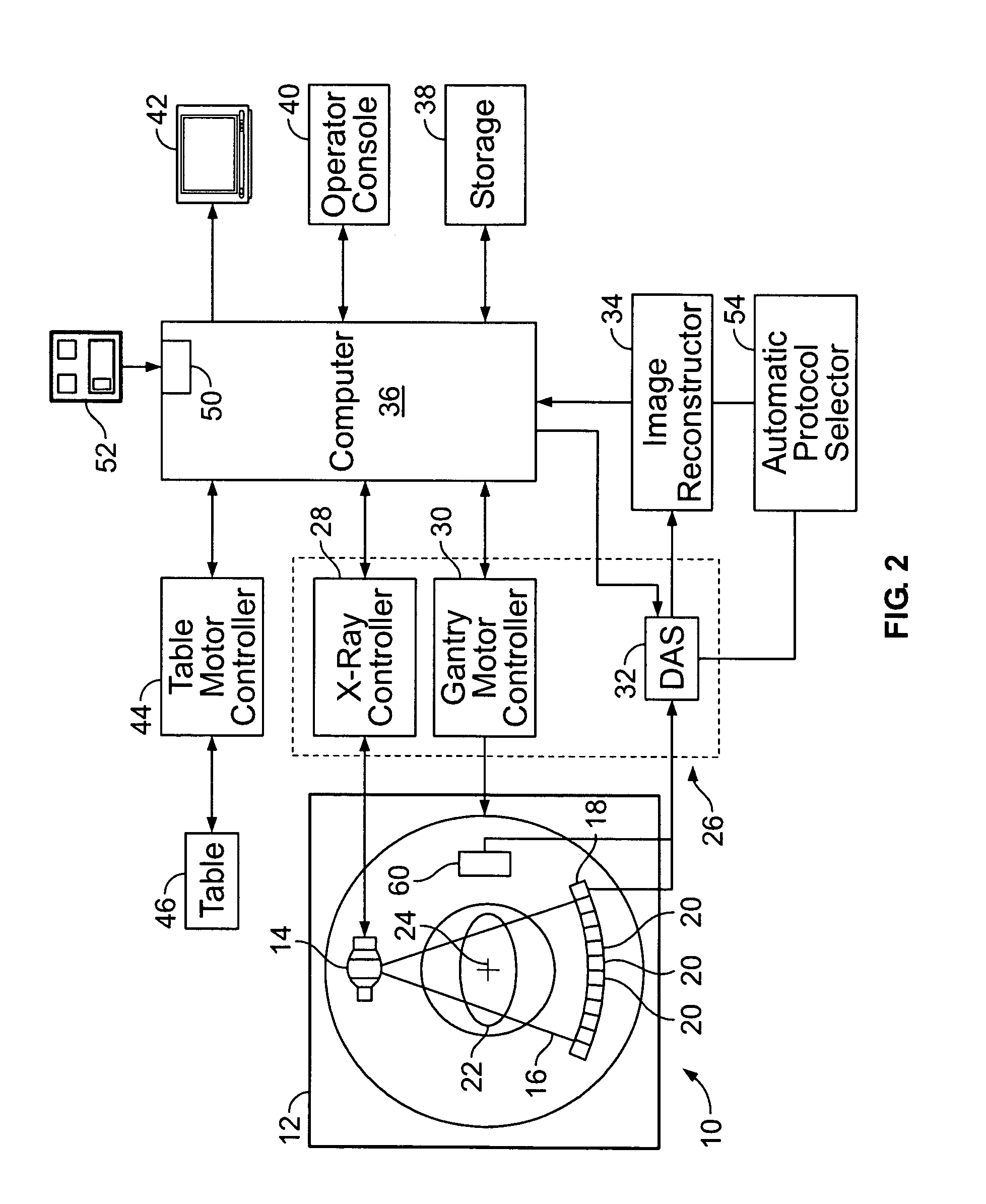
[0014]There is herein provided a system and method for correcting Positron Emission Tomography (PET) data. The apparatus and methods are illustrated with reference to the figures wherein similar numbers indicate the same elements in all figures. Such figures are intended to be illustrative rather than limiting and are included herewith to facilitate explanation. of an exemplary embodiment of the apparatus and methods of the invention.

[0015]At least some multi-modality systems have different fields of views (FOVs) for the different modalities. For example, a CT / PET system may have a CT FOV which is smaller than a PET FOV, and under some scanning conditions, portions of a patient may extend beyond the region measured by a CT detector, which may lead to image artifacts and an incomplete representation of the imaged object. Some known methods have been published that address the artifact reduction but not the imaging of the portion of the patient that is outside the CT FOV.

[0016]In such...


PUM


Abstract

A method for correcting emission data from Positron Emission Tomography (PET) or SPECT includes generating a plurality of computed tomography (CT) image data, selecting a portion of the CT image data, processing the selected CT data to generate a plurality of attenuation correction (CTAC) factors at the appropriate energy of the emission data, weighting the CTAC factors to generate an emission attenuation correction map wherein the weights are determined based on the axial location and the slice thickness of the CT data and the axial location and the slice thickness of the PET or SPECT data, and utilizing the generated attenuation correction map to generate an attenuation corrected PET image.

Description

BACKGROUND OF THE INVENTION[0001]This invention relates generally to image processing and image reconstruction based on acquired raw image data. In particular, improving the emission image quality in combined imaging systems such as CT-PET and CT-SPECT which use a CT image for multiple purposes including diagnostic review, fusion review with the PET, and calibration and correction of the PET data for effects including attenuation and scatter.[0002]Raw image data from various diagnostic medical systems, such as Computed Tomography (CT), Positron Emission Tomography (PET) systems, and Single Photon Computed Tomography (SPECT) systems are acquired for diagnostic purposes. The CT PET and SPECT systems are configured to support numerous scanning and reconstruction modes. The associated reconstruction algorithms are complex and computationally intensive. Users of the diagnostic medical systems desire an improvement in image quality, to minimize the time required to generate images based o...

Claims


Application Information

Patent Timeline
Patent Type & Authority Patents(United States)
IPC IPC(8): G01T1/166
CPCA61B6/032A61B6/037G01T1/1611
Inventor WOLLENWEBER, SCOTT DAVIDLONN, ALBERT H. R.STEARNS, CHARLES WILLIAMMIESBAUER, DIANE MARIELITTLEJOHN, KATHRYN MARY
Owner GENERAL ELECTRIC CO
Why Eureka
Try Eureka